ScholarWorks @ Georgia State University
ScholarWorks @ Georgia State University
Biology Dissertations Department of Biology
Summer 8-12-2014
Benzene and Beyond: Mechanisms of Novel Anaerobic Aromatic
Benzene and Beyond: Mechanisms of Novel Anaerobic Aromatic
Degradation Pathways in Geobacter daltonii
Degradation Pathways in Geobacter daltonii
Alison Kanak
Follow this and additional works at: https://scholarworks.gsu.edu/biology_diss
Recommended Citation Recommended Citation
Kanak, Alison, "Benzene and Beyond: Mechanisms of Novel Anaerobic Aromatic Degradation Pathways in Geobacter daltonii." Dissertation, Georgia State University, 2014.
https://scholarworks.gsu.edu/biology_diss/143
MECHANISMS OF NOVEL ANAEROBIC AROMATIC DEGRADATION PATHWAYS IN
GEOBACTER DALTONII
by
ALISON KANAK
Under the Direction of Kuk-Jeong Chin
ABSTRACT
Petroleum spills causes contamination of drinking water with carcinogenic aromatic
compounds including benzene and cresol. Current knowledge of anaerobic benzene and cresol
degradation is extremely limited and it makes bioremediation challenging. Geobacter daltonii
strain FRC-32 is a metal-reducing bacterium isolated from radionuclides and
hydrocarbon-contaminated subsurface sediments. It is notable for its anaerobic oxidation of benzene and its
unique ability to metabolize p-, m-, or o-cresol as a sole carbon source. Location of genes
involved in aromatic compound degradation and genes unique to G. daltonii were elucidated by
the start of the genome. Of particular note, G. daltonii has two copies of the bss genes, which are
responsible for the first step in anaerobic toluene oxidation. This bacterium is unique among the
family Geobacteraceae and other toluene degraders in this aspect. The subunits have 74%
identity to one another. The remaining genes in each operon are not identical. BssA was
upregulated when G. daltonii was grown on benzene and toluene while the grlA was upregulated
during growth on m-cresol. Toluene was accumulated during degradation of benzene by cell
lysate. Cells grown with benzene and toluene exhibited a similar protein profile compared to
cells grown with benzoate. These results indicate that benzene is converted to toluene and further
degraded via the toluene pathway.
Both the bss and grl operons were predicted to have sigma54-dependent promoters. This
was confirmed using 5' RACE and sequence analysis. E. coli transformed with the bss operon
were able to grow in the presence of toluene but lost this capability when sigma 54 was knocked
out. Growth was restored with complementation of sigma 54. The sigma 54-dependent signaling
system bamVW was upregulated in the presence of all aromatic compounds tested. These results
suggest that the bss operon is regulated via sigma 54-dependent mechanisms. This study
significantly contributes to anaerobic aromatic gene regulation which is crucial in effective oil
spill bioremediation.
MECHANISMS OF NOVEL ANAEROBIC AROMATIC DEGRADATION PATHWAYS IN
GEOBACTER DALTONII
by
ALISON KANAK
A Dissertation Submitted in Partial Fulfillment of the Requirements for the Degree of
Doctor of Philosophy
in the College of Arts and Sciences
Georgia State University
Copyright by Alison Elizabeth Kanak
MECHANISMS OF NOVEL ANAEROBIC AROMATIC DEGRADATION PATHWAYS IN
GEOBACTER DALTONII
by
ALISON KANAK
Committee Chair: Kuk-Jeong Chin
Committee: Adam Wilson
Eric Gilbert
Frank Cruz
Electronic Version Approved: 7-21-14
Office of Graduate Studies
College of Arts and Sciences
Georgia State University
ACKNOWLEDGEMENTS
First, I would like to thank my advisor, Dr Kuki Chin, for all of her help and guidance
during my research. I also thank my committee for serving as invaluable sources of wisdom and
advice while completing this work. To my husband, Jason Kanak, there are not words that
express my gratitude for your support and understanding throughout my graduate career. None of
it could have been completed without your help. You held me through the tears, celebrated with
my triumphs, and never minded late nights and weekends spent in the lab. Thank you so much
for your years of support. Finally, thank you to my fellow PhD student, Ryan Perry. You have
TABLE OF CONTENTS
ACKNOWLEDGEMENTS ... iiv
LIST OF TABLES ... vi
LIST OF FIGURES ... vii
1 INTRODUCTION... 1
2 MATERIALS AND METHODS ... 18
3 RESULTS ... 366
4 DISCUSSION ... 622
LIST OF TABLES
Table 1.Primers used in this study ... 31
LIST OF FIGURES
Figure 1 ... 9
Figure 2 ... 10
Figure 3………..10
Figure 4………..11
Figure 5………..12
Figure 6………..12
Figure 7………..15
Figure 8………..16
Figure 9………..38
Figure 10………39
Figure 11………42
Figure 12………49
Figure 13………50
Figure 14………52
Figure 15………53
Figure 16………....55
Figure 17………56
Figure 18………57
Figure 19………58
Figure 20………59
Figure 21………60
1 INTRODUCTION
Bioremediation and significance of the present study
Widespread use of petroleum in developed countries often results in the contamination of
natural environments, as occurred after the 2010 Deepwater Horizon oil spill (DWH) off the
Gulf of Mexico coast. The explosion resulted in the loss of 11 lives as well as the release of an
estimated 6.7x10⁵ mT of Macondo well 252 Crude oil into local waters and caused an estimated
$100-$200 million of lost revenue in local fisheries (1). The DWH is considered the largest
accidental marine oil spill in the history of petroleum industry. This contamination results in a
desperate need for finding economic means for its removal. Benzene, toluene, ethylbenzene, and
xylene isomers (BTEX), cresols and polycyclic aromatic hydrocarbons (PAHs) are found in high
quantities in contaminated sites, including the DWH site (2-4). Due to their higher water
solubility than other organic compounds monoaromatics spread to and pollute nearby
groundwater and sediments. Since groundwater is often a vital source for obtaining drinking
water, technologies to remove petroleum hydrocarbons from polluted areas are critically
necessary.
There are currently multiple methods by which BTEX can be eliminated from
groundwater including physical, chemical, and biological removal (5). Bioremediation is an
environmentally sound approach that is both more efficient and less costly than other more
acerbic methods (6). The continued pollution of our environment has tangible costs to human
health as many human diseases have preventable environmental contributors. Bioremediation is
the neutralization of environmental contaminants via the utilization of living organisms. These
organisms range from bacteria to archaea to plants with the strategies employed being as diverse
bioreactors. In situ bioremediation relies on stimulation of the natural environment to promote
degradative processes. Techniques include bioaugmentation, or the introduction of an organism
to a contaminated site for remediation, and biostimulation, the stimulation of growth conditions
of organisms already present at the contaminated site. Processes can involve the biodegradation
of contaminants as well as the adsorption of contaminants into cells for later removal. While
other strategies exist for remediating contaminated sites bioremediation provides a natural
approach. Because of this it requires less equipment, labor, or energy which translates to it being
less costly than other remediation approaches. Bioremediation is also appealing as it creates few
waste byproducts. Bioremediation is by no means a novel approach to remediating
contaminants, however it is evolving as new technologies are developed for remediating more
and more challenging pollutants (7).
Microorganisms or microbial products are frequently used in bioremediation strategies to
degrade waste or concentrate it for removal. In the case of organic pollutants microbes are able
to catabolize the waste, taking it from a high energy substance to a low energy one. This can
occur through novel xenobiotic pathways or also through co-metabolism in which natural
microbial catabolic enzymes degrade wastes as well. Microbes are also capable of concentrating
metals for more easy removal. Microbes extract metals from the environment both via direct
transport and secretion of metal-sequestering compounds such as siderophores. The
siderophore-metal complex is then absorbed into the cell and utilized. The focus of this work is to understand
an organism present at contaminated sites so as to stimulate its growth and natural degradative
Anaerobic benzene-oxidizing microorganisms
Because a lot of the contaminants needing to be removed are buried deep within the soil,
understanding anaerobic degradation of organic pollutants has become much more important in
recent years. Anaerobic degradation of benzene involves reduction of the very stable benzene
ring, which is a more complex process than oxygenation. It is known that benzene is rapidly
degraded and completely oxidized to carbon dioxide under iron-reducing, sulfate-reducing, and
methanogenic conditions (3, 8, 9). No universal pathway has currently been demonstrated in
anaerobic benzene degradation. Recent studies have shown that in the archaeon Ferroglobus
placidus benzene is first activated via carboxylation and conversion to benzoate while in the
anaerobe Geobacter metallireducens benzene is activated via hydroxylation and conversion to
phenol (10, 11).
Family Geobacteraceae and anaerobic respiration
Under iron-reducing conditions the most prevalent family of bacteria found are the family
Geobacteraceae (12). For example, they were found to represent 85% of the active
microorganisms oxidizing acetate in an Fe(III)-reducing rice paddy soil (13). Geobacter species
are part of the family Geobacteraceae within the Bacteria domain, phylum Proteobacteria, class
Deltaproteobacteria, and order Desulfuromonadales. These bacteria are Gram-negative,
non-spore-forming, curved rod-shaped cells capable of motility (14-18). Twenty seven published
species of the family Geobacteraceae have been reported to date (19). Many of the members of
this family are obligate anaerobes capable of heavy metal reduction with oxidation of acetate
These organisms play an important role in the biogeochemical cycling and the microbial
ecology of sediments by reducing insoluble Fe(III) and releasing soluble Fe(II). Geobacter
species have also been found to play a role in the reduction of U(VI) in sediments resulting in the
differential migration of this species and other radionuclides. Because of their ability to couple
oxidation of organic compounds to the reduction of Fe(III), Geobacter species occupy key niches
in anaerobic environments. Another factor could be their “remarkably low” maintenance energy
requirement. This allows them to live in such varied environments as petroleum-contaminated
aquifers, groundwater contaminated with landfill leachate, sediments contaminated with organic
acids and radionuclides such as uranium and technetium as well as environments contaminated
with arsenates and chlorinated compounds. These organisms can also be found on the surface of
electrodes. In addition to iron, some of the family Geobacteraceae can also reduce Mn(IV),
U(VI), Tc(VII), Pu(VI), elemental sulfur and humic substances. Some are able to utilize
hydrogen or ethanol as electron donors in addition to small organic acids or aromatic compounds
(13).
In order to study the physiology and genetics of the family Geobacteraceae it is
imperative to obtain pure cultures. The goal is to have isolates that are good representatives of
this group that are abundant in the environment of interest. The first species discovered was
Geobacter metallireducens, and was isolated from freshwater sediment by Lovley and Philips
(1988) (11). This organism has served as the focus for much Geobacter study and subsequent
knowledge. Geobacter sulfurreducens is another model organism that has been extensively
studied physiologically and genetically and was isolated from hydrocarbon-contaminated surface
sediments (10, 13). Coppi and colleagues established a genetic system for G. sulfurreducens
genetically manipulated. Because of this it has been the source of most of the knowledge about
physiology and gene regulation, in family Geobacteraceae. G. sulfurreducens was also the first
to be found to utilize hydrogen as an electron donor. One laboratory strain of G. sulfurreducens,
KN400, was designed to study growth of these organisms on electrodes. This organism produces
a greater amount of current than others and also reduces Fe(III) oxides much more rapidly. This
strain is also motile, whereas other lab strains have lost this ability (13).
One of the unique features of Geobacter species is their ability to utilize a wide variety of
electron acceptors. Soluble electron acceptors such as nitrate, fumarate, and chlorinated
compounds can be reduced intra-cellularly. These organisms are also capable of utilizing a
number of external electron acceptors such as soluble (ferric citrate) and insoluble iron
(ferrihydrite) and manganese. Fe(III) is the most common electron acceptor for Geobacter
species and it can be present in the form of crystalline hydroxides and phyllosilicates within clay
fractions. Fe(III) oxides are rarely encountered in the environment and so serve as poor electron
acceptors during lab cultivation. Smaller particle sizes of Fe(III) oxides may accelerate
reduction. In addition to insoluble Fe(III) compounds Geobacter species can also be cultivated
using soluble chelated forms of Fe(III) such a ferric citrate. However, these cells appear to have
different physiologies than their insoluble Fe(III)-reducing counterparts. Geobacter species have
also been found to be able to pass electrons to graphite electrodes, thus using them as terminal
electron acceptors and harvesters of electricity. Geobacters were the first organisms found to be
able to sustain growth coupling oxidation of organic matter with electron transfer to electrodes.
These organisms are so adept at this process that they are frequently the most abundant
organisms found after starting with mixed inoculum. Geobacter species are also able to transfer
by the interspecies hydrogen transfer between the syntrophic species or possible the interspecies
transfer of electrons (13).
Acetate is a key extracellular intermediate of the degradation of organic matter
anaerobically. It is a conserved ability for Geobacter species to be able to oxidize acetate as an
electron donor and carbon source via the TCA cycle. In addition to acetate these organisms can
also oxidize other short fatty acids, alcohols, hydrogen, and aromatic compounds as electron
donors. Geobacter species are frequently found in petroleum-contaminated environments where
aromatic hydrocarbons are being removed. G. metallireducens was the first Geobacter species
found to degrade aromatic compounds such as toluene and has also been implicated in the
anaerobic degradation of other aromatic compounds as well. Common to any Geobacter species
capable of degrading any aromatic compound is the ability to also degrade benzoate. Pathways
involved in anaerobic aromatic degradation have been predominantly studied in G.
metallireducens. All monoaromatic pathways converge on a Benozyl-CoA intermediate.
Geobacter species utilize a class II benzoyl Co-A reductase that is not ATP-dependent like
others are. The eight gene cluster BamBCDEFGHI is believed to code for this reductase and this
protein is believed to be membrane-bound. Additional reactions further oxidize benzoyl CoA to
3-hydroxypimelyl-CoA and eventually acetyl-CoA and CO2. The pathways for anaerobic
toluene and phenol degradation elucidated in G. metallireducens are similar to that in the
facultative anaerobic bacterium Thauera aromatica (13, 24).
Geobacter species utilize complex gene regulatory pathways in order to optimally
respond to their local environment. Bacteria rely on the use of multiple sigma factors as an
upstream means of regulating gene expression with each sigma factor recognizing different
differ from other bacteria is the heavy use of the “alternative sigma factor”, sigma 54. Deletion
of this gene is lethal to Geobacter species and so is implicated in regulating a wide variety of
Geobacter genes. The presence of 151 putative transcription factors and an uncommonly large
number of two-component signaling cascades demonstrates the diversity of signals perceived by
G. sulfurreducens. The transcription factor HgtR, for example, is induced when hydrogen is the
sole electron donor and represses genes involved in classical central metabolism such as citrate
synthase. HgtR is involved in the sigma 54 regulatory networks and is thought to be an
enhancer binding protein for this sigma. The genes BamVW are an example of a two-component
regulatory system that is also part of the sigma 54 network. BamVW are coded in one operon,
not always observed in Geobacter species, and are induced in the presence of aromatic
compounds such as benzoate and p-cresol. Most of the domains in the sensor proteins of a
two-component system in Geobacters are unique or do not share much homology with other bacterial
systems (13).
Geobacter species are motile. It aids in the acquisition of external nutrients in sediments
and chemotaxis. Interestingly, Geobacter species have multiple chemotaxis regulatory systems
whereas E. coli only has one. Some of these systems regulate flagellar motility while others are
responsible for regulating extracellular protein expression and cytochromes. These bacteria code
a very high number of chemoreceptor genes as well which are randomly located throughout the
genome (13).
Geobacter daltonii strain FRC-32
A member of the genus Geobacter, Geobacter daltonii strain FRC-32, was isolated from
Oak Ridge Field Research Center (ORFRC), Oak Ridge, Tennessee (25). Like other
Geobacteraceae, cells of G. daltonii are Gram-negative, non-spore-forming and curved rods.
Due to the large number of cytochromes present, also characteristic of its genus, G. daltonii
cultures take on a distinctive pink color during cultivation and form biofilms when growth
conditions are optimal. Its optimal growth temperature is 30°C and growth is best when cultures
are not agitated.
G. daltonii’s genome is larger than those of other Geobacter species at 4.3 kb in length
(22). This could be due to gene duplications present within its genome. For example, G. daltonii
encodes multiple copies of benzoate oxidation genes as well toluene oxidation genes. The
organization of aromatic degradation genes in G. daltonii is similar to G. metallireducens in that
both group these genes together into aromatic islands. Both of these organisms also have their
toluene degradation genes farther away from the large island. However, whereas G.
metallireducens has its aromatics island close to the midpoint of the genome G. daltonii’s
aromatics island is close to the start of the genome. According to 16S rRNA gene sequence
analysis, G. daltonii is most closely related to G. uraniireducens, however, it is unique from this
organism in its ability to degrade aromatic compounds (22).
Anaerobic oxidation of aromatic compounds
Benzoate is the simplest of the aromatic compounds that G. daltonii can degrade. The
pathway for its degradation has been studied extensively in anaerobes. Benzoate is first
activated to Benzoyl CoA via an ATP-dependent benzoate-CoA ligase (26). This reaction also
releases AMP and PPi. The ligase is coded by the bamY gene in G. metallireducens and G.
pathway proceeds. This pathway involves linearization of the stable aromatic ring and
subsequent beta oxidations. While dearomatization is achieved in other anaerobes by the
benzoyl CoA reductase this enzyme has not yet been identified in Geobacteraceae (27) and
therefore some alternate strategy must be employed. Following dearomatization is a series of
beta oxidation reactions resulting in the production of an aliphatic C7-dicarboxyl-CoA compound
(26) (Fig. 1).
Figure1. Anaerobic degradation pathway of aromatic compound benzoate (adapted from Carmona et al. (26)
Phenol is a substrate formed by various natural processes as well as environmental
contamination. The pathway involved in degradation of phenol has been determined in G.
metallireducens. Phenol is first converted to phenylphosphate by the phenylphosphate synthase
(pps). This enzyme is made of three subunits and transfers a phosphoryl group from ATP to the
hydroxyl group of phenol. Next, phenylphosphate carboxylase (ppc) catalyzes the conversion of
phenylphosphate to 4-hydroxybenzyl alcohol (Fig. 2). The pps and ppc operons are both found
Figure 2. Anaerobic degradation pathway of phenol (adapted from Carmona et al.) (26)
The first step of anaerobic toluene degradation is the enzymatic addition of toluene’s
methyl group to the double bond of fumarate, resulting in benzylsuccinate biosynthesis (28).
This step is catalyzed by a radical enzyme called benzylsuccinate synthase (bss). An acetyl CoA
group is then added to the hydroxyl group to form benzylsuccinyl CoA. This compound is then
further oxidized to E-phenylitaconyl CoA and Hydroxymethylphenyl succinyl CoA until
eventually being oxidized again to form benzoyl CoA. This compound then undergoes beta
oxidation and ring cleavage (Fig. 3) and eventually becomes CO2 and acetyl CoA via traditional
anaerobic mechanisms (29).
Figure 3. Anaerobic degradation pathway of toluene (adapted from Carmona et al.) (26).
Multiple primary activation steps have been proposed for benzene degradation.
Conversion to benzoate via carboxylation (30, 31), to phenol via hydroxylation (30), and to
[image:20.612.83.541.475.571.2]whether one of these pathways is used during anaerobic benzene oxidation by G. daltonii
transcript levels were measured for key genes in each pathway. This approach has been
successful previously with other metabolic pathways in other Geobacter species both in pure
culture and in the environment (33, 34).
It has been reported that all three cresol isomers can be degraded anaerobically in
contaminated environments (35). Also, apparent from previous studies is that each of these
isomers is degraded by different metabolic pathways. In G. metallireducens p-cresol is first
converted to p-hydroxybenzyl alcohol and then eventually is degraded via the classical benzoate
[image:21.612.112.464.350.424.2]degradation pathway (36). This same pathway is also observed in denitrifyers (Fig. 4) (7).
Figure 4. Anaerobic degradation pathway of p-cresol (adapted from Carmona et al.) (26)
The accumulation of 3-hydroxybenzoate degradation intermediates during the anaerobic
degradation of m-cresol is also observed in denitrifying bacteria and this compound is then
Figure 5. Anaerobic degradation pathway of 3-hydroxybenzoate as part of the proposed
m-cresol degradation pathway (adapted from Carmona et al.) (26)
The o-cresol pathway also does not appear to funnel into the benzoate degradation
pathway like the p-cresol pathways does. Under methanogenic conditions a consortia produces
metabolites indicative of carboxylation para to the hydryoxyl group followed by subsequent
dehydroxylation and formation of methylbenzoic acid (37) and under denitrifying conditions a
Pseudomonas-like strain S100 proceeds through a 3-methylbenzoyl CoA intermediate (Fig. 6)
(38, 39).
Figure 6. Anaerobic degradation pathway of 3-methylbenzoate as part of the proposed degradation pathway of o-cresol (Adapted from Juarez et al.) (39)
No organism to date has been reported to be capable of degrading all three cresol
Transcriptional Regulation in bacteria and G. daltonii
The initiation of transcription in bacteria requires the recognition of a specific promoter
and the melting of that region to provide a single stranded DNA template for RNA synthesis.
The presence of multiple sigma factors within bacteria lends a level of transcriptional control as
these sigma factors direct the RNA polymerase to appropriate promoters with great specificity.
While both sigma 70 and sigma 54 direct RNA polymerase, this is the extent of similarity in their
actions (Fig. 7A). Sigma 70 typically contacts its target promoter after it has already bound the
polymerase while sigma 54 binds to promoters before recruiting the polymerase. Sigma 70 does
not need additional accessory proteins to initiate transcription while sigma 54 is reliant on
accessory proteins, termed enhancer binding proteins, to initiate transcription in a manner similar
to eukaryotic RNA transcription. These proteins aid in the start of transcription by facilitating
the opening of the DNA at the promoter. While this is achieved spontaneously by the sigma 70
holoenzyme, sigma 54 binds its promoter so tightly that formation of an open complex can only
occur by hydrolyzing ATP (38). These activators bind upstream of the target promoter, typically
around 80-150bp away. These regions are termed upstream activator sequences or enhancer
sites. Because these regions are so far away from the promoter, DNA bending is required for the
activator to interact with the holoenzyme. To accomplish this integration host factor is often
recruited. The location of the promoter sequence also differs for each sigma factor. Rather than
identifying the classical -10/-35 promoter associated with sigma 70, sigma 54 recognizes a
-12/-24 promoter sequence. There is also a much lower degree of sequence conservation in the sigma
54 promoter. A consensus of TGGCA—N7—TGC(t/a)(t/a) is recognized for sigma 54 with GG
at -24 and GC at -12 being the most critical sites to maintain function (40-43). Mutation of ether
composition of the sigma factors differs from each other also. Whereas sigma 70 has 4
conserved regions, sigma 54 only has three. Region I is typically involved in binding of the -12
region of the promoter as well as the activator protein. Region II is highly variable amongst
bacterial species both in length and necessity for sigma activity. Region III is responsible for
promoter recognition and binds to the majority of the promoter, including the -24 element.
The effector binding proteins typically have three domains to their structure. The
N-terminal region is termed the “regulatory” domain and plays a part in signal perception. The
central domain is the location of the ATPase activity of the activator as well as binding to sigma
54. This region is critical to the activator function. Finally, the C-terminal domain contains a
helix-turn-helix responsible for DNA binding. While the central domain is conserved across
activator proteins, either of the flanking regions varies amongst bacteria. The enhancer binding
protein typically functions as hexamer or heptamer ring (38) (Fig. 7B).
Also, known as the “alternate” sigma factor, sigma 54 is known to regulate genes
involved in alternative metabolisms, and has been found to regulate aromatics related genes in
Geobacter species as well as Pseudomonas putida (44, 45). It also plays a role in the regulation
of virulence, biofilm formation, and quorum sensing genes in other bacteria (41, 46).
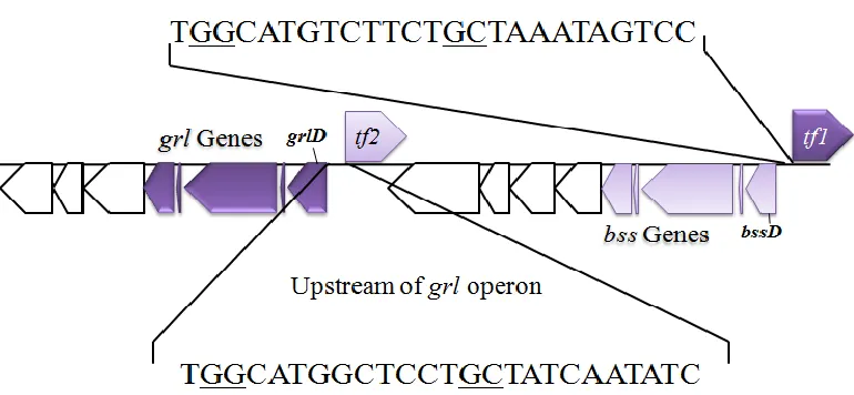
Immediately upstream of the operons encoding the bss genes in G. daltonii and oppositely
oriented are genes annotated as a “Sigma 54-specific transcriptional regulator” (Geob_2441 and
Geob_2451; Fig. 8). Due to their proximity and orientation, these regulators may be responsible
for regulating the transcription of the bss operons. The G. daltonii genome also contains the
genes bamVW, which have been demonstrated to be activated during degradation of toluene and
p-cresol in G. metallireducens (44). The py promoter recognized by bamV was also
played by sigma 54 in other aromatics pathways and the proximity of sigma-54 type
transcriptional regulators, it is possible this sigma is involved in regulating transcription of the
bss operons.
A
Figure 7. A. Diagram showing differences between the classical sigma 70 and the
alternative sigma 54. B. Diagram showing action of effector binding proteins, IHF, and sigma
54 (adapted from Bush et al.) (38)
Figure 8. Diagram showing coding and upstream regions of bss
(Geob_2442-Geob_2450) and grl (Geob_2431-Geob_2441) genes in G. daltonii. Dark purple arrows are grl
genes and light purple arrows are bss genes.
Aims of this study
Aim 1: How does G. daltonii activate the benzene ring for further degradation?
Before the stable benzene ring can be cleaved open for further utilization it must be
activated via the addition of a functional group. Multiple primary activation steps have been
proposed for benzene degradation. The anaerobic mechanism of activation of the stable benzene
ring was different between G. metallireducens and F. placidus, which are two known anaerobic
[image:26.612.117.502.241.419.2]benzene oxidation it is uncertain which mechanism will be utilized by G. daltonii when
oxidizing benzene anaerobically. The present study aimed to determine how G. daltonii
activates benzene and which degradation pathway will subsequently be used. In order to
determine which pathway is utilized by G. daltonii this study 1) Examined expression of genes
uniquely critical to the phenol, benzoate, and toluene pathways during growth of G.
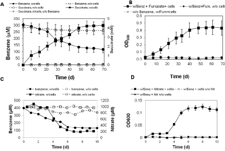
daltonii on benzene, 2) Measured formation of downstream metabolites formed during
anaerobic degradation of benzene by G. daltonii, and 3) Compared protein profiles of cells
grown using toluene, benzoate, or benzene as the sole carbon source.
Aim 2: Are the bss operons in G. daltonii regulated via sigma 54 dependent mechanisms?
The bss operons in G. daltonii are preceded by genes annotated as sigma 54 type
transcriptional regulators. Analysis of the upstream regions of bss with bioinformatics programs
also fails to identify the classical -35/-10 sequence of TTGACA/TATAAT typically observed in
promoters regulated by the vegetative sigma, sigma 70. Sigma 54 has been demonstrated to help
regulate expression of toluene degradation in Pseudomonads as well as benzoate degradation in
G. metallireducens. Because other aromatic operons are controlled via sigma 54 dependent
mechanisms, it is possible that the toluene degradation genes in G. daltonii are as well. In order
to determine the role of sigma 54 in regulation of bss the present study: 1) Defined the
promoter regions of bss1 and 2) Tested whether the absence of sigma 54 halts transcription
2 MATERIALS AND METHODS
Chemicals, gases and source of bacterium
All chemicals used in this study were of analytical grade and obtained from
Sigma-Adlrich (St. Louis, MO), Acros Organics (New Jersey, US) and Thermo Fisher Scientific
(Waltham, MA).
Gases including N2 (99.999%), CO2 (99.995%), N2/CO2 (80:20 [vol:vol]) and H2
(99.999%) were supplied by Airgas (Radnor, PA).
Geobacter daltonii strain FRC-32 (22) was originally enriched and isolated from
anoxic subsurface sediments which were contaminated with radionuclides and hydrocarbon
waste. The strain is deposited in the Deutsche Sammlung von Mikroorganismen und Zellkulturen
(DSMZ) under number DSM 22248 and the Japan Collection of Microorganisms (JCM) under
number JCM 15807. The strain has been subcultured in the laboratory since its isolation, and
was kindly provided for the present study from Dr. Kostka laboratory (Georgia Institute of
Technology, Atlanta, GA).
Cultivation technique
Medium preparation and cultivation of G. daltonii strain FRC-32 were carried out as
Medium composition and preparation
Preparation of stock solutions
Trace element -SL 10
HCl (25%; 7.7 M) 10 mL
FeCl2 x 4 H2O 1.5 g
ZnCl2 70 mg
MnCl2 x 4 H2O 100 mg
H3BO3 6 mg
CoCl2 x 6 H2O 190 mg
CuCl2 x 2 H2O 2 mg
NiCl2 x 6 H2O 24 mg
Na2MoO4 x 2 H2O 36 mg
Deionized water 990 mL
First, FeCl2 was dissolved in HCl, then was diluted in deionized water. Remaining salts
were dissolved and the final volume was made up to 1 L. The solution was bubbled with N2 gas
and headspace was filled with N2 gas. Solutions were stored in serum bottles and sealed with
Vitamin solution
Biotin 2 mg
Folic acid 2 mg
Pyridoxine-HCl 10 mg
Thiamine-HCl x 2 H2O 5 mg
Riboflavin 5 mg
Nicotinic acid 5 mg
D-Ca-pantothenate 5 mg
Vitamin B12 0.1 mg
p-Aminobenzoic acid 5 mg
Lipoic acid 5 mg
Salts were dissolved in deionized water (ca. 800 mL) and adjusted to a final volume of
1000 mL. Solutions were bubbled with N2 gas and anaerobically filter-sterilized through
nitrocellulose membrane (pore size 0.2 µm) and transferred to sterile anaerobic serum bottles.
Bottles were sealed with butyl rubber stoppers and aluminum caps and stored in the dark at 4°C.
Sodium bicarbonate solution
NaHCO3 2.5 g
Salt was dissolved in water (ca. 40 mL) and a final volume of 50 mL was adjusted. The
solution was bubbled with CO2 gas, sealed with a butyl rubber stopper and aluminum cap,
autoclaved, and stored at room temperature.
Selenite-Tungstate solution
Na2SeO3 x 5 H2O 3 mg
Na2WO4 x 2 H2O 4 mg
NaOH 0.5 g
Distilled water 1 L
Salts were dissolved in water (ca. 800 mL) and final volume was adjusted to 1L. The
solution was bubbled with N2 gas, sealed with a butyl rubber stopper and aluminum cap,
autoclaved, and stored at 4°C.
Cysteine hydrochloride solution
Cysteine hydrochloride 17.56 g
Distilled water 100.0 mL
Salts were dissolved in water (ca. 80 mL) and final volume was adjusted to 100 mL.
The solution was bubbled with N2 gas, sealed with a butyl rubber stopper and aluminum cap,
Preparation of freshwater medium
Salt Solution
KH2PO4 0.2 g
NH4Cl 0.25 g
NaCl 1.0 g
MgCl2 x 6 H2O 0.4 g
KCl 0.5 g
CaCl2 x 2 H2O 0.1 g
* Fumarate (NaC4H4O4) 1.6 g
* Nitrate (NaNO3) 0.085 g
Distilled water 930 mL
* Either fumarate or nitrate was added as electron acceptor according to specific
experimental purpose unless ferric citrate was required as electron acceptor.
Salts were dissolved in water and transferred to serum bottles. The solution was bubbled
with N2/CO2 anaerobically (80%:20% [vol:vol]) and pressurized to 5-10 psi. The serum bottles
were sealed with butyl rubber stopper and aluminum cap, and were autoclaved. Solutions were
Trace element (1 ml), sodium bicarbonate (50 ml), vitamin (10 ml), selenite-tungstate (1
ml), and cysteine hydrochloride (2 ml) solutions are added to the sterile, cooled salt solution (930
mL) in the sequence as indicated. Final pH of the medium was adjusted to 7.2-7.3.
Preparation of organic substrates
Aromatic compound solution
Benzene 108.5 µL
Toluene 90 µL
Benzoate 0.1mL
For the preparation of organic substrates, 10 mL of 2,2,4,4,6,8,8-Heptamethylnonane (HMN,
Acros Organics only) was bubbled with N2 gas and the serum bottle was sealed with a butyl
rubber stopper, aluminum cap, and laboratory tape. This solution was then autoclaved. The
appropriate volume of HMN was removed anaerobically and replaced with an equal volume of
organic substrate. Solutions were vortexed for 20 min to dissolve the organic solutes. Solutions
were stored at room temperature in the dark. The concentration of the stock solution of benzene,
toluene and benzoate was 100 mM, 100 mM and 1M, respectively.
Cresol solution
o-cresol 100 µL
p-cresol 0.1 g
A volume of 10 mL of distilled water was bubbled with N2 gas, autoclaved, and the serum bottle
was sealed with a butyl rubber stopper and aluminum cap. Solutions were then autoclaved.
Appropriate volume of water was removed and replaced with the cresol compound. Solutions
were vortexed for 10 min to dissolve all cresol evenly. The concentration of the stock solution of
o-cersol, p-cersol and m-cresol was 100 mM, 100 mM and 100 mM, respectively.
Preparation of iron medium
Ferric citrate 13.7g
Deionized water 1.0 L
Approximately 50% volume of water was heated to near boiling. Ferric citrate was added
and allowed to dissolve. The solution was then cooled to room temperature and pH adjusted to
6.0 using NaOH. The solution was bubbled with N2/CO2 anaerobically (80:20 [vol:vol]). The
serum bottles were sealed with butyl rubber stopper and aluminum cap, and were autoclaved.
Solutions were stored at room temperature.
Trace element (1 ml), sodium bicarbonate (50 ml), vitamin (10 ml), selenite-tungstate (1
ml), and cysteine hydrochloride (2 ml) solutions are added to the sterile, cooled salt solution (930
mL) in the sequence as indicated.
Cultivation
Cells were grown at 30°C in the dark with minimal agitation in serum bottles sealed with
(48) was prepared under an atmosphere of N2/CO2 (80/20 [vol:vol]) and pressurized to 5-10 psi.
Cells were grown with either1 mM benzene, 1 mM toluene, 1 mM p-, o-, or m-cresol as a sole
carbon source, and either 10 mM fumarate or 1 mM nitrate as the sole electron acceptor.
Physiological adaptations
G. daltonii strain FRC-32, which has been initially grown on acetate as carbon source and
fumarate as electron acceptor, was adapted to various organic substrates such as benzoate,
toluene, benzene, p-, o-, or m-cresol and different electron acceptors such as fumarate, nitrate or
ferric citrate. Cells were transferred for at least 5 passages to allow adaptation to the respective
adaptation conditions.
Monitoring growth
The growth of G. daltonii was monitored by measuring optical density changes at 600
nm, loss of aromatic compounds, succinate formation (in cultures grown on fumarate as electron
acceptor), loss of nitrate (in cultures grown on nitrate as electron acceptor), and Fe(II) formation
(in cultures grown on ferric citrate as electron acceptor).
Growth was monitored carefully to determine accurate time points (middle log phase)
for harvesting the cells for total RNA extraction and other experiments. Incubation and
measurements of substrate and products were carried out as described above.
Genomic analysis of anaerobic aromatic compound degradation genes
The genomes of Geobacter metallireducens (NC_007517) and G. daltonii (NC_011979)
Biotechnology Information (NCBI; http://blast.ncbi.nlm.nih.gov/Blast.cgi) (49). The relative
locations and similarities of aromatics degradation genes were compared as well as similarities
between these genes amongst species using the KEGG database (50) and BLAST of the NCBI
non-redundant protein database. Genes within the aromatic-degrading gene islands in the G.
daltonii genome were analyzed by BLAST, LAlign, and ClustalW (51, 52) for homology to
other organisms and functional assessment. Genes without similarities to other Geobacteraceae
species or known aromatic-degrading prokaryotes were notated as “unique”. These genes were
categorized into functional segments based on proximity to one another within the genome.
These segments were analyzed for GC content using a script in Python (https://www.python.org)
coded for this study. Using a Python script that searched for locations of classical sequences the
genome was screened for the presence of insertion sequences, inverted repeats, and transposons.
Cell lysis and analysis of cellular lysate activity
All procedures were carried out anaerobically in an anaerobic chamber (Coy Laboratory,
Grass Lake, MI). G. daltonii cultures in middle log phase were centrifuged at 4500 rpm for 20
min at 2°C. Resulting pellet was resuspended in 200 µL of lysis buffer consisting of 50 mM
HEPES pH7.4, 150 mM NaCl, 10% glycerol, 1 mM EDTA, 1 mM benzamidine, and 1 mM DTT
and agitated with glass beads for 1 min. After centrifugation lysate supernatant was collected
and analyzed for enzymatic activity.
Anaerobic lysis buffer, 9.8 mL, was stored in 20 mL serum bottles sealed with butyl
rubber stoppers and pressurized to 5 psi. Aromatic substrates, either benzene or toluene, were
added to the lysis buffer to a final concentration of 1 mM and the solution was shaken vigorously
Reactions were halted by exposure to oxygen. Aromatic metabolites were then extracted using
an equal volume of ethyl acetate and the aqueous phase was tested for these metabolites by
HPLC.
Analyticial methods
Quantification of organic substrates
Cells were harvested anaerobically from middle log phase cultures and lysed (see Lysate
section above). Forty microliters of cell lysates were separated in a Supelco LC-PAH column,
25cm x 4.6mm filled with 5µm silica particles using a Beckman Gold High Performance Liquid
Chromatograph (HPLC) (Pasadena, CA). Samples were eluted using acetonitrile in water (60:40
[vol:vol]) at a flow rate of 0.7 mL/min for 20 min. Benzene, toluene, p-cresol, and m-cresol
were detected at 254 nm and o-cresol was detected at 270 nm. Karat 62 software was used to
analyze chemical peaks.
Quantification of fumarate, nitrate and succinate
Cells (1 mL) were harvested anaerobically from middle log phase cultures and filtered
using Nalgene 0.2 µm PTFE syringe filters. Cell filtrate (0.5 mL) was injected into a Dionex
ICS-2000 Ion Chromatograph (IC) (Thermo Scientific Dionex, Canton, GA) equipped with
anionic resin column and eluted with 5.6% 1M KOH at a flow rate of 1.5mL/min for 20 min.
Detection of lysate metabolites
In order to determine metabolites of benzene degradation Thin Layer Chromatography
(TLC) was performed. Silica TLC plates, 0.25 cm F254 (Sorbent Technologies, Norcross, GA)
were spotted with products of lysate reactions and eluted with 1:10 ethyl acetate: glacial acetic
acid and then visualized with iodine.
Separation of total cellular protein by SDS-PAGE
Cells were lysed by boiling in TruSep SDS Sample Buffer (NuSep, French Forest
Australia) for 20 min. Proteins were separated in a 4%-20% gradient SDS Nusep pre-cast
polyacrylamide gel and visualized under ultraviolet light.
Total RNA extraction
Total RNA was extracted from G. daltonii cultures in middle log phase using the method
described by Chin and colleagues (53) with some modifications. The complete procedure was
performed at 2°C or on ice. A volume of 100 mL of cells were harvested by centrifugation for 25
min and the cell pellets were resuspended in 0.6 mL of TM buffer [50 mM Tris-HCl (pH 7.0), 20
mM MgCl2 in DEPC-treated water]. The suspension was transferred to a tube containing 0.5 g
of 0.1 mm diameter glass beads. The tubes were beaten by ballistic cell destruction for 1 minute
at 2,500 rpm using a mini-bead beater (Biospec Products; Barthlesville, OK). Cell debris and
glass beads were concentrated by centrifugation for 3 min. The pellet was resuspended in 0.6 ml
phenol-saturated lysis buffer [50 mM Tris/HCl (pH 7.0), 50 mM EDTA, 1% (w/v) sodium
dodecyl sulfate (Life Technologies-Ambion), and 6% water-saturated phenol. After an additional
and the supernatant was pooled with the supernatant of the first round of bead-beating. A volume
of 0.6 ml phenol (pH 4.3) was added to the pooled supernatant, the tubes were vortexed for 40
seconds and exposed to centrifugation for 5 min. The supernatant was extracted with
phenol-chloroform-isoamylalcohol (pH 7.8-8.2) [25:24:1 (v/v/v)] and phenol-chloroform-isoamylalcohol [24:1
(v/v)]. The aqueous phase was transferred to a fresh 2 ml tube containing 10% volume of 3 M
sodium acetate (pH 5.2), and filled with 100% cold ethanol up to a final volume of 2 ml.
Samples were incubated at -80°C for 1 hour and then centrifuged for 1 hour. The
supernatant was added to 0.5 mL 70% ethanol and spun for 10 min. Following removal of
ethanol the remaining pellet was dried by desiccation, resuspended in 20 µL of nuclease-free
water and treated with TURBO DNase (Life Technologies-Ambion, Grand Island, NY)
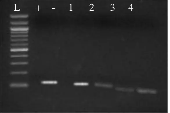
according to manufacturer’s instructions to remove any DNA. DNA contamination was checked
with agarose gel electrophoresis following reverse transcription-polymerase chain reaction
(RT-PCR) by performing control experiments in which no reverse transcriptase was added to the
isolated RNA before the PCR step. RNA concentration was determined by absorption at 260 nm
with a Biophotometer (Eppendorf, Hamburg, Germany) and NanoDrop 2000 UV-Vis
Spectrophotometer (Thermo Fisher Scientific, Wilmington, DE). Purified RNA was stored at
-80°C.
Reverse Transcription-Polymerase Chain Reaction (RT-PCR)
PCR primers were designed using the PrimerQuest software (Integrated DNA
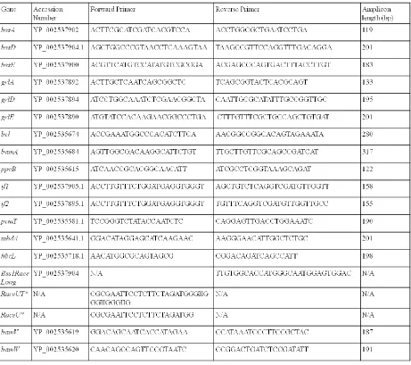
Technologies, Coralville, IA). The primers used in this study are detailed in Table 1.
Degenerate primers directed at citrate synthase genes were used to identify DNA contamination
volumes of 20 L containing final concentrations of 20 mM Tris-HCl, 10 mM KCl, 10 mM
NH4SO4, 2 mM MgSO4, 0.1% TritonX, 200 M of each dNTP, 0.1 mL of 10X BSA, 1 M of
cDNA was used. The following program was used: 95°C for 5 min followed by 43 cycles of
96°C for 40 sec, 52°C for 1 min, 72°C for 30 sec and a final incubation of 72°C for 10 min.
Amplification products were visualized on a 1.5% agarose gel and stained with ethidium
bromide.
The cDNA synthesis was carried out in 0.2 mL thin-walled PCR tubes with total reaction
volumes of 20 L containing final concentrations of 200 M of each dNTP, 1 M of
gene-specific reverse primers, 2U Multiscribe reverse transcriptase (Life Technologies-Applied
Biosystems, Grand Island, NY), and 10 M RNase inhibitor, and 500 ng of total RNA. The
Table 1: Primers used in this study
*primers RaceU and RaceUT were kindly provided by the Wilson laboratory, Georgia
State University, Atlanta, GA.
Cloning and sequencing
One Shot Top10 E. coli (Life Technologies Invitrogen, Carlsbad, CA) were transformed
with the pCR2.1Topo vector containing the amplicon of interest. Cells were thawed for a few
were incubated at 37°C while shaking vigorously for 1 hour. Cells were spread on
antibiotic-selective plates and grown overnight at 37°C. Clones were grown in the presence of X-gal and
IPTG and selected by color.
Those colonies which were white were picked for PCR verification of insert.
These cells were then inoculated in 2 mL of LB media and grown overnight at 37°C shaking
vigorously. The vector was then isolated from cells. Briefly, tubes were centrifuged at
10,000rpm for 5 min at room temperature. Supernatant was discarded and the pellet was
resuspended in 100µL chilled GTE buffer (50 mM Glucose, 25 mM Tris-HCl, 10 mM EDTA).
Also, 200 µL of 0.2 M NaOH and 1% SDS was added as well as 150 µL potassium acetate (100
mL 5 M potassium acetate, 57.5 mL glacial acetic acid, 142.5 mL distilled water). Tubes were
incubated on ice for 10 min and occasionally mixed by inversion. Tubes were then centrifuged
for 20 min at 20,000 rpm at room temperature and the supernatant was recovered. To the
supernatant 280µL of 100% chilled Isopropanol was added and these were mixed by inversion.
Samples were incubated at -20°C for 30 min and centrifuged for 15min at 13000 rpm at room
temperature. Supernatant was discarded and the pellet was washed with cold 75% ethanol then
centrifuged for 10 min at 13000 rpm at room temperature. The ethanol was discarded and pellet
was dried by desiccation. This pellet was then resuspended in nuclease-free water. The sample
was then sequenced by the Georgia State University Department of Biology Core Facilities to
Real-time Reverse Transcription-PCR quantification (qPCR)
cDNAs were amplified with gene specific primers and the resulting amplicons were
purified, quantified, and prepared for serial dilution then stored at -20°C. These dilutions were
used as calibration standards for real-time PCR. qPCR reactions were carried out in 0.2 mL
thin-walled optical PCR tubes with total reaction volumes of 20 L. The protocol with SYBR
Green PCR Master Mix (Life Technologies-Applied Biosystems, Grand Island, NY) was
followed per manufacturer’s instructions. Primers were carefully designed to prevent formation
of primer dimers during amplification and these cycles were followed by a dissociation curve to
verify no dimers or non-specific amplification was present. If more than a single peak was
observed on the dissociation curve, the concentration of primer and MgCl2 was optimized. An
amount of primer and MgCl2 that forms no primer dimers and gives optimal amplification was
used for qPCR assays of all the samples. The temperature profile used was as follows: an initial
activation step at 50°C for 5 min and denaturation at 98°C for 40 s, followed by 40 cycles of
denaturation at 98°C for 40 s, annealing at primer specific temperature (Table 1) for 32 sec, and
elongation at 65°C for 32 s, with a final extension step at 65°C for 10 min.
The size of the PCR products was checked with agarose gel electrophoresis, and the
specificity of PCR products was verified by sequence analysis of the clone library. qPCR
analysis of the cDNA was carried out with the Applied Biosystems 7500 Real-Time PCR system
(Life Technologies, Grand Island, NY) using 7500 Real-Time PCR System Sequence Detection
Software (Version 1.3.1). The precision as well as the reproducibility of quantification were
carefully optimized, and PCR products were checked for their correct lengths as described
Identification and analysis of Sigma 54-dependent genes
Sigma 54 promoter identification
Regions upstream of the bss and grl operons were analyzed for the presence of the
sigma54 promoter consensus using the PromScan (http://molbiol-tools.ca/promscan/) software.
Rapid amplification of 5' cDNA ends (5' RACE)
RNA was treated with 0.1 M DTT and incubated at 42°C for 2 min to eliminate
secondary structure. First strand synthesis was carried out using Reverse Transcriptase as
described previously. Primers reverse complimentary to the region near the start sight of bssD
were used for first strand synthesis. See Table 1 for primer sequences. cDNA was purified by
incubating with 1 M NaOH at 65°C for 20 min and cleaned with a GeneJet PCR Purification kit
(Thermo Scientific-Fermentas, Pittsburgh, PA) per manufacturer’s instructions. DNA was tailed
by incubating 5 pmol DNA with Terminal Transferase (New England Biolabs, Ipswitch, MA),
10x TdT Buffer (New England Biolabs, Ipswitch, MA), 2.5 mM CoCl2, and 2 mM dCTP for 30
min at 37°C. The reaction was halted by incubating at 70°C for 10 min. For second strand
synthesis the RACEUT and Race1 long primers were used with the following recipe: 20 mM
TrisHCl, 10 mM KCl, 10 mM NH4SO4, 2 mM MgSO4, 0.1% TritonX, 200 M of each dNTP,
0.1 mL of 10X BSA, 1 M of each primer, and 2U Ampli Taq Gold polymerase (Life
Technologies-Applied Biosystems, Grand Island, NY). Reactions were carried out in
thin-walled PCR tubes with the following program: 95°C for 5 min followed by 43 cycles of 96°C for
40 sec, 54°C for 1 min, 72°C for 30 sec and a final incubation of 72°C for 10 min. This step was
concentrations for future steps. Amplification products were separated on a 1.5% agarose gel
and stained with ethidium bromide. Products were cleaned with a GeneJet PCR Purification kit
(Thermo Scientific Fermentas, Pittsburgh, PA) per manufacturer’s instructions and sequenced.
Sigma 54 regulation of bss genes
The bbsD, bssA, bssB, bssC, and Geob_2441 were inserted into the pCR Topo2.1 vector
(Life Technologies-Invitrogen, Carlsbad, CA) following amplification with AmpliTaq Gold
polymerase (bss1). RpoN-E. coli strains were purchased from the E. coli Genetic Stock Center
at Yale University (New Haven, CT). One Shot Top10 E. coli (Life Technologies Invitrogen,
Carlsbad, CA) and RpoN- were transformed with the bss1 vector. Cells were thawed for a few
minutes on ice and incubated on ice for 5 min with vector. Cells were then heat shocked at 42°C
cells were incubated at 37°C while shaking vigorously for 1 hour. Cells were spread on
antibiotic-selective plates and grown overnight at 37°C. Clones were grown in the presence or
3 RESULTS
Growth characteristics of G. daltonii strain FRC-32
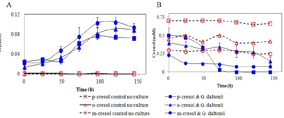
G. daltonii FRC-32 can grow with various aromatic compounds including benzoate,
toluene, benzene, m-, o-, and p-cresol. Toluene and benzene were toxic when amended at a high
concentration (2 mM). Therefore, these aromatic hydrocarbons were supplied in a hydrophobic
carrier phase with 2,2,4,4,6,8,8-Heptamethylnonane (HMN) to allow continuous supply with low
concentrations (1 mM) because the direct addition to the culture was known to be inhibitory for
growth (54). Also, cultures were tested for growth on toluene and benzene without HMN as a
carrier in order to test if G. daltonii utilizes HMN as substrate. Good growth with toluene and
benzene without HMN was observed (data not shown).
Various electron acceptors such as fumarate, nitrate and ferric citrate were tested for G.
daltonii cultivation. G. daltonii was grown using 1 mM nitrate as an electron acceptor. Higher
concentrations, up to 5 mM, were tested. However, these were all toxic and inhibitory to growth.
Ferric citrate was tested as an electron acceptor at either 60 mM or 30 mM. Growth was
observed when benzoate was used as the substrate (data not shown). However, significant
growth was not observed when toluene or benzene were provided to the culture with ferric citrate
as the electron acceptor. This is possibly due to the stressful nature of the combination of
aromatic hydrocarbons and ferric citrate. G. daltonii culture was able to only grow using one of
these at a time. Molecular genetic experiements for the present study were performed on cultures
grown with 10 mM fumarate as an electron acceptor.
After establishing the growth conditions, G. daltonii FRC-32 was adapted to the
curve of the adapted cultures was then monitored by measuring increases in optical density,
substrate loss and product formation in order to determine optimal time points for harvesting
cultures. At mid-log phase cells were harvested for further genomic and metabolite analyses, so
that misleading results in the gene expression study originating from growth phase effects were
avoided.
These cultures grew to an average OD600 of 0.1-0.3. This growth required 7-60 days
depending upon the substrate provided. While G. daltonii is able to utilize multiple electron
acceptors, it did not correspond with equal energy output. For example, more energy, and thus
more rapid growth, was accomplished when this organism was grown utilizing nitrate as a sole
electron acceptor than Fe(III) or even fumarate.
Genomic analysis of anaerobic aromatic compound degradation genes
In order to identify which regions of the G. daltonii genome might code for enzymes
involved in anaerobic degradation of aromatic compounds, particularly benzene, genes within
the reported aromatic compound-degrading gene island of G. metallireducens (55) were
compared to genes within G. daltonii using BLAST analysis. As observed in the G.
metallireducens genome (Fig. 9A), many genes predicted to play a role in aromatic compound
degradation clustered into a large aromatic degrading gene island from Geob_0095 to
Geob_0255 in the G. daltonii genome (Fig. 9B). This island spans a region of 165 genes and
roughly 194 kbp. While most of the genome has a GC content of 53.5%, this region has a GC
content of 56.3%. In addition to the main aromatic degrading gene island, both organisms have
Geob_2420-Geob_2451 in G. daltonii and has a GC content of 56.65% and spans a region of 32
genes and approximately 39 kbp.
Figure 9: Locations of predicted aromatic compound degradation genes in Geobacter species. Genes predicted to play a role in aromatic compound degradation in G. metallireducens and G. daltonii were mapped with black bars indicating presence of genes associated with aromatic compound degradation.
Because anaerobic benzene oxidation is such a unique characteristic of G. daltonii, its
genome was scanned for genes that shared no homology to other anaerobic aromatic- degrading
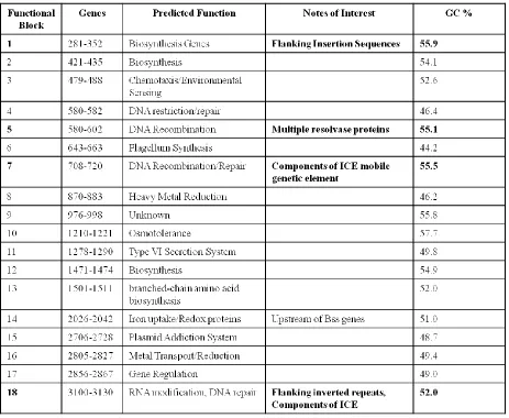
bacteria. Once identified, these genes were grouped into functional blocks (Fig. 10) based on
physical proximity within the genome, and functions were predicted if possible based on
Figure 10: Location of “unique” genes in the G. daltonii genome. Genes that are predicted to have “hypothetical” or “unknown” function that are proximal to one another.
Table 2. Predicted function of the unique genesa
a
[image:49.612.76.539.267.647.2]One of the largest segments, segment 1 is located close to the large aromatic-degrading
gene island close to the start of the genome. At 55.9% GC, this region has a different GC
content from the rest of the genome. This region contains multiple glycosylases and glycosyl
transferases as well as dehydratases and cyclases. Because of these observations, this region is
predicted to encode genes involved in biosynthesis pathways. Also unique to this region is that it
is flanked by insertion sequences which could be used for genetic movement. This bares interest
as it has been speculated that large mobile genetic elements, sometimes called ICE regions, are
the mechanism by which the ability to degrade aromatics spread from one organism to another.
An example of this is Tn4371, a 55 kbp region that confers the ability to degrade biphenyls to its
host (56). Another region in G. daltonii with characteristics similar to ICE/integrated
transposons is segment 7. This region has genes for DNA recombination as well as competence
proteins and a recombinase. Segment 15 is an anomaly in that most of the genes in this region
seem to code for a toxin/antidote system. This block also has a vastly different GC content from
the rest of the genome at 48.7%. Finally, segment 18 is a region with mostly hypothetical
proteins. However, it is flanked by inverted repeats and transposons as seen with ICEs. None of
the regions of unique genes fell within close proximity to either of the aromatic degrading gene
islands identified.
The large aromatic-degrading gene cluster from Geob_0095-Geob_0253 includes genes
for utilization of phenol, which is a proposed intermediate in anaerobic oxidation of benzene.
The first step in the anaerobic degradation of phenol is the addition of a phosphate group from
ATP by the enzyme phenylphosphate synthase (pps). Phenylphosphate is carboxylated with CO2
the carboxyl group by an unknown enzyme, and the hydroxyl group of 4-hydroxybenzoyl-CoA is
eliminated by reduction to form benzoyl CoA (57). The remainder of the pathway consists of
reduction and opening of the aromatic ring, catalyzed by the bam gene products, beta-oxidation
to acetyl-CoA, and oxidation to CO2 through the TCA cycle. Expression levels of pps and ppc
genes have been examined to detect activation of this pathway within anaerobic
phenol-degrading organisms and could be used to determine whether they are involved in degradation of
benzene by G. daltonii (58).
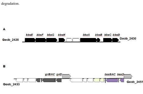
The smaller aromatic-degrading gene cluster from Geob_2420-Geob_2451 contains
genes involved in toluene oxidation, another proposed route for anaerobic benzene degradation.
The first step in anaerobic toluene degradation is the enzymatic addition of fumarate to toluene’s
methyl group, resulting in the formation of R-benzylsuccinate (59). The enzyme benzylsuccinate
synthase is a duplex of three subunits (, β and γ), forming a heterohexamer (60); it is activated
by an accessory protein, BssD, which generates a glycyl radical within the 98 kDa alpha subunit
(61). Bss must be maintained in strictly anaerobic conditions as O2 will cause irreversible
cleavage of the protein (62). Subsequent steps are catalyzed by the bbs (beta oxidation of
benzylsuccinate) gene products (Fig. 11A). Coenzyme A is transferred from succinyl-CoA to
the alpha-carboxyl group of R-benzylsuccinate to form R-benzylsuccinyl-CoA. This compound
is oxidized to E-2-benzylidenesuccinyl-CoA, hydrated to
[2-hydroxy(phenyl)methyl]succinyl-CoA, oxidized to benzoylsuccinyl-[2-hydroxy(phenyl)methyl]succinyl-CoA, and cleaved thiolytically to succinyl-CoA plus
benzoyl-CoA. Benzoyl-CoA undergoes reduction and opening of the aromatic ring, catalyzed by the bam
gene products, followed by beta oxidation and entry into the TCA cycle as acetyl-CoA which is
benzene could be an evidence for use of the toluene pathway during anaerobic benzene
[image:52.612.68.537.95.404.2]degradation.
Figure 11: bbs and bss gene clusters in G. daltonii. Organization of anaerobic toluene
oxidation genes within the genome of G. daltonii. White arrows indicate genes without predicted aromatic compound degradation functions according to NCBI annotations. Yellow arrows indicate genes predicted to be responsible for Bss formation and activation. Green arrows indicate similar predicted sigma 54-dependent transcriptional regulators. Spaces within arrows indicate predicted non-coding regions between genes and/or operons.
Of particular interest is the observation that G. daltonii has two adjacent operons
encoding glycl radical-like genes (Fig. 11B). No other anaerobic toluene-oxidizing prokaryotes
at present have been identified which code two copies of Bss. Also, it is interesting that these
copies lie within completely independent operons, each with its own divergently transcribed
gene encoding a transcriptional regulator and with different genes downstream. These copies
only share 76% amino acid identity to one another. The bbs genes, which are responsible for the