© 2017, IRJET | Impact Factor value: 5.181 | ISO 9001:2008 Certified Journal
| Page 1400
Robust system for patient specific classification of ECG signal using PCA
and Neural Network
Vishwajeeta Patil
1, Prof S. N. Patil
21
Student, Dept of Electronics engineering, Budhgaon college, Maharashtra, India
2Professor, Dept of Electronics engineering, Budhgaon college, Maharashtra, India
---***---Abstract -
For ECG data classification, we have beenproposed many system. It is very important that detection of ECG arrhythmias for the treatment of patients during diagnosing the heart disease at the early stage. Human eye is poorly suited to detect the morphological variation of ECG signal also difficult for doctors to analyse long ECG records in the short period of time ,so we required an effective diagnostic system . We proposed a generic and patient specific classification system designed for robust and accurate detection of ECG heartbeat patterns. In the system, feature extraction uses morphological feature which are projected onto a lower dimensional feature space using Principal Component Analysis (PCA) and temporal feature from ECG data. Artificial neural network ANNs is used for pattern recognition. ANN is powerful tools for pattern recognition , as it having the capability to learn complex and nonlinear surfaces. The performance of ECG pattern classification strongly depends on the characterization power of the features extracted from the ECG data and the design of the classifier. Nonstationary ECG signal is efficiently analysed by the TI-DWT due to its time–frequency localization properties. PCA is the optimal linear transformation, It has been used for data compression, redundancy and dimensionality reduction, In PCA it project the input pattern vectors onto a lower dimensional feature space so that retains the maximum amount of energy . The proposed classification system can adapt significant interpatient variation in ECG patterns by training the network structure, and thus achieves higher accuracy over larger datasets.
Key Words: ECG, PCA, ANN, TI-DWT
1. INTRODUCTION
The recorded ECG waveform shows the time evolution of the heart’s electrical activity, which is made of different electrical depolarization–repolarization patterns of system will be developed in modules, for patient specific classification of ECG data, These modules are combined and training will be given to the system. For training and performance evaluation of the proposed patient specific ECG classifier we used MIT/BIH database. The database contains both timing information and beat class information that verified by independent experts.
The heart. Any change in the morphological pattern or disorder of heart rate is an indication of an arrhythmia,
which could be detected by analysis of the recorded ECG waveform. Classification of ECG beats is a challenging problem as the morphological and temporal characteristics of ECG signals show significant variations for different patients and under different temporal and physical conditions.
For automatic detection and classification of ECG heartbeat patterns many algorithms have been presented but ECG classifier systems based on past approaches have not performed well in practice because of their important common drawback of having an inconsistent performance when classifying a new patient’s ECG waveform. This makes system unreliable to be widely used clinically, and causes severe degradation in their accuracy and efficiency for larger databases.
Pattern of ECG classification strongly depends on the characterization power of the features extracted from the ECG data and the design of the classifier.ECG signal can be used to decompose an according to scale by using wavelet transform, thus allowing separation of the relevant ECG waveform morphology descriptors from the noise, interference, baseline drift, and amplitude variation of the original signal.
The dimension of the input morphological feature vector is reduced by projecting it onto a lower dimensional feature space using Principal Component Analysis (PCA) in order to significantly reduce redundancies in such a high-dimensional data space. .
Pattern recognition is done by using ANN as they have the capability to learn complex, nonlinear surfaces among different classes, and such ability can therefore be the key for ECG beat recognition and classification.
2. METHODOLOGY
© 2017, IRJET | Impact Factor value: 5.181 | ISO 9001:2008 Certified Journal
| Page 1401
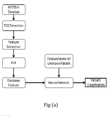
and finally it given to Artificial Neural Network (ANN) for patient classification Proposed ECG classification system is given below in fig (a)
Fig (a)
2.1 ECG Data
Here MIT/BIH arrhythmia database is used for training and performance evaluation of the proposed patient specific ECG classifier. The MIT/BIH arrhythmia database contains annotation for both timing information and beat class information verified by independent experts.
Fig (b): diagram of ECG signal
Fig (c): Real time ECG signal
2.2 ECG Detection module
The ECG is detected using Eigenvector method. It is used for estimating frequencies and powers of signals from noise-corrupted signal. The Eigen vector associated with the minimum Eigen value of the estimated autocorrelation matrix is used to calculate the PSD.
These methods are based on an Eigen decomposition of the correlation matrix of the noise-corrupted signal
2.3 Feature Extraction module
After ECG detection using above method the morphological and temporal feature will be extracted from ECG data. Wavelet transform is used to extract morphological information from the detected ECG signal. We can also use translation- invariant dyadic wavelet transform (TIDWT) for effectively extraction of the morphological information from ECG data
2.4 Dimension reduction using PCA module
The wavelet-based morphological features in the training set are post processed using PCA to reduce dimensionality (and redundancy) of input feature vectors are reduced by using PCA so the wavelet-based morphological features in the training set are post processed using PCA
2.5 Feature Database
The feature database consisting morphological and temporal features of healthy and diseased persons can be created for training of Neural Network.
2.6 ECG data classification module
ANNs having ability to learn complex, nonlinear surfaces among different classes, and such ability can therefore it is used for ECG beat recognition and classification .Therefore ANNs are used for the classification of ECG data from each individual patient in the database. ANN compares features of test case patient with the features stored in database to classify patient as normal or diseased heart patient.
3. EMPIRICAL ANALYSIS
[image:2.595.62.273.145.370.2] [image:2.595.42.284.460.636.2]© 2017, IRJET | Impact Factor value: 5.181 | ISO 9001:2008 Certified Journal
| Page 1402
The same wavelet function has already been successfully applied to QRS detection in ,achieving a 99.8% QRS detection rate for the MIT/BIH arrhythmia database.
After extraction of features , processed using PCA to reduce dimensionality (and redundancy) of input feature vectors. PCA, also known , is a well-known statistical method. It has been used for data analysis, data compression, redundancy and dimensionality reduction, and feature extraction .Let F
be a feature matrix of size K × N, whose rows are wavelet features of size 1 × N , each belonging to one of K heartbeats in the training data. First, the covariance matrix of this feature matrix is computed as
= E
where m is the mean pattern vector. From the Eigen decomposition of , which is a K × K symmetric and positive-definite matrix, the principal components taken as the Eigenvectors corresponding to the largest Eigenvalues are selected, and the morphological feature vectors are then projected onto these principal components
ANNs are used for the classification of ECG data from each individual patient in the database. ANN particularly focus drawn on automatic design of the multilayer perceptron’s (MLPs). This evolutionary operator makes the proposed
system generic.The optimum dimension found corresponds
to a distinct ANN architecture where the network parameters (connections, weights, and biases) can be resolved from the positional optimum to reached on that dimension
4. EXPERIMENTAL RESULTS
We are taking three types of ECG signal, including normal ECG and ECG signal of diseases like Hanipas and Breithman Here all ECG signal are going to different process such as detection of ECG signal, feature extraction using TI-DWT, dimensionality reduction using PCA so we get feature database and finally data classification process by using ANN.
4. 1 Stepwise procedures on different ECG Signal
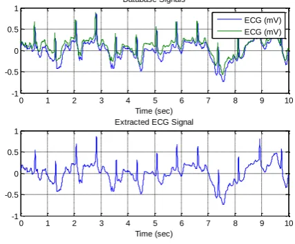
Step1: Extraction of ECG signal
In step first data base signal and extracted ECG signal as database signal contains contains noise, baseline drift and amplitude variation which can be removed by using TI-DWT
Step2: Detection of QRS on ECG signal
Step second indicates detection of QRS on ECG signal and pulse train of the QRS on the ECG signal
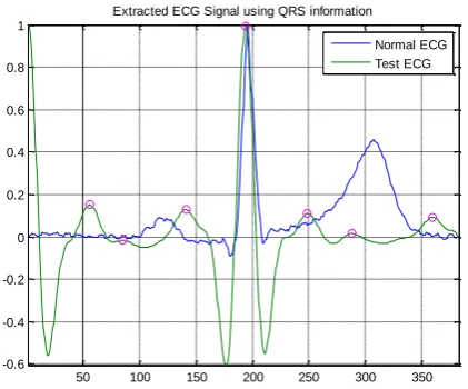
Step3: Extraction of ECG signal using QRS information
Step third indicate extracted ECG signal using QRS information
Now all this steps can be represented as shown in figures below
(a) Stepwise procedure for detection of Normal
ECG signal
0 1 2 3 4 5 6 7 8 9 10
-1 -0.5 0 0.5 1
Time (sec) Database Signals
ECG (mV) ECG (mV)
0 1 2 3 4 5 6 7 8 9 10
-1 -0.5 0 0.5 1
Time (sec) Extracted ECG Signal
Fig (d):
Extraction of ECG signal500 1000 1500 2000 2500
-0.5 0 0.5 1
QRS on Filtered Signal
500 1000 1500 2000 2500
-0.5 0 0.5
Pulse train of the found QRS on ECG signal
[image:3.595.326.540.231.408.2]© 2017, IRJET | Impact Factor value: 5.181 | ISO 9001:2008 Certified Journal
| Page 1403
50 100 150 200 250 300 350 -0.6
-0.4 -0.2 0 0.2 0.4 0.6 0.8 1
Extracted ECG Signal using QRS information
Normal ECG Test ECG
Fig (f):Extraction of ECG signal using QRS information
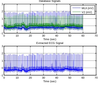
(b) Stepwise procedure for detection of Breithman
ECG signal
0 10 20 30 40 50 60 70
-1 0 1 2 3
Time (sec) Database Signals
MLII (mV) V2 (mV)
0 10 20 30 40 50 60 70
-1 0 1 2 3
Time (sec) Extracted ECG Signal
Fig (g):
Extraction of ECG signal0.2 0.4 0.6 0.8 1 1.2 1.4 1.6 1.8 2
x 104 -0.5
0 0.5 1
QRS on Filtered Signal
0.2 0.4 0.6 0.8 1 1.2 1.4 1.6 1.8 2
x 104 0
1 2
Pulse train of the found QRS on ECG signal
Fig (h):Detection of QRS on ECG signal
Fig(i):Extraction of ECG signal using QRS information.
(c) Stepwise procedure for detection of Hanipas
ECG signal
0 2 4 6 8 10 12
-1 0 1 2
Time (sec) Database Signals
MLII (mV) V1 (mV)
0 2 4 6 8 10 12
-1 0 1 2
Time (sec) Extracted ECG Signal
Fig (j):
Extraction of ECG signal500 1000 1500 2000 2500 3000 3500 -0.5
0 0.5 1
QRS on Filtered Signal
500 1000 1500 2000 2500 3000 3500 -0.5
0 0.5 1
Pulse train of the found QRS on ECG signal
[image:4.595.58.266.101.276.2] [image:4.595.328.535.102.269.2] [image:4.595.336.538.359.519.2] [image:4.595.65.541.360.788.2] [image:4.595.69.258.369.534.2]© 2017, IRJET | Impact Factor value: 5.181 | ISO 9001:2008 Certified Journal
| Page 1404
50 100 150 200 250 300 350 -0.6
-0.4 -0.2 0 0.2 0.4 0.6 0.8 1
Extracted ECG Signal using QRS information
Normal ECG Test ECG
Fig (l):Extraction of ECG signal using QRS information
5.
CLASSIFICATION PERFORMANCE
We performed classification experiments on 15 records of the MIT/BIH arrhythmia database ,in which 10 records used as traning database and 5 records used as testing database according to procedure as we load neural network we get 80% to 90% accuracy
6.
CONCLUSION
Many works have been proposed for ECG data classification. Detection of ECG arrhythmias is necessary for the treatment of patients for diagnosing the heart disease at the early stage. It is very difficult for doctors to analyze long ECG records in the short period of time and also human eye is poorly suited to detect the morphological variation of ECG signal, hence imposing the need for an effective diagnostic system. This system is useful for physician to take decision for heart condition of a patient in life threatening condition ,so it is very useful to medical field.
7.REFERENCES
[1] Turker Ince ,Serkan Kiranyaz and Moncef Gabbouj,“A Generic and Robust System for Automated Patient-Specific Classification of ECG Signals,” IEEE Trans.on biomed. Eng.,vol.56,no.5, May 2009.
[2] Elif Derya U beyli, “Eigenvector Methods for Automated Detection of ECG Changes in Partial Epileptic Patients IEEE Trans. On Information Technology In Biomedicine”, Vol. 13, No. 4, July 2009
[3] L. Y. Shyu, Y. H. Wu, and W. C. Hu, “Using wavelet transform and fuzzy neural network for VPC detection from the holter ECG,” IEEE Trans.biomed.Eng., vol. 51, no. 7, pp. 1269–1273, Jul. 2004..
[4] O. T. Inan, L. Giovangrandi, and G. T. A.Kovacs, “Robust neural-network based classification of premature ventricular contractions using wavelet transform and timing interval features,” IEEE Trans.biomed.Eng., vol. 53, no. 12, pp. 2507– 2515, Dec. 2006.
[5] D. A. Coast, R. M. Stern, G. G. Cano, and S. A. Briller, “An approach to cardiac arrhythmia analysis using hidden Markov models,” IEEE Trans.biomed.Eng., vol. 37, no. 9, pp. 826–836, Sep. 1990.
[6] S.Osowski, L. T.Hoai, and T.Markiewicz, “Support vector machine based expert system for reliable heartbeat recognition,” IEEE Trans.biomed.Eng., vol. 51, no. 4, pp. 582– 589, Apr. 2004.
[7] Y. Hu, S. Palreddy, and W. J. Tompkins, “A patient-adaptable ECG beat classifier using a mixture of experts approach,” IEEE Trans.biomed.Eng., vol. 44, no. 9, pp. 891– 900, Sep. 1997.
[8] P. de Chazal and R. B. Reilly, “A patient-adapting heartbeat classifier using ECG morphology and heartbeat interval features,” IEEE Trans.biomed.Eng., vol. 53, no. 12, pp. 2535–2543, Dec. 2006.
[9] P. de Chazal, M. O’Dwyer, and R. B. Reilly, “Automatic classification of heartbeats using ECG morphology and heartbeat interval features,” IEEE Trans.biomed.Eng., vol. 51, no. 7, pp. 1196–1206, Jul. 2004.
[10] R. Silipo and C. Marchesi, “Artificial neural networks for automatic ECG analysis,” IEEE Trans.Signal Process vol. 46, no. 5, pp. 1417–1425, May 1998.
[11] S. Osowski and T. L. Linh, “ECG beat recognition using fuzzy hybrid neural network,” IEEE Trans.biomed.Eng., vol. 48, no. 11, pp. 1265–1271, Nov. 2001.
[12] T. Ince, S. Kiranyaz, and M. Gabbouj, “Automated patient-specific classification of premature ventricular contractions,”i proc. IEEE Int.Conf.EMBS,2008,pp.5474– 5477.
[13] S. Pittner and S.V.Kamarthi, “Feature extraction from wavelet coefficients for pattern recognition tasks,” IEEE Trans. Pattern Anal.Mach., vol. 21, no. 1, pp. 83–88, Jan. 1999.
[14] J. Kennedy and R. Eberhart, “ Particle swarm optimization,” in Proc.IEEE Int. Conf. Neural Netw., vol. 4, Perth, W.A., Australia, 1995, pp. 1942– 1948.
[image:5.595.56.267.114.289.2]