Normalizing glycosphingolipids restores
function in CD4
+
T cells from lupus patients
Georgia McDonald, … , Terry Butters, Elizabeth C. Jury
J Clin Invest.
2014;
124(2)
:712-724.
https://doi.org/10.1172/JCI69571
.
Patients with the autoimmune rheumatic disease systemic lupus erythematosus (SLE) have
multiple defects in lymphocyte signaling and function that contribute to disease
pathogenesis. Such defects could be attributed to alterations in metabolic processes,
including abnormal control of lipid biosynthesis pathways. Here, we reveal that CD4
+T
cells from SLE patients displayed an altered profile of lipid raft–associated
glycosphingolipids (GSLs) compared with that of healthy controls. In particular,
lactosylceramide, globotriaosylceramide (Gb3), and monosialotetrahexosylganglioside
(GM1) levels were markedly increased. Elevated GSLs in SLE patients were associated
with increased expression of liver X receptor
b
(LXR
b
), a nuclear receptor that controls
cellular lipid metabolism and trafficking and influences acquired immune responses.
Stimulation of CD4
+T cells isolated from healthy donors with synthetic and endogenous
LXR agonists promoted GSL expression, which was blocked by an LXR antagonist.
Increased GSL expression in CD4
+T cells was associated with intracellular accumulation
and accelerated trafficking of GSL, reminiscent of cells from patients with glycolipid storage
diseases. Inhibition of GSL biosynthesis in vitro with a clinically approved inhibitor
(N-butyldeoxynojirimycin) normalized GSL metabolism, corrected CD4
+T cell signaling and
functional defects, and decreased anti-dsDNA antibody production by autologous B cells in
SLE patients. Our data demonstrate that lipid metabolism defects contribute to SLE
pathogenesis and suggest that targeting GSL biosynthesis restores T cell function in SLE.
Research Article
Find the latest version:
Research article
Normalizing glycosphingolipids restores
function in CD4
+
T cells from lupus patients
Georgia McDonald,1 Shantal Deepak,1 Laura Miguel,1 Cleo J. Hall,1 David A. Isenberg,1
Anthony I. Magee,2 Terry Butters,3 and Elizabeth C. Jury1
1Centre for Rheumatology Research, Department of Medicine, University College London, Rayne Building, London, United Kingdom. 2Section of Molecular Medicine, National Heart and Lung Institute, Imperial College London, South Kensington, London, United Kingdom.
3Oxford Glycobiology Institute, Department of Biochemistry, University of Oxford, Oxford, United Kingdom.
Patients with the autoimmune rheumatic disease systemic lupus erythematosus (SLE) have multiple defects
in lymphocyte signaling and function that contribute to disease pathogenesis. Such defects could be
attrib-uted to alterations in metabolic processes, including abnormal control of lipid biosynthesis pathways. Here,
we reveal that CD4
+T cells from SLE patients displayed an altered profile of lipid raft–associated
glycosphin-golipids (GSLs) compared with that of healthy controls. In particular, lactosylceramide,
globotriaosylcera-mide (Gb3), and monosialotetrahexosylganglioside (GM1) levels were markedly increased. Elevated GSLs in
SLE patients were associated with increased expression of liver X receptor
β
(LXR
β
), a nuclear receptor that
controls cellular lipid metabolism and trafficking and influences acquired immune responses. Stimulation
of CD4
+T cells isolated from healthy donors with synthetic and endogenous LXR agonists promoted GSL
expression, which was blocked by an LXR antagonist. Increased GSL expression in CD4
+T cells was
associ-ated with intracellular accumulation and accelerassoci-ated trafficking of GSL, reminiscent of cells from patients
with glycolipid storage diseases. Inhibition of GSL biosynthesis in vitro with a clinically approved
inhibi-tor (N-butyldeoxynojirimycin) normalized GSL metabolism, corrected CD4
+T cell signaling and functional
defects, and decreased anti-dsDNA antibody production by autologous B cells in SLE patients. Our data
demonstrate that lipid metabolism defects contribute to SLE pathogenesis and suggest that targeting GSL
biosynthesis restores T cell function in SLE.
Introduction
The mechanisms underlying the immunopathogenesis of the auto-immune rheumatic disease systemic lupus erythematosus (SLE) remain uncertain; however, both the disease and its treatment result in a substantially increased risk of cardiovascular disease, suggesting that a defect in lipid metabolism contributes to the dis-ease process (1). In support of this concept, patients are character-ized by dyslipidemia and defects in lymphocyte plasma membrane lipid rafts that result in increased cell stimulation (2, 3).
Glycosphingolipids (GSLs) are essential for many cellular pro-cesses and are composed of a ceramide backbone embedded in the outer leaflet of the plasma membrane and a sugar moiety that projects into the extracellular space (4). GSLs are enriched predom-inantly in lipid rafts, regions in the plasma membrane that coordi-nate the interaction of key signaling molecules that facilitate lym-phocyte activation and function (2, 5). Furthermore, differential GSL expression influences a range of T cell functions including TCR-mediated signaling (6–8), apoptosis (9), and recycling and endocytosis of membrane signaling and receptor molecules (4).
The control of plasma membrane GSL levels is tightly regu-lated. De novo biosynthesis is catalyzed by enzymes that promote sequential molecular changes from ceramide to generate unique GSL categories including globo-, asialo-, and a-series GSLs (Fig-ure 1A and ref. 10). Vesicular trafficking of newly synthesized lip-ids to the plasma membrane and subsequent lysosomal and/or late endosomal degradation are also integral to the maintenance of healthy GSL levels (11). Alterations to these processes can lead
to a plethora of clinical manifestations, including the lysoso-mal storage diseases (LSDs) Niemann-Pick type C (NPC), Fabry disease, and Gaucher disease (12). However, very little is known about the effect of altered GSL expression on T cell function in human health and autoimmunity.
CD4+ T cells from SLE patients are characterized by many
abnor-malities including: increased levels of raft-associated GSLs and cholesterol; defects in the lipid raft location and function of key TCR signaling molecules; accelerated recycling of TCR-associated proteins; and increased cell death and defects in mitochondrial function and autophagy (2, 3, 13). Given that GSLs mediate many of these cellular processes (4, 12), it is possible that changes in GSL expression could contribute to SLE pathogenesis. Intriguingly, manipulation of membrane lipids by in vitro culture with ator-vastatin (known to reduce cholesterol biosynthesis) can normalize membrane GM1 expression, phosphorylation of LCK and ERK, and production of IL-10 and IL-6 in T cells from SLE patients (14). This effect suggests that targeting membrane lipids could control or alter immune cell activation and may be an important therapeu-tic approach for autoimmune disease.
Here, we show that CD4+ T cells from SLE patients had a
dis-rupted GSL profile that was associated with accelerated GSL trafficking and accumulation in intracellular compartments. We found that elevated GSL expression could be recapitulated in healthy T cells by in vitro stimulation with synthetic (GW3965) or potential endogenous liver X receptor β (LXRβ) agonists (oxidized LDL and serum), suggesting that T cell defects in SLE patients could be driven, in part, by dyslipidemia. Inhibition of GSL expres-sion in vitro using the clinically approved inhibitor N-butyldeoxy-nojirimycin (NB-DNJ) (15) modified GSL metabolism and T cell Conflict of interest: The authors have declared that no conflict of interest exists.
Citation for this article:J Clin Invest. 2014;124(2):712–724. doi:10.1172/JCI69571.
research article
function to resemble that observed in T cells from healthy donors. Thus, our findings suggest that defects in lipid metabolism con-tribute to the immunopathogenesis of SLE and that targeting lipid biosynthesis pathways could be a novel therapeutic strategy for the treatment of SLE.
Results
Dysregulated GSL expression in CD4+T cells from patients with SLE. Our
recent findings show that changes in the composition and organi-zation of lipids in the plasma membrane can influence T cell func-tion (16). In order to characterize total T cell GSL composifunc-tion,
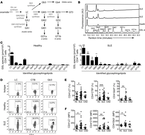
lip-Figure 1
Altered GSL profile in T cells from patients with SLE. (A) Scheme showing GSL biosynthesis pathways, indicating some of the enzymes con-trolling biosynthesis. Cellular lipids were isolated from negatively selected CD4+ T cells from 40 SLE patients and 15 healthy donors by
chloro-form-methanol extraction. The total cellular GSL profile was analyzed by HPLC following glycanase digestion to release the GSL sugar head groups. (B) Representative qualitative HPLC plots showing the position of known GSL standards and GSL species in 1 healthy control and 2 SLE patients. (C) Cumulative quantitative data for each GSL species identified on the HPLC plots. GSL expression was calculated from the peak HPLC areas after applying an experimentally derived response factor (18) by relating the area of the HPLC peak to the cell number of the sample. Two-tailed Mann-Whitney U test; **P = 0.008; *P ≤ 0.05. Expression of surface GSL was determined in ex vivo PBMCs from 58 SLE patients, 36 healthy donors, and 10 patients with OADs (Sjögren’s syndrome and RA). Cells were stained using fluorescently labeled antibodies against CD4-v450, LC-PE-Cy5, Gb3-FITC, or CTB-FITC and analyzed by flow cytometry. (D) Representative flow cytometric dot plots showing staining with appropriate controls (percentage of CD4+GSLhi T cells and GSL MFI of total CD4+ T cells is shown). Cumulative data of percentage of
[image:3.585.49.535.82.536.2]research article
ids were extracted from negatively isolated CD4+ T cells from SLE
patients and healthy donors and analyzed by HPLC (ref. 17 and Fig-ure 1B). Quantitative analysis of the HPLC plots revealed (18) that T cells from SLE patients had a profoundly altered GSL profile, with a significantly increased expression of lactosylceramide (LC), GA2, Gb3, GM2, GD1a, and GM1 compared with that of T cells from healthy donors (Figure 1C). This represented an increase in several different GSL species (including globo-, a-, and asiolo-series) when assessed according to their position in the GSL biosynthesis pathway (Supplemental Figure 1A; supplemental material available online with this article; doi:10.1172/JCI69571DS1).
We determined GSL expression in CD4+ T cell plasma
mem-branes by flow cytometry using the available GSL-specific anti-bodies. We found that expression of LC, Gb3 (using anti-CD77 antibody but also binding to Shiga toxin B; data not shown), and GM1 (binding to cholera toxin B [CTB]) was substantially increased in CD4+ T cells from SLE patients compared with that
in cells from healthy donors and from patients with other auto-immune disease (OAD) (Figure 1, D–F). We observed no change in the expression of GM3 or GM2 (data not shown). Culture of
T cells from healthy donors with a potent inhibitor of GSL synthe-sis (D-PDMP) decreased cell-surface GSL expression, confirming specificity of the anti-GSL antibodies (Supplemental Figure 1B).
Interestingly, we found that expression of GM1 was low in all sam-ples, as measured by HPLC, compared with its detection by CTB binding and flow cytometry. This has been observed previously and is attributed to CTB binding to related structures including GM3 and GM2 (T. Butters, unpublished observations and ref. 19). In light of this finding, we continued to use CTB binding as a surrogate marker of GSL expression rather than a specific marker for GM1.
GSL expression was not influenced by disease activity, as assessed by the British Isles Lupus Assessment Group (BILAG) global score (Sup-plemental Figure 1C), or by treatment with prednisolone or hydroxy-chloroquine (Supplemental Figure 1, D and E). Since hydroxychloro-quine is known to affect lipid homeostasis (20), we cultured healthy T cells with this drug for 72 hours at a concentration equivalent to that detected in vivo, but did not observe an effect on plasma mem-brane LC, CTB, or Gb3 expression (Supplemental Figure 1F).
[image:4.585.58.537.83.409.2]Thus, we show that GSL expression in both total cellular and plasma membrane compartments was profoundly altered in ex
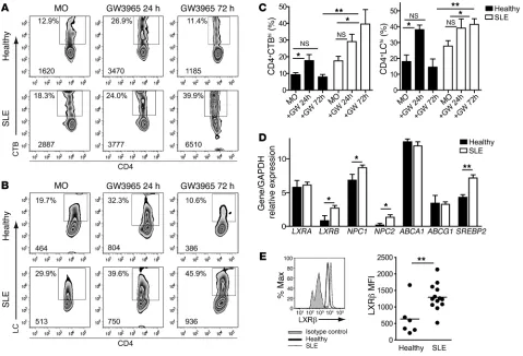
Figure 2
Increased GSL expression is associated with defective GSL homeostasis in T cells from SLE patients. PBMCs from 8 healthy donors and 8 SLE patients were stimulated for 24 and 72 hours with or without 1 μM GW3965 (GW). Cells were stained for CD4-APC and CTB-FITC or LC-FITC. Representative flow cytometric plots for (A) CTB binding and (B) LC expression after a 24- and 72-hour culture and (C) cumulative data. MO, medium only. One-way ANOVA, *P ≤ 0.05; 2-tailed Student’s t test, **P ≤ 0.003. RNA extracted from negatively isolated CD4+ T cells from 10 SLE
research article
vivo CD4+ T cells from patients with SLE compared with that seen
in healthy donors and disease controls.
Increased GSL expression is associated with defective GSL homeostasis in T cells from SLE patients. Increased GSL expression is associated with T cell activation (2, 21). We confirmed this observation in T cells from healthy donors; however, an analysis of GSL expression in functional CD4+ T cell subsets revealed that increased GSL
expres-sion was not restricted to activated T cells in SLE patients (Supple-mental Figure 2, A–E). The association between cell activation and increased GSL expression was further confirmed when we either rested (no stimulation) or TCR stimulated CD4+ T cells in vitro.
In contrast to our observation that T cells from healthy donors upregulated CTB binding and LC and Gb3 expression in response to TCR stimulation, T cells from patients with SLE had a dysregu-lated pattern of GSL expression in both resting and TCR-activated cells (Supplemental Figure 2F). Further, we detected no evident changes in GM3 expression levels (data not shown). Together, these results suggest that T cell activation was not solely responsi-ble for the increased GSL expression we observed in patients.
GSL expression can be driven by mechanisms other than TCR stimulation. GSLs are derived from ceramide (Figure 1A), whose production is mediated by proinflammatory cytokines and an altered serum lipid environment, among other stimuli (22). We
tested whether these stimuli translated into increased GSL expres-sion in CD4+ T cells from healthy donors by stimulating cells with
TNF-α and IL-6, which are significantly increased in the serum of SLE patients (Supplemental Figure 3A and ref. 1), or with syn-thetic agonists of nuclear receptors that regulate cellular lipid homeostasis, such as liver X receptor (LXR) (GW3965) and PPARγ
(23). Surprisingly, we found that stimulation with recombinant human TNF-α (rhTNF-α), IL-6, and PPARγ agonist had an insig-nificant effect on GSL expression (Supplemental Figure 3B and data not shown). In contrast, GW3965 stimulation significantly increased LC expression and CTB binding in CD4+ T cells from
healthy donors after a 24-hour culture (Figure 2, A–C). We found that this effect was transient, with LC expression and CTB binding returning to ex vivo levels by 72 hours, suggesting that GSL expres-sion was strictly controlled. In contrast, CD4+ T cells from SLE
patients displayed a sustained increased expression of GSL after GW3965 stimulation for 72 hours (Figure 2, A–C).
A role for LXR in defective CD4+ T cell GSL homeostasis was
supported by our finding that LXRB and its target genes NPC1
and NPC2, but not ABCA1 or ABCG1, were upregulated in ex vivo CD4+ T cells from SLE patients compared with those from healthy
[image:5.585.61.527.80.330.2]donors, as assessed by quantitative PCR (qPCR) (Figure 2D). We found that SREBP2, a gene that can indirectly affect LXR expression
Figure 3
Upregulation of LXRβ by oxysterol and TCR stimulation. (A) CD4+ T cells from 3 healthy donors and 3 SLE patients were cultured for 18
hours with GW3965 or CM and analyzed by Western blotting for LXRβ expression. (B) Cumulative data. Paired and 2-tailed Student’s t test; *P ≤ 0.05. (C) CD4+ T cells from 5 healthy donors and 5 SLE patients cultured for 18 hours with GW3965 or CM were assessed by qPCR for
LXRB expression. Cumulative results comparing LXRB with GAPDH control. Paired and 2-tailed Student’s t tests; *P ≤ 0.05. PBMCs from a healthy donor were cultured for 72 hours (all plus IL-2) with serum from 5 heterologous healthy donors (HC serum), 5 SLE patients (SLE serum), or with CM only (MO). (D) Cumulative data. One-way ANOVA; *P = 0.05. (E) CD4+ T cells from 5 healthy donors were cultured for 72
research article
(24), was also upregulated in T cells from SLE patients. Increased
LXRB expression was associated with significantly increased LXRβ
protein levels, as assessed by flow cytometry (Figure 2E) and West-ern blotting (Figure 3, A and B). We detected no significant change in the levels of glycolipid synthase enzymes (shown in Figure 1A), including glucosylceramide synthase, GM3 synthase, and Gb3 syn-thase (data not shown).
LXRβexpression in CD4+T cells is upregulated by oxysterol and TCR
stimu-lation. Most work examining the function of LXRβ has been described in macrophages, where it is known to be stimulated by increased cel-lular or serum oxysterols (25). To ascertain whether this was also the case in CD4+ T cells, we stimulated them with the synthetic oxysterol
agonist GW3965. GW3965 activation increased LXRβ protein and gene expression levels in T cells from both healthy donors and SLE patients (Figure 3, A–C). This effect was recapitulated by culture with serum from SLE patients (as a potential source of endogenous oxysterols) compared with cells cultured with serum from healthy individuals or with medium alone (Figure 3D). Furthermore, we observed that culture with oxidized LDL or extended culture with serum from SLE patients increased GSL expression in CD4+ T cells
from healthy donors (Figure 3, E and F). This suggests that chronic stimulation by potential endogenous LXR agonists in the serum of SLE patients could contribute to increased T cell GSL biosynthesis.
TCR stimulation also increases GSL expression (refs. 2, 21, and Supplemental Figure 2F), and we found that in vitro TCR stimu-lation significantly upregulated LXRβ gene and protein expression in both healthy and SLE patients (Supplemental Figure 3, C and D, and data not shown).
Finally, to confirm that oxysterol and TCR stimulation of LXRβ influenced GSL expression, we costimulated CD4+ T cells
from SLE patients and healthy donors with an LXR antagonist (5-chloro-N-2′-n-penthylphenyl-1,3-dithiophthalimide, 5CPPSS-50) (2, 26). 5CPPSS-50 blocked LXRβ upregulation in response to GW3965 and SLE serum (Supplemental Figure 3, E and F), but had only an insignificant effect following TCR stimulation (data not shown). Similarly, 5CPPSS-50 reversed increased GSL expres-sion in response to GW3965 and SLE serum, but not TCR stimu-lation (Figure 3, G and H, and data not shown).
[image:6.585.70.512.85.379.2]Together, these results support the hypothesis that increased GSL expression in T cells from SLE patients is driven, in part, by
Figure 4
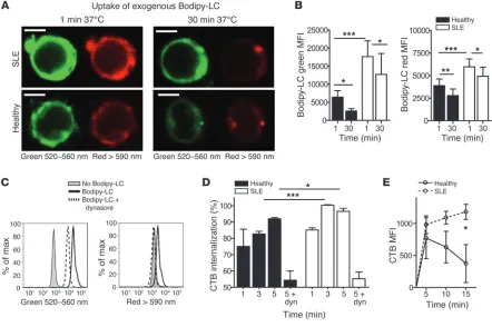
Increased recycling of GSLs in T cells from SLE patients. CD4+ T cells from 6 SLE patients and 6 healthy donors were incubated with Bodipy-LC
on ice, then washed and cultured for up to 30 minutes at 37°C to allow endocytosis before analysis by confocal microscopy and flow cytometry. (A) Representative confocal microscopy images of Bodipy-LC at 1 and 30 minutes. Bodipy-LC was excited at 450 to 490 nm and viewed at 520 to 560 nm (green) or >590 nm (red). Scale bars: 5 μM. (B) Quantitative flow cytometric data showing Bodipy-LC emissions at 520 to 560 nm (left) and >590 nm (right). Paired Student’s t test, *P = 0.05 and **P = 0.005; 2-tailed Student’s t test, ***P ≤ 0.05. (C) Experiment in B was repeated ± dynasore to inhibit endocytosis. Representative histograms. (D) CD4+ T cells from 5 healthy donors and 5 SLE patients were labeled with
CTB-FITC. Cells were washed and incubated at 37°C for 1, 3, or 5 minutes to allow endocytosis of CTB-stained lipids. Cells were washed in neutral or acidic PBS, and lipid internalization was calculated as a ratio of the two. As a control, cells were preincubated with dynasore (dyn). Two-tailed Student’s t test; *P = 0.04; ***P = 0.0005. (E) Surface GSLs on CD4+ T cells from 5 healthy donors and 5 SLE patients were blocked with
research article
LXRβ stimulation via altered serum lipids and T cell activation. They also suggest that the homeostatic mechanisms controlling balanced GSL expression levels were dysregulated in CD4+ T cells
from SLE patients, resulting in an accumulation of cellular GSL.
GSL recycling and turnover is altered in T cells from SLE patients com-pared with those from healthy controls. Stimulation of LXR is known to directly control expression of NPC1 and NPC2 proteins, which regulate cellular GSL transport and recycling (27). Since expression of these genes was also increased in CD4+ T cells from
patients with SLE (Figure 2D), we investigated whether abnormal GSL trafficking contributed to their increased surface expression in SLE patients. We used a fluorescently labeled exogenous probe,
Bodipy-LC, which emits fluorescence at differential wavelengths depending on its cellular location (520–560 nm [green] when incorporated into the plasma membrane and cytosol or >590 nm [red] when concentrated into intracellular compartments includ-ing endosomes or lysosomes) (28). At baseline, T cells from SLE patients had incorporated significantly more Bodipy-LC into both the plasma membrane and cytosol (green fluorescence) and intracellular compartments (red fluorescence) compared with healthy controls (1-minute time point Figure 4A, left panels, and Figure 4B). After 30 minutes, T cells from SLE patients retained higher levels of Bodipy-LC, whereas these levels were significantly reduced in T cells from healthy controls (Figure 4A, right
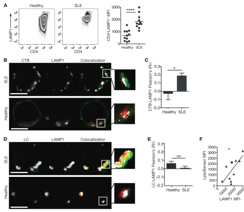
pan-Figure 5
Increased LAMP1 expression and altered lipid colocalization to functionally normal lysosomes in SLE CD4+ T cells. (A) PBMCs from 11 healthy
donors and 11 SLE patients were surface stained for CD4-APC before fixing, permeabilizing, and staining for intracellular LAMP1-PE (CD107a-PE). Representative dot plots and cumulative data. Two-tailed Student’s t test; ****P = 0.00001. (B–E) CD4+ T cells from 6 SLE patients and 3
[image:7.585.48.540.79.507.2]research article
els, and Figure 4B). Although T cells from SLE patients incorpo-rated and retained significantly more Bodipy-LC than those from healthy donors, the rate of reduction of this probe in the plasma membrane, cytosol, and intracellular compartments was compara-ble over time (Supplemental Figure 4A).
We found that the uptake of Bodipy-LC was an active process, since inhibition of clathrin-independent endocytosis (known to be responsible for GSL internalization; ref. 28) using the dynamin inhibitor dynasore or the Src kinase inhibitor PP2 inhibited the probe’s uptake and reduced its incorporation into intracellular compartments (Figure 4C and data not shown, respectively).
We next investigated both the internalization of endogenous GSLs from the plasma membrane into intracellular compart-ments and their recycling from intracellular compartcompart-ments back to the plasma membrane. We observed that GSL internalization was prompt in CD4+ T cells from both healthy donors and SLE
patients. However, we found that GSL internalization was signifi-cantly more rapid at the 3- and 5-minute time points in T cells from SLE patients compared with those from healthy donors (Fig-ure 4D and data not shown for LC).
Recycling of GSLs back to the plasma membrane was also rapid in T cells from both SLE patients and healthy donors. However, we observed that GSL recycling was more sustained over a 15-minute time course in T cells from SLE patients than in those from healthy donors in whom GSL recycling declined over time (Figure 4E and data not shown for LC).
Thus, elevated cellular GSL levels were associated with acceler-ated and sustained GSL internalization and recycling dynamics in CD4+ T cells from SLE patients, leading to a net increase in plasma
membrane expression.
Increased retention of cellular GSLs in CD4+T cells from SLE patients.
[image:8.585.57.531.82.328.2]Since cellular GSL expression levels are controlled by lysosomal and/or late endosomal degradation (11), we investigated whether GSL degradation was also defective in T cells from SLE patients compared with those from healthy donors. First, we observed that the expression of the lysosomal marker lysosomal-associated membrane protein 1 (LAMP1) was significantly increased in T cells from SLE patients compared with those from controls (Figure 5A). We confirmed by confocal microscopy that expression of LAMP1, LC, and CTB binding was elevated in T cells from SLE patients (Fig-ure 5, B and D). We observed that CTB distribution was more clus-tered in T cells from patients compared with those from healthy and mirrored the pattern of LAMP1 distribution. This was associ-ated with increased CTB and LAMP1 colocalization in SLE patients compared with that observed in healthy donors (Figure 5, B and C). We detected no differences in LC or LAMP1 colocalization (Figure 5, D and E); however, the overall levels of colocalization between GSLs and LAMP1 were low in the T cells from both healthy donors and SLE patients (Figure 5, C and E). On close examination, the distribution pattern for GSLs and LAMP1 suggested subcompart-mentalization of the lyso somes between protein-rich (LAMP1) and lipid-rich (CTB/LC) regions (Figure 5, B and D, enlarged insets).
Figure 6
Inhibition of GSL biosynthesis modifies GSL recycling in T cells from SLE patients. (A) PBMCs from 24 SLE patients and 11 healthy donors were TCR stimulated for 72 hours with or without 10 μM NB-DNJ. Cells were surface stained using fluorescently labeled antibodies against CD4-v450, LC-PE-Cy5, or CTB-FITC and assessed by flow cytometry. Cumulative data showing expression of CTB and LC. Two-tailed Student’s t test; *P ≤ 0.05. (B) CD4+ T cells from 5 SLE patients and 5 healthy donors were cultured for 72 hours with or without NB-DNJ. Internalization of
endoge-nous GSLs after a 5-minute incubation was assessed as described in Figure 4D. Cumulative data showing the effect of NB-DNJ treatment on CTB internalization. Two-tailed Student’s t test; *P ≤ 0.05. CD4+ T cells from 3 SLE patients and 3 healthy donors were cultured for 72 hours with or
with-out NB-DNJ. Uptake of Bodipy-LC was assessed as described in Figure 4, A and B. (C) Line graphs showing uptake of Bodipy-LC by flow cytometry. Paired Student’s t-test for SLE samples; *P ≤ 0.01. (D) Representative confocal images showing merged green and red Bodipy-LC accumulation in T cells after a 30-minute incubation. Scale bars: 5 μM. (E) CD4+ T cells isolated from 4 patients with SLE were cultured for 72 hours with or without
research article
Figure 7
NB-DNJ modifies TCR-associated defects in T cells from patients with SLE. PBMCs from 5 SLE patients and 5 healthy donors were cultured for 7 days with NB-DNJ. Cells were labeled with anti-CD3/CD28 (1 μg/ml) and cross-linking anti-mouse IgG on ice before culturing at 37°C for 1 and 5 minutes. Cells were fixed and labeled with fluorescent barcoding reagents and stained for CD4-APC and intracellularly stained for the phosphorylated signaling proteins pTCR-ζ, pERK, pAKT, and pNF-κB. (A) Cumulative results from three separate experiments. Two-tailed Stu-dent’s t test; *P ≤ 0.05 or paired t test; **P = 0.001. PBMCs from 24 SLE patients and 11 healthy donors were TCR stimulated for 72 hours with anti-CD3/CD28 (1 μg/ml) with or without 10 μM NB-DNJ. Cell culture supernatants were collected. Cells were surface stained with CD4-v450 and intracellularly stained for Ki67-PE and assessed by flow cytometry. (B) Representative flow cytometric dot plots (C) and cumulative results showing Ki67 expression as the percentage change from unstimulated controls. Two-tailed Student’s t test; *P ≤ 0.05. Cell culture supernatants from 12 SLE patients and 10 healthy donors collected from B were analyzed by CBA. Representative CBA plots (D) and cumulative data (E) are shown. Two-tailed Student’s t test and paired t test; *P ≤ 0.05. (F) CD4+ T cells isolated from 4 SLE patients were pretreated with NB-DNJ, then
research article
No significant differences were noted in early endosome antigen 1 expression or colocalization with LC or CTB in T cells from healthy donors and SLE patients (Supplemental Figure 4, B–D).
We tested whether lysosomes in T cells from SLE patients were defective and unable to process excess levels of intracellular GSLs. Using a probe to detect lysosomal acidity (LysoSensor Green DND-189), we found a significant positive correlation between LAMP1 and LysoSensor staining in T cells from SLE patients, sug-gesting that although the lysosomes were increased in number, they were functionally normal (in terms of acidity) (Figure 5F).
Therefore, the results imply that the normal mechanisms con-trolling GSL expression were overwhelmed by increased GSL bio-synthesis in T cells from SLE patients. This culminated in acceler-ated GSL trafficking and the accumulation of GSLs in the plasma membrane and intracellular compartments, despite apparently normal lysosome function.
Inhibition of GSL biosynthesis reduced GSL expression and trafficking in CD4+T cells from patients with SLE. GSL accumulation, which
interferes with GSL trafficking and lysosomal degradation, is also a feature of patients with LSDs (12). Blocking GSL synthe-sis using specific inhibitors, including NB-DNJ (a reversible com-petitive inhibitor of glucosylceramide synthase; ref. 29), rectifies defective GSL homeostasis and is used therapeutically in patients (15). Therefore, we assessed the in vitro effect of NB-DNJ on T cells from SLE patients. Culture for 72 hours in the presence of NB-DNJ diminished TCR-induced upregulation of CTB bind-ing and LC and GB3 expression in T cells from SLE patients to the levels observed in T cells from healthy donors (Figure 6A and data not shown for GB3). This was accompanied by a significantly reduced rate of internalization of endogenous GSL (Figure 6B and
data not shown for LC) and reduced uptake of exogenous Bodipy- LC by T cells from SLE patients (Figure 6C). Furthermore, culture with NB-DNJ altered the intracellular distribution of internalized Bodipy-LC in T cells from SLE patients in a pattern that we con-sistently observed in T cells from healthy donors (Figure 6D) and was associated with a reduced expression of LAMP1 in T cells from SLE patients (Figure 6E).
NB-DNJ moderated TCR-associated defects in T cells from patients with SLE. Since culture with NB-DNJ was able to restore a healthy pat-tern of GSL expression and trafficking in T cells from patients with SLE, we assessed whether it could also reverse some of the in vitro lipid raft–associated defects associated with these T cells (2, 3). We cultured PBMCs with and without NB-DNJ (10 μM) for 7 days to ensure that endogenous GSL levels were reduced. We then stimulated CD4+ T cells with anti-CD3/28 antibodies and
used flow cytometry to measure the phosphorylation of key sig-naling molecules. As described previously, sigsig-naling molecule phosphorylation patterns are altered in T cells from SLE patients compared with those from healthy donors (2, 3). However, we found that TCR-mediated phosphorylation of TCR-ζ, ERK, and NF-κB was partially restored in NB-DNJ–treated T cells from SLE patients, while we detected no significant effect on AKT phospho-rylation (Figure 7A). The gating strategy for the analysis is shown in Supplemental Figure 5, A and B.
[image:10.585.42.343.83.384.2]As expected, moderation of T cell signaling by NB-DNJ was associated with the partial normalization of some T cell func-tions. This included reduced proliferation, as measured by Ki67 expression (Figure 7, B and C) and thymidine incorporation (Sup-plemental Figure 5C), and altered regulation of IL-6, IL-10, and TNF-α production (Figure 7, D and E). In addition, we observed
Figure 8
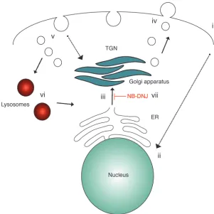
Proposed model for altered GSL expression and metabolism in CD4+ T cells from SLE patients. We
propose that several components of SLE pathol-ogy, including dyslipidemia and T cell hyperactivity (i), elicit upregulation of the nuclear receptor LXRβ, and hence its downstream target genes NPC1 and
NPC2 (ii). Enhanced activation of LXRβ induces increased de novo GSL biosynthesis (iii). CD4+ T
cells from SLE patients are characterized by accel-erated GSL trafficking and accumulation of GSL in the plasma membrane (iv) and intracellular compart-ments (v), despite higher levels of functionally nor-mal lysosomes (vi). Inhibition of GSL biosynthesis with NB-DNJ reduced cellular GSL levels and mod-ified cell function in CD4+ T cells from SLE patients
research article
acterized by increased recycling of TCR-associated molecules, including CD4 and CTLA-4, which affects their activation and inhibitory functions, respectively (37).
Therefore, an altered plasma membrane GSL profile could influ-ence the balance between positive and negative signals transduced during activation and contribute to the characteristic T cell dys-function seen in SLE patients. This proposition is supported by work showing that in vitro cross-linking of GSLs elicits different functional responses in CD4+ T cells from healthy donors compared
with those from SLE patients (E. Jury, unpublished observations). The presence of anti-GSL antibodies in the serum of a small per-centage of SLE patients has been reported previously (38). However, anti-GSL antibodies were not detected in our patient cohort (E. Jury, unpublished observations). Nevertheless, it remains uncertain how differential GSL expression alters T cell function in vivo, although it seems likely that GSLs play an essential role in modifying signals transduced via TCR and costimulatory molecule activation.
Our results imply that more than one factor influences increased GSL levels in patients. In addition to TCR stimulation, activation of lipid biosynthesis pathways using an LXR agonist induced a sus-tained increase in GSL levels in CD4+ T cells from SLE patients, but
not in T cells from healthy controls. This was associated with an increased ex vivo expression of LXRβ, but not LXRα, and some LXRβ
target genes in T cells from SLE patients. To date, little is known about the interaction between TCR-stimulation and lipid homeo-stasis. Tontonoz et al. showed that TCR stimulation downregulates some LXRβ target genes involved in cholesterol efflux (39). Crucially, this suggests that the functional capacity of immune cells can be reg-ulated by mechanisms controlling lipid homeostasis (25, 39).
The results of our study point to an unrecognized function for LXRβ stimulation in the control of T cell GSL expression. Contrary to previous reports examining LXRβ expression in macrophages, we found that GW3965 and oxLDL stimulation upregulated LXRβ
protein and mRNA expression in human T cells (Figure 3 and ref. 40). Since differences in LXR stimulation exist between human and murine macrophages and between different cell types (41), this upregulation could possibly be due to an unrecognized human T cell–specific LXR enhancer. However, we cannot rule out a role for LXRα in lipid-mediated T cell function, although no difference in ex vivo (Figure 2D) or GW3965-stimulated LXRα expression (data not shown) was found in T cells from healthy donors and SLE patients.
Since levels of cellular cholesterol and GSL are closely associated (12, 42), it seems likely that alterations in cholesterol homeosta-sis could also contribute to changes in GSL expression in T cells from SLE patients. Two primary features of LSDs are an aberrant trafficking of GSLs coupled with cholesterol accumulation and an inability of lysosomes to degrade excess glycolipids (12). A similar phenotype is observed in T cells from SLE patients, since choles-terol measured by filipin binding is also elevated (2). Interestingly,
SREBP2 was upregulated in ex vivo T cells from SLE patients. SREBP2 activates cholesterol synthesis, indirectly activates LXR, and can transactivate NPC1/2 genes (24, 43). Thus, it is clear that more work is needed to fully understand the complex interactions in human T cells that occur between LXRβ, SREBP2, and associ-ated target genes to maintain GSL homeostasis, lipid raft stability, and lipid raft function in health and autoimmunity.
The exact nature of the potential LXR agonists present in the serum or cells of patients needs to be determined. Dyslipidemia is common in SLE patients, and several studies have reported reduced levels of HDL, increased levels of triglycerides, and increased LDL that apoptosis, as measured by annexin V binding, was reduced
(data not shown).
Finally, we found that NB-DNJ reduced the capacity of T cells from SLE patients to drive anti-dsDNA antibody production by autologous B cells (Figure 7F). Thus, we show that NB-DNJ, a suc-cessful, clinically approved therapy for LSDs, could rectify some of the defects associated with abnormal T cell function in SLE patients.
Discussion
We report a number of findings in support of our hypothesis that alterations in the control of lipid metabolism underlie abnormal T cell function in patients with SLE. First, we demonstrate a pro-found alteration in the GSL profile of T cells from SLE patients. Second, we show that defects in GSL biosynthesis were driven, in part, by stimulation of the lipid-responsive nuclear receptor LXRβ. Finally, we reveal that NB-DNJ, a successful, clinically approved therapy for patients with LSDs, normalized GSL expression and rectified some of the signaling and functional abnormalities char-acteristic of T cells from SLE patients. A scheme outlining the pro-posed mechanism for altered GSL expression in T cells from SLE patients is shown in Figure 8.
The consequences of GSL accumulation have been studied exten-sively in experimental models and in patients with LSDs. These diseases result from mutations in single proteins involved in the control of GSL trafficking or metabolism (12). Patients are charac-terized by severe neurological, renal, and cardiac deficiencies attrib-uted to GSL accumulation in organs and tissues as well as by multi-ple immunological defects (30). Unlike most of the inherited LSDs, which are characterized by the accumulation of a specific class of GSL, we found that the accumulation of GSLs in CD4+ T cells from
SLE patients was not restricted to a specific GSL species and that the precise GSL profile varied between patients. This observation suggests that the increased GSL expression was not associated with a specific defect in synthase enzyme expression or activity. However, although we did not detect any significant change in the expression of key GSL synthase enzymes by Western blot analysis, this does not rule out the possibility of a defect in enzyme activity or in one of the other enzymes that control GSL expression (31).
There is a growing appreciation that GSL heterogeneity con-tributes to membrane protein compartmentalization in resting and activated T cells. Within T cells, the distribution of GSLs is heterogeneous; for example, GM1 and GM3 are distributed asym-metrically toward the leading edge or the uropod of T cells, respec-tively (32). The essential proximal TCR signaling molecules LCK and LAT reside in different lipid raft subsets that fuse upon TCR stimulation (33), and the interaction of TCR-associated signaling molecules (including LCK, LFA1, and PI3K) with GM3 or GM1 induces differential recruitment of downstream signaling targets that affect T cell function (34, 35). Moreover, recent reports show that CD4+ and CD8+T cells exclusively require a-series and o-series
GSLs, respectively, for TCR-mediated activation (8, 36). The reduc-tion of cellular GSL levels by pharmacological means or by disrup-tion of GM3 synthase activity can inhibit the differentiadisrup-tion of CD4+ T cells into Th17 cells (7) and reduce cytokine production in
activated T cells during viral infection (31).
char-research article
qPCR
RNA was extracted from CD4+ T cells with TRIzol (Invitrogen) according
to the manufacturer’s instructions. Sample concentration and purity were determined by spectrophotometric analysis. RT-PCR and qPCR were per-formed using primers for LXRB, NPC1, NPC2, ABCA1, ABCG1, SREBP2, and the housekeeping gene GAPDH (QIAGEN).
Cell cultures
T cell activation. PBMCs or CD4+ T cells were cultured in complete medium
(CM) (RPMI 1640 containing L-glutamine and NaHCO3 [Sigma-Aldrich]
with 10% FCS [Lonza], and 100 U/μg/ml penicillin-streptomycin [Sigma-Aldrich] at 37°C in 5% CO2); 1 μg/ml anti-CD3 (HIT-3A); and
1 μg/ml anti-CD28 (CD28.2) (eBioscience). Cells were cultured as above with or without GSL biosynthesis inhibitors (100 μM D-PDMP; Sigma-Aldrich or 10 μM NB-DNJ).
LXR agonist experiments. PBMCs were cultured in CM with or without 1 μM GW3965 (Sigma Aldrich) for 24 hours and 72 hours.
Serum experiments. PBMCs were cultured in CM or RPMI and 10% serum from SLE patients or healthy donors plus IL-2 (0.5 ng/ml) for 24 hours, 3 days, 7 days, or 10 days. The CM was replenished every 3 days.
Antagonist experiments. PBMCs or CD4+ T cells were cultured with 1 μM
GW3965, anti-CD3/28 (each at 1 μg/ml), or serum from SLE patients or healthy donors with or without an LXR antagonist (10 μM 5C PPSS-50; Wako) for 24 and 72 hours in CM.
T and B cell cocultures. PBMCs were cultured with or without 10 μM NB-DNJ in CM for 24 hours. CD4+ T cells were then negatively isolated and
resuspended in CM with autologous B cells negatively isolated from a freshly thawed aliquot. T and B cells were cocultured for 7 days with or without anti-CD3/CD28 (1 μg/ml each) and with or without NB-DNJ (10 μM). Supernatants were removed on day 7 and analyzed by ELISA (Euro Diag-nostica) for the presence of anti-IgG dsDNA antibodies.
Flow cytometry
All surface stains were performed in 1% FCS and 0.01% sodium azide in PBS on ice. All data were collected on an LSRII (BD) and analyzed with FlowJo software (Tree Star Inc.).
GSL expression. PBMCs were stained with either CTB-FITC (Sigma-Aldrich), LC-PE-cyano dye 5 (Cy5), LC-FITC (LifeSpan Biosciences Inc.), or CD77-FITC (Gb3-FITC), followed by CD4-APC or CD4-BD Horizon V450 (BD Biosciences).
Assessment of Ki67, LAMP1 (CD107a), and LXRβexpression. Cells were surface stained for 60 minutes before fixation and permeabilization (Fix/Perm Buf-fer; eBioscience) then stained intracellularly with anti-Ki67–PE (BD Biosci-ences), anti–CD107a-PE (eBioscience), or purified rabbit anti-human LXRβ
IgG (Abcam), followed by anti-rabbit IgG FITC (Sigma-Aldrich). In addition, the appropriate isotype or fluorescence-minus-one controls were used.
Detection of phosphorylated signaling proteins and fluorescent barcoding. PBMCs from SLE patients and healthy donors were stimulated and labeled with fluorescent cell barcoding dye according to the manufactur-er’s instructions (BD Biosciences). The procedure is described in detail in the Supplemental Methods.
Lysosomal pH analysis. PBMCs were surface stained with CD4-APC (BD Biosciences), washed, and resuspended in LysoSensor Green DND-153 (Molecular Probes) for 30 minutes at 37°C in 5% CO2.
Measurement of GSL trafficking
Internalization of Bodipy-LC. CD4+ T cells from SLE patients and healthy
donors were either labeled ex vivo, cultured overnight in CM with or with-out 10 μM NB-DNJ, or preincubated with 50 μM dynasore (Invitrogen) or 50 μM Src kinase inhibitor PP2 (Calbiochem). Samples were incubated with 10 μg/ml Bodipy-LC (Invitrogen) on ice for 30 minutes, then at 37°C
oxidation, which can be associated with increased disease activity and proinflammatory cytokine production (1, 44). Thus, it seems likely that defects in GSL expression in SLE patients could be driven by serum dyslipidemia. Indeed, many patients included in the present study had altered lipid levels (Supplemental Table 1).
The ability of inhibitors of GSL biosynthesis to restore defects in lipid (GSL and cholesterol) homeostasis and to inhibit T cell pro-liferation, cytokine production, and differentiation has been iden-tified previously in cell lines and animal models (7, 45). We found that NB-DNJ treatment of CD4+ T cells from patients with SLE
modified their T cell function in response to in vitro TCR stim-ulation (Figure 7). Critically, this was achieved by reducing GSL levels and modifying GSL recycling and accumulation, effects also seen in cells from patients with LSDs who were treated with GSL reduction therapies (12, 46). Interestingly, we found that NB-DNJ increased TNF-α production in T cells from SLE patients, but decreased its production in T cells from healthy donors. The role of TNF-α in human SLE is controversial; it can cause tissue dam-age via its proinflammatory activity or be beneficial by dampening autoimmune responses. Such heterogeneity of effect could be due to changes in membrane lipids that influence immune cell func-tion (5). Blocking TNF-α therapeutically has not been accepted in clinical practice (47). There are several precedents for the thera-peutic control of lipids, examples of which include HIV infection (48), cancer (49), and atherosclerosis (50). Thus, a growing body of evidence supports the hypothesis that correct cellular lipid metabolism regulates the capacity of T cells to correctly respond to microenvironmental stimuli and that therapeutic targeting of such molecules could influence immune cell activation and help regulate abnormal immune responses in autoimmunity.
In summary, we provide evidence of a profound dysregulation in both cellular and surface GSL expression in CD4+ T cells from SLE
patients. This dysregulation was due to increased GSL biosynthe-sis driven, at least in part, by serum dyslipidemia and T cell hyper-activity in SLE patients. Abnormal GSL levels and a range of func-tional cellular defects were rectified by targeting GSL biosynthesis with a clinically approved inhibitor. Our findings reveal a role for defects in lipid metabolism in SLE pathogenesis and highlight what we believe to be a novel treatment strategy for the disease.
Methods
Patients and controls
Peripheral blood was obtained from SLE patients attending the lupus clinic at University College Hospital (London, United Kingdom). Supplemental Table 1 provides a summary of the healthy donors included in this study as well as the treatments and clinical characteristics of the SLE patients.
PBMC and CD4+ T cell isolation
PBMCs were isolated from heparinized blood using Ficoll-Paque PLUS (GE Healthcare) gradient centrifugation. PBMCs (107 cells/ml) were resuspended
in FCS (Biosera) and 10% DMSO (Sigma-Aldrich) and frozen until use. CD4+
T cells were isolated by negative selection (MACS; Miltenyi Biotec).
Lipid extraction, GSL glycan labeling, and HPLC analysis
Lipids were extracted from CD4+ T cells (107 cells) using
research article
using a 2-tailed Student’s t test, a paired Student’s t test, or 1-way ANOVA as specified. P values less than 0.05 were considered significant. Data were tested for normal distribution using the Kolmogorov-Smirnov test with GraphPad Prism software.
Study approval
Ethical approval (00/0241, 11/H0808/19) was obtained for this study from the ethics committee of the University College London Hospitals National Health Service Trust (London, United Kingdom). All participating patients and healthy donors provided written informed consent.
Acknowledgments
Arthritis Research UK supports E.C. Jury (18106) and G. McDon-ald (19607). A University College London Hospital (CRDC) project grant (GCT/2008/EJ) and Lupus UK pilot study award supported L. Miguel. A.I. Magee was supported by MRC grant G0700771. T.D. Butters thanks the Glycobiology Institute of the University of Oxford for support. We thank P. Kabouridis, F. Flores-Borja, and I. Pineda Torra for their critical evaluation of the manuscript.
Received for publication March 15, 2013, and accepted in revised form October 24, 2013.
Address correspondence to: Elizabeth Jury, Centre for Rheuma-tology Research, University College London, Rayne Building, Lon-don W1CE 6JF, United Kindgom. Phone: 44.0.20.3108.2154; Fax: 44.0.20.3108.2152; E-mail: [email protected].
for 1 to 30 minutes. Cells were fixed and analyzed by confocal microscopy and flow cytometry. Detailed procedures for Bodipy-LC labeling and detec-tion, internalization of endogenous plasma membrane GSLs, and recycling of endogenous GSLs to the cell surface from intracellular compartments are described in the Supplemental Methods.
Analysis of cell culture supernatants
Detection of cytokines from purified CD4+ T cell culture supernatants
was performed by cytokine bead array (CBA) analysis (BD Bioscience) according to the manufacturer’s instructions. Samples were analyzed on a FACSArray Bioanalyzer (BD).
Quantitative immunoblots
CD4+ T cells (107 cells) were lysed and analyzed by Western blotting using
mouse anti-LXRβ (Abcam) or anti-actin mAb (Sigma-Aldrich). Adobe Photo-shop was used for quantification of the protein bands (Adobe Systems Inc.).
Confocal microscopy
CD4+ T cells were fixed, permeabilized, and blocked before staining for
lysosomes (anti-LAMP1 IgG1 and anti-IgG1 Alexa 555), early endosomes (EEA1 IgG and IgG-Alexa 633), and either LC IgM and anti-IgM Alexa Fluor 488 or CTB-FITC. Detailed microscopy procedures are described in the Supplemental Methods.
Statistics
All values are expressed as the absolute mean ± SEM. Analysis for signifi-cance was performed with GraphPad Prism software (GraphPad Software)
1. Rahman A, Isenberg DA. Systemic lupus erythema-tosus. N Engl J Med. 2008;358(9):929–939. 2. Jury EC, Flores-Borja F, Kabouridis PS. Lipid rafts
in T cell signalling and disease. Semin Cell Dev Biol. 2007;18(5):608–615.
3. Moulton VR, Tsokos GC. Abnormalities of T cell signaling in systemic lupus erythematosus. Arthritis Res Ther. 2011;13(2):207.
4. Degroote S, Wolthoorn J, van Meer G. The cell biology of glycosphingolipids. Semin Cell Dev Biol. 2004;15(4):375–387.
5. Simons K, Toomre D. Lipid rafts and signal transduction. Nat Rev Mol Cell Biol. 2000;1(1):31–39. 6. Garofalo T, et al. Association of GM3 with Zap-70 induced by T cell activation in plasma mem-brane microdomains: GM3 as a marker of micro-domains in human lymphocytes. J Biol Chem. 2002; 277(13):11233–11238.
7. Zhu Y, et al. Lowering glycosphingolipid levels in CD4+ T cells attenuates T cell receptor signaling,
cytokine production, and differentiation to the Th17 lineage. J Biol Chem. 2011;286(17):14787–14794. 8. Nagafuku M, et al. CD4 and CD8 T cells require
different membrane gangliosides for activation. Proc Natl Acad Sci U S A. 2012;109(6):E336–E342. 9. Morales A, Colell A, Mari M, Garcia-Ruiz C,
Fer-nandez-Checa JC. Glycosphingolipids and mito-chondria: role in apoptosis and disease. Glycoconj J. 2004;20(9):579–588.
10. Gault CR, Obeid LM, Hannun YA. An overview of sphingolipid metabolism: from synthesis to break-down. Adv Exp Med Biol. 2010;688:1–23. 11. Tettamanti G. Ganglioside/glycosphingolipid
turn-over: new concepts. Glycoconj J. 2004;20(5):301–317. 12. Platt FM, Boland B, van der Spoel AC. The cell
biology of disease: lysosomal storage disorders: the cellular impact of lysosomal dysfunction. J Cell Biol. 2012;199(5):723–734.
13. Fernandez D, Perl A. Metabolic control of T cell activation and death in SLE. Autoimmun Rev. 2009; 8(3):184–189.
14. Jury EC, Isenberg DA, Mauri C, Ehrenstein MR.
Atorvastatin restores Lck expression and lipid raft-associated signaling in T cells from patients with systemic lupus erythematosus. J Immunol. 2006;177(10):7416–7422.
15. Butters TD, Dwek RA, Platt FM. Imino sugar inhibitors for treating the lysosomal glycosphin-golipidoses. Glycobiology. 2005;15(10):43R–52R. 16. Miguel L, et al. Primary human CD4+ T cells
have diverse levels of membrane lipid order that correlate with their function. J Immunol. 2011; 186(6):3505–3516.
17. Neville DC, et al. Analysis of fluorescently labeled glycosphingolipid-derived oligosaccharides follow-ing ceramide glycanase digestion and anthranilic acid labeling. Anal Biochem. 2004;331(2):275–282. 18. Alonzi DS, Neville DC, Lachmann RH, Dwek RA,
Butters TD. Glucosylated free oligosaccharides are biomarkers of endoplasmic-reticulum alpha-glu-cosidase inhibition. Biochem J. 2008;409(2):571–580. 19. Kuziemko GM, Stroh M, Stevens RC. Cholera toxin binding affinity and specificity for gangliosides determined by surface plasmon resonance. Biochem-istry. 1996;35(20):6375–6384.
20. Poole B, Ohkuma S. Effect of weak bases on the int-ralysosomal pH in mouse peritoneal macrophages. J Cell Biol. 1981;90(3):665–669.
21. Zech T, et al. Accumulation of raft lipids in T-cell plasma membrane domains engaged in TCR sig-nalling. EMBO J. 2009;28(5):466–476.
22. Bikman BT, Summers SA. Ceramides as modula-tors of cellular and whole-body metabolism. J Clin Invest. 2011;121(11):4222–4230.
23. Kidani Y, Bensinger SJ. Liver X receptor and perox-isome proliferator-activated receptor as integrators of lipid homeostasis and immunity. Immunol Rev. 2012;249(1):72–83.
24. Horton JD, et al. Combined analysis of oligonucle-otide microarray data from transgenic and knock-out mice identifies direct SREBP target genes. Proc Natl Acad Sci U S A. 2003;100(21):12027–12032. 25. Zelcer N, Tontonoz P. Liver X receptors as
inte-grators of metabolic and inflammatory signaling.
J Clin Invest. 2006;116(3):607–614.
26. Noguchi-Yachide T, Miyachi H, Aoyama H, Aoyama A, Makishima M, Hashimoto Y. Structural develop-ment of liver X receptor (LXR) antagonists derived from thalidomide-related glucosidase inhibitors. Chem Pharm Bull (Tokyo). 2007;55(12):1750–1754. 27. Goldman SD, Krise JP. Niemann-Pick C1 functions
independently of Niemann-Pick C2 in the initial stage of retrograde transport of membrane-impermeable lysosomal cargo. J Biol Chem. 2010;285(7):4983–4994. 28. Sharma DK, Choudhury A, Singh RD, Wheatley
CL, Marks DL, Pagano RE. Glycosphingolipids internalized via caveolar-related endocytosis rap-idly merge with the clathrin pathway in early endo-somes and form microdomains for recycling. J Biol Chem. 2003;278(9):7564–7572.
29. Platt FM, Neises GR, Dwek RA, Butters TD. N-butyl-deoxynojirimycin is a novel inhibitor of glycolipid biosynthesis. J Biol Chem. 1994;269(11):8362–8365. 30. Castaneda JA, Lim MJ, Cooper JD, Pearce DA.
Immune system irregularities in lysosomal storage disorders. Acta Neuropathol. 2008;115(2):159–174. 31. Moore ML, et al. Cutting Edge: Oseltamivir
decreases T cell GM1 expression and inhibits clear-ance of respiratory syncytial virus: potential role of endogenous sialidase in antiviral immunity. J Immu-nol. 2007;178(5):2651–2654.
32. Gomez-Mouton C, et al. Segregation of leading-edge and uropod components into specific lipid rafts during T cell polarization. Proc Natl Acad Sci U S A. 2001;98(17):9642–9647.
33. Schade AE, Levine AD. Lipid raft heterogeneity in human peripheral blood T lymphoblasts: a mech-anism for regulating the initiation of TCR signal transduction. J Immunol. 2002;168(5):2233–2239. 34. Barbat C, et al. p56lck, LFA-1, and PI3K but not
SHP-2 interact with GM1- or GM3-enriched micro-domains in a CD4-p56lck association-dependent manner. Biochem J. 2007;402(3):471–481. 35. Sorice M, et al. Association between GM3 and
research article
36. Inokuchi J, Nagafuku M, Ohno I, Suzuki A. Hetero-geneity of gangliosides among T cell subsets. Cell Mol Life Sci. 2013;70(17):3067–3075.
37. Jury EC, et al. Abnormal CTLA-4 function in T cells from patients with systemic lupus erythematosus. Eur J Immunol. 2010;40(2):569–578.
38. Galeazzi M, et al. Anti-ganglioside antibodies in a large cohort of European patients with systemic lupus erythematosus: clinical, serological, and HLA class II gene associations. European Concerted Action on the Immunogenetics of SLE. J Rheumatol. 2000;27(1):135–141.
39. Bensinger SJ, et al. LXR signaling couples sterol metabolism to proliferation in the acquired immune response. Cell. 2008;134(1):97–111.
40. Laffitte BA, et al. Autoregulation of the human liver X receptor alpha promoter. Mol Cell Biol. 2001; 21(22):7558–7568.
41. Torocsik D, Szanto A, Nagy L. Oxysterol signaling links cholesterol metabolism and inflammation via the liver X receptor in macrophages. Mol Aspects Med. 2009;30(3):134–152.
42. Lingwood CA. Glycosphingolipid functions. Cold Spring Harb Perspect Biol. 2011;3(7):a004788. 43. Gevry N, Schoonjans K, Guay F, Murphy BD.
Cho-lesterol supply and SREBPs modulate transcrip-tion of the Niemann-Pick C-1 gene in steroido-genic tissues. J Lipid Res. 2008;49(5):1024–1033. 44. Svenungsson E, Gunnarsson I, Fei GZ, Lundberg IE,
Klareskog L, Frostegard J. Elevated triglycerides and low levels of high-density lipoprotein as markers of disease activity in association with up-regulation of the tumor necrosis factor alpha/tumor necrosis factor receptor system in systemic lupus erythema-tosus. Arthritis Rheum. 2003;48(9):2533–2540. 45. Glaros EN, et al. Glycosphingolipid accumulation
inhibits cholesterol efflux via the ABCA1/apo lipo-protein A-I pathway: 1-phenyl-2-decanoylamino- 3-morpholino-1-propanol is a novel cholesterol efflux accelerator. J Biol Chem. 2005;280(26):24515–24523. 46. Sillence DJ, et al. Glucosylceramide modulates
membrane traffic along the endocytic pathway. J Lipid Res. 2002;43(11):1837–1845.
47. Zhu LJ, Yang X, Yu XQ. Anti-TNF-alpha therapies in systemic lupus erythematosus. J Biomed Biotech-nol. 2010;2010:465898.
48. Puri A, et al. An inhibitor of glycosphingolipid metabolism blocks HIV-1 infection of primary T-cells. AIDS. 2004;18(6):849–858.
49. Hakomori S, Zhang Y. Glycosphingolipid antigens and cancer therapy. Chem Biol. 1997;4(2):97–104. 50. Ehrenstein MR, Jury EC, Mauri C. Statins for