Journal of Chemical and Pharmaceutical Research, 2016, 8(4):313-317
Research Article
ISSN : 0975-7384
CODEN(USA) : JCPRC5
Chronological assessment of Isoniazid and Rifampicin induced hepatotoxicity
in Wistar Albino Rats
Sherry Joseph Martin and Evan Prince Sabina
*School of Bio Sciences and Technology, VIT University, Vellore - 632014, India
_____________________________________________________________________________________________
ABSTRACT
The anti-tuberculosis drugs Isoniazid (INH) and Rifampicin (RIF) are known to be hepatotoxic agents. Wistar albino rats have been used as a model for INH and RIF induced hepatotoxicity. We have in the present study looked at the chronological progression of liver injury (AST, ALT, ALP), oxidative damage (Super Oxide Dismutase, Catalase, Glutathione peroxidase, Lipid peroxidation) and immune response (IL-6, IL-10 and CD3 levels) to INH
and RIF induced liver injury on the 7th, 14th, 21st and 28th days of treatment. Our study has shown that biochemical
changes precede changes in the immunological parameters. It is also seen that INH and RIF induce hepatotoxicity at a slower pace than other known liver toxins as seen by its progression. An initial pro-inflammatory response with infiltration of T cells, macrophages and natural killer T cells followed by an anti-inflammatory response as seen by increase in CD3 and IL-10 levels. This has shown that Wistar albino rats provide a robust model for immune changes during INH and RIF induced liver injury.
Key words: Isoniazid; Rifampicin; Hepatotoxicity; Cytokines; Antioxidants
_____________________________________________________________________________________________
INTRODUCTION
Isoniazid (INH) and Rifampicin (RIF) which are the mainstay of drugs used in the treatment of tuberculosis are known to cause hepatic injury in some patients on long term treatment[1]. Treatment with INH alone is prone to cause lesser incidence of hepatotoxicity than concomitant treatment with RIF[2]. It has been shown that the metabolism of INH by cytochrome P450 (CYP) enzymes leads to the formation of hepatotoxic metabolites such as hydrazine and acetyl hydrazine[3]. RIF is a known to increase the activity of CYP enzymes thereby increasing the production of INH toxic metabolites[4,5]. This may be the reason for the synergistic effect of INH and RIF in causing hepatotoxicity. It is also known that production of reactive oxygen species and loss of oxidant/antioxidant balance in the hepatocytes is a major cause of hepatic inflammation and injury due to INH and RIF[6]. Previous studies have proven that there is depletion of antioxidants (enzymatic and non-enzymatic) in the liver cells and increase in lipid peroxidation due to reactive oxygen species[6]. This leads to leakage of intracellular enzymes such as Alanine transaminase (ALT), Aspartate transaminase (AST), and Alkaline phosphatase (ALP) that serve as indicators of liver injury.
______________________________________________________________________________
biochemical parameters such as ALT, AST, ALP and for antioxidant measurements (Catalase, Superoxide dismutase, Glutathione-s-transferase, Lipid peroxidation). Recent studies have shown a higher involvement of the immune mechanisms of the animals in the induction and control of liver injury as indicated by the measurements of Interleukins (IL-6, IL-10, TNF-α) and transcription factors such as NF-κB[8,9]. It is also thought that the infiltration of T-cells and Natural killer (NK) cells play a major role in the progression of liver injury. The changes in such immunological parameters occur at a chronologically differing pace as compared to biochemical parameters. Therefore we proposed to study the changes in these immunological parameters in comparison to the biochemical and antioxidant measurements on the 7th, 14th, 21st and 28th days of INH and RIF treatment. This would provide us insight into the correlation between immunological, biochemical and antioxidant measurements.
EXPERIMENTAL SECTION
Female Wistar albino rats were used for this study and obtained from the animal house, VIT University, Vellore. The mean weight of the animals was 149 ± 14 g. Animals were housed in a climate controlled environment at 27°C with a 12-h dark-light cycle. The rats were fed with commercially available pelleted feed from Hindustan Lever Ltd. (Mumbai, India) and had a continuous supply of water for drinking. The experimental procedure was approved by the ethical committee (VIT/IAEC/VIIIth/09) of VIT University, Vellore, India.
Drugs
INH and RIF were obtained from Novus Life Sciences Pvt. Ltd., Mumbai, India. INH was dissolved in saline while RIF was suspended in saline.
Experimental Design
The rats were divided into five groups of six animals each. The first group served as the control and represented the 0th day without induction of toxicity. The animals of groups II to V were administered INH and RIF (50 mg/kg b.w./d) daily. The animals of group II were sacrificed on day 7, followed by group III on day 14, group IV on day 21 and group V on day 28. After 24 h of the last dosage, the rats were decapitated; trunk blood collected and liver procured for biochemical, histopathological, and antioxidant studies.
Biochemical Analysis
Commercially available kits were used to measure the liver marker enzymes Alanine Transaminase (ALT), Aspartate Transaminase (AST), Alkaline Phospatase (ALP) according to the manufacturer’s instruction in the serum samples. Lipid Peroxidation (LPO) [10], Superoxide Dismutase (SOD) [11], Catalase (CAT) [12] and Glutathione Peroxidase (GPx) [13] were measured to assess the antioxidant levels in the rodent livers.
Cytokine levels
The levels of the cytokines IL-6 and IL-10 in the serum of the experimental animals were measured using commercial ELISA kits manufactured by Sigma-Aldrich, India.
Immunohistochemistry
Immunohistochemical analysis was carried out on 5µm sections of liver tissue. After deparaffinizing and rehydration the sections were cooked for 20 min in antigen retrieval buffer. Guinea pig polyclonal anti-CD3 antibody (Dako antibody diluent, 1/100 dilution, New Delhi, India) was used as primary antibody and secondary antibody conjugated with horseradish peroxidase was then added and incubated. Super sensitive (SS) Link label IHC detection system (Biogenex Ltd., Hyderabad, India) was used with haematoxylin as a counterstain.
Statistics
The mean and standard deviation (S.D) were calculated from the data obtained. Statistical analysis was carried out by one way ANOVA.
RESULTS AND DISCUSSION
Progression of biochemical markers of liver injury
Table 1 Chronological effect of INH and RIF on Liver Marker Enzymes in experimental rats
Parameters Control (0 Day) 7th Day 14th Day 21st Day 28th Day
AST (U/L) 80.53±4.11 89.45±3.29 119.65±3.54a*b* 149.21±5.11 a*b*c* 202.15±4.02 a*b*c*
ALT (U/L) 51.89±2.69 65.54±3.58 86.88±3.15a*b* 95.59±4.77 a*b* 112.20±3.22 a*b*c*
ALP (U/L) 86.96±3.22 99.49±3.89 120.33±4.21a*b* 168.32±3.99 a*b*c* 189.77±3.46 a*b*c*
Notes: N=6 animals in each group. The values are expressed as mean ± S.D. Comparisons indicated by lower case letters were made as follows: a – group I vs. group II, III, IV, V; b – group II vs. group III, IV , V; c – group III vs. group IV, V; d – group IV vs. group V. Statistical analysis
was carried out by one way ANOVA
Changes in antioxidant parameters
INH and RIF treatment resulted in impaired antioxidant defense system which was evident from the depletion of antioxidants (SOD, CAT, GPx) and elevated levels of lipid peroxidation (LPO) as seen in Table 2. These followed a linear progression on the 7th, 14th, 21st and 28th days.
Table 2 Chronological effect of INH and RIF on antioxidant levels in experimental rats
Parameters Control (0 Day) 7th Day 14th Day 21st Day 28th Day
SOD (Units/mg protein) 128.22±9.65 121.11±9.54 105.21±10.01a* 91.12±7.21a*b* 72.58±8.59a*b*c*
CAT (Units/min/mg protein) 95.47±5.11 83.72±3.21 78.54±3.78a* 66.81±2.77a*b* 48.45±3.20a*b*c*
GPx (µg of GSH utilized/min/mg protein) 55.12±2.22 48.78±1.52 42.21±1.58a* 35.56±1.10a*b* 25.47±2.69a*b*c*
LPO (nmol/mg protein) 1.28±0.09 1.89±0.11 2.03±0.08a* 2.40±0.09a*b* 3.11±0.12a*b*c*
Notes: N=6 animals in each group. The values are expressed as mean ± S.D. Comparisons indicated by lower case letters were made as follows: a – group I vs. group II, III, IV, V; b – group II vs. group III, IV , V; c – group III vs. group IV, V; d – group IV vs. group V. Statistical analysis
was carried out by one way ANOVA
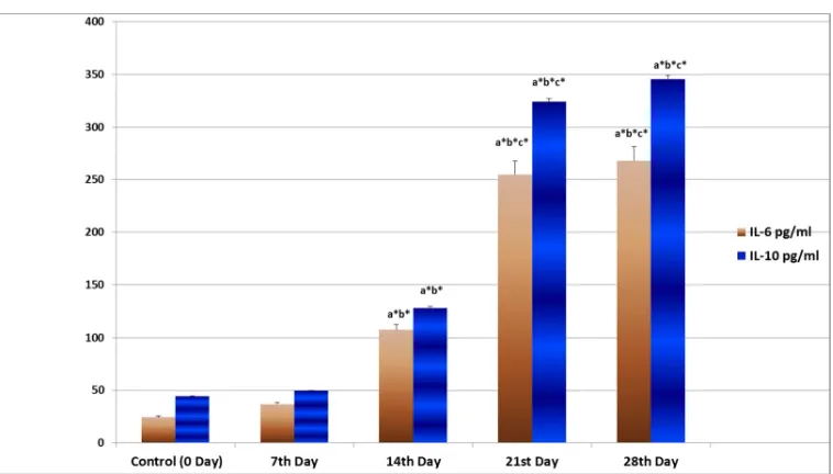
Cytokine levels
[image:3.595.110.489.414.630.2]The levels in IL-6 and IL-10 show an increase after the 21st day with a marginal increase before that as shown in Figure 1.
Figure 1 Time based effect of INH and RIF on IL-6 and IL-10 levels in experimental rats
Notes: N=6 animals in each group. The values are expressed as mean ± S.D. Comparisons indicated by lower case letters were made as follows: a – group I vs. group II, III, IV, V; b – group II vs. group III, IV , V; c – group III vs. group IV, V; d – group IV vs. group V. Statistical analysis
was carried out by one way ANOVA
Levels of CD3
______________________________________________________________________________
Figure 2 Immunohistochemical staining of liver sections for CD3 A) Group 1 (normal control): Negative for CD3; B) Group II (7th Day):
Negative for CD3; C) Group III (14th Day): 3% positive for CD3; D) Group IV (21st Day): 10% positive for CD3; E) Group V (28th Day): 10% positive for CD3
The induction of deleterious hepatic changes has been seen while studying rats as a model for INH and RIF induced hepatotoxicity previously. Various studies have shown that a dosage of 50 mg/kg b.w./day of both INH and RIF given for 28 days induces hepatotoxicity in the rodents. Toxic changes in the liver have been deduced by measuring biochemical variations in blood and liver of the rodents (AST, ALT, ALP and antioxidants). These changes reflect liver damage and inflammation but are not a measure of the immunological changes in the hepatic environment. We have looked at chronological changes in pro and anti-inflammatory cytokines (IL-6 and IL-10) and T-cell infiltration (measured by number of CD3 cells) into the liver in response to the inflammation caused by INH and RIF treatment. These changes when studied along with the biochemical data show the differential pace of biochemical and immunological indices.
Our study has shown that the levels of markers of hepatic injury and the levels of antioxidants in the liver show a linear increase along the 7th, 14th, 21st, and 28th day mark. This is an indication that the damage to hepatocytes is progressive and reaches significant proportions by the 28th day and plateau off on further treatment. The increase in cytokine and CD3 levels though show a lag till the 14th day and then show a significant increase from the 21st day. Though not linear in progression, this can be correlated with the low levels of hepatic injury markers till the 14th day, thereby showing less cytokine production and T-cell infiltration until the biochemical changes reach a significant level.
CONCLUSION
Our results have therefore shown that the immune response to INH and RIF mediated liver injury sets in after the initial liver injury and also that it takes more than 20 days to set in at a significant level in comparison to other hepatic toxins that show more immediate responses. This has shown that Wistar albino rats provide a robust model for immune changes during INH and RIF induced liver injury.
Acknowledgement
The authors would like to thank
1. VIT University, Vellore - 632014, India for providing the necessary facilities for conducting experiments and analyzing results.
2. Council of Scientific and Industrial Research (CSIR), New Delhi, India for providing Senior Research Fellowship grant.
REFERENCES
[1] Saukkonen JJ; Cohn DL; Jasmer RM; Schenker S; Jereb JA; Nolan CM; Peloquin CA; Gordin FM; Nunes D; Strader DB; Bernardo J; Venkataramanan R; Sterling TR; ATS(American Thoracic Society) Hepatotoxicity of Antituberculosis Therapy Subcommittee. Am J Respir Crit Care Med.,2006, 174, 935–952.
[2] Gangadharam PRJ. Am Rev Respir Dis.,1986, 133(6), 963–965. [3] Delaney J; Timbrell JA. Xenobiotica.,1995, 25(12), 1399–1410.
[4] Inui N; Akamatsu T; Uchida S; Tanaka S; Namiki N; Karayama M; Chida K; Watanabe H . Clin Pharmacol The.,2013, 94(6), 702–708.
[5] Skakun NP; Shman’ko VV. Antibiot Meditsinskaia Biotekhnologiia Antibiot Med Biotechnol Minist Meditsinskoĭ
Promyshlennosti SSSR.,1985, 30(3), 185–189.
[6] Attri S; Rana SV; Vaiphei K; Sodhi CP; Katyal R; Goel RC; Nain CK; Singh K. Hum Exp Toxicol.,2000,19(9), 517–22.
[7] Pal R; Rana SV; Vaiphei K; Singh K. Clin Chim Acta.,2008,389(1-2), 55–60. [8] Ziglam HM; Daniels I; Finch RG. J Chemother.,2004,16(4), 357–361. [9] Baskaran UL; Sabina EP. Cell Biol Toxicol2015, 31(4-5), 211–219. [10] Ohkawa H; Ohishi N; Yagi K. Anal Biochem.,1979,95(2), 351–358. [11] Marklund S; Marklund G. Eur J Biochem FEBS.,1974,47(3), 469–474. [12] Sinha AK. Anal Biochem.,1972,47(2), 389–394.
[13] Rotruck JT; Pope AL; Ganther HE; Swanson AB; Hafeman D; Hoekstra WG. Science.,1973,179(4073), 588– 590.
[14] Possamai LA; Thursz MR; Wendon JA; Antoniades CG. J Hepatol.,2014,61(2), 439–45.