Journal of Chemical and Pharmaceutical Research, 2015, 7(1):390-394
Research Article
CODEN(USA) : JCPRC5
ISSN : 0975-7384
Removal of dental plaque formation using bioactive compounds
from sea weeds
Devangana Bhuyan, Madhubanti Mullick, Anamika Das and Jabez W. Osborne*
School of Biosciences and Technology, VIT University, Vellore, Tamil Nadu, India
_________________________________________________________________________________________
ABSTRACT
Biofilms are a consortium of various microorganisms which is a major source for the formation of dental plaques. In this study, biofilm formation on orthodontic brackets and its inhibition was observed. Dental plaque formation is a common problem among the general population in developing as well as developed countries. In the present study, samples of the tooth swabs were taken from people using braces. The samples were inoculated onto LB agar plates and were purified based on their difference in colony morphology. These isolates were maintained in LB agar slants. Further, the isolates were characterized using morphological and biochemical methods. Gram’s staining method revealed the presence of rod shaped bacteria. These strains were used for biofilm formation using test tube method. The formation of biofilm was estimated qualitatively and quantitatively. Sea weeds (Chaetomorpha sp.), known for its production of bioactive compounds were selected. The bioactive compounds were extracted using the solvents-hexane and ethyl acetate. The inhibition activity of both the solvents against biofilm formation was analyzed. The hexane solvent extract was found potentially effective in the inhibition of biofilms on orthodontic brackets in comparison to the ethyl acetate extract.
Key words: Biofilm, Test tube method, Solvent extraction, bioactive compounds (sea weeds), Inhibition.
_____________________________________________________________________________________________
INTRODUCTION
The inclination for bacteria to become surface-bound is extremely ubiquitous in diverse ecosystems and it shows a strong survival and selective advantage for surface dwellers over their free ranging counterparts [1]. The main reason for this is the tendency of nutrients to accumulate near the solid surfaces in aquatic ecosystems. The affinity of bacteria to colonize over surfaces is a double-edged sword, however, that can either prove to be beneficial or potentially destructive [2].
Costerton et al., defined biofilm as “a structural community of bacterial cells enclosed in a self-produced polymeric matrix and adherent to an inert or living surface”. There are three basic components of a biofilm: microbes, glycocalyx and surface. When any one of these three components is removed the biofilm does not develop. A functional degree of organization or cooperatively exists within biofilms to allow maximum interaction with the environment without compromising cell survival or exhausting available resources. A diverse number of microorganisms are capable of generating biofilms, but only bacteria are considered in this study [3].
bacterial adhesion, a very distinct process; an integral part in biofilm formation, is divided into two stages: the primary or docking stage and the secondary or locking phase [5].
When a biofilm is composed of heterogeneous species, the metabolic by-products of one organism may serve to support the growth of another, while the adhesion of one species might provide ligands allowing the attachments of others [6; 7; 8]. Conversely, the competition for nutrients and accumulation of toxic byproducts generated by primary colonizers can limit the species diversity within a biofilm [8]. When a cell switches to biofilm mode of growth; it undergoes a phenotypic shift in behavior in which large shifts of genes are differentially regulated. Growth potential of any bacterial biofilms is limited by the availability of nutrients in the immediate environment, the perfusion of biofilm those nutrients to cells within the biofilms. There is also the existence of an optimum hydrodynamic flow across the biofilm that favors its growth. Other factors that control biofilm maturation include internal pH, oxygen perfusion, carbon source and osmolarity. Recent evidences suggest that the primary development, maturation and breakdown of a biofilm might be regulated at the level of population density dependent gene expression controlled by cell-to-cell signaling molecules such as acylated homoserine lactones.
Biofilms present on the teeth of most animals as dental plaques, which may ultimately result in tooth decay or gum disease. Dental plaque is a film-like deposit on the surface of a tooth consisting of a mixture of mucous, bacteria, food particles etc. It is formed by colonizing bacteria that try to attach themselves onto the smooth surface of a tooth or orthodontic brackets (dental braces). In the present study, the microorganisms are isolated from a tooth swab and assessed for the formation of biofilm and inhibition of dental plaque formation using Chaetomorpha sp.
EXPERIMENTAL SECTION
Sample Collection
The isolation of the microbes for formation of plaques on orthodontic brackets was carried out. The bacteria forming plaques on orthodontic brackets were collected using swabs from individual using permanent orthodontic brackets (dental braces). The sea weed samples for solvent for the study was obtained from the marine sources from Rameshwaram, India. The type of sea weed used in this study is Chaetomorpha sp. The media used in this study were Luria Bertani Media, Trypticase Soy Broth [9].
Isolation of the microbes
The teeth swabs were streaked (quadrant streaking) onto Luria Bertani media and was kept for incubation for 24 h. The pure bacterial colonies were isolated were further subcultured and maintained in LB media.
Characterization of the isolates
The isolates colony morphology was studied. They were morphologically and biochemically characterized by Gram’s staining, Catalase and Oxidase tests [10].
Formation of Biofilm
The isolated and subcultured bacteria were then used for the formation of biofilm using the test tube method. A qualitative analysis of biofilm formation was determined as previously described by Christensen [11]. 10 ml of autoclaved Trypticase Soy Broth (TSB) was inoculated with the isolates obtained from teeth swab and incubated at 37 ̊ C for 24 hours. The tubes were decanted, washed with PBS (pH 7.3) and dried. The dried tubes were stained with 0.1% crystal violet for one minute. The excess stain was removed by washing with running deionized/distilled water. The tubes were then dried in an inverted position and were observed for biofilm formation. Biofilm formation was considered positive when a visible film lined the wall and the bottom of the tube. Ring formation at the liquid interface was not an indication of the biofilm formation. The tubes were examined and the amount of biofilm formation was scored as 0- absent, 1- weak, 2- moderate or 3- strong. The experiments were performed in triplicates and were repeated thrice. The biofilm formed was used in the detection of the activity of bioactive compound in its effective inhibition.
Extraction of bioactive compounds from Chaetomorpha sp.
Coating of bioactive compounds
The extraction of bioactive compounds was performed and the extracted compound was used for coating on orthodontic brackets. The orthodontic brackets were made into individual pieces and the pieces were taken and immersed in beakers containing the precipitate of bioactive compounds, obtained by the solvent extraction method using the two compounds. This setup was kept undisturbed for 2-3 days. The coated orthodontic brackets were taken out and analyzed for inhibition property against biofilm formation.
Biofilm studies on orthodontic brackets
The biofilm studies on orthodontic brackets were done by taking both treated brackets, that is, the one coated with one bioactive compound and untreated brackets and the one without any coating [13]. The brackets considered for the studies were as follows:-
i)Orthodontic bracket + Broth + bacteria = Positive control
ii)Orthodontic bracket + coating with Hexane solvent + Broth +bacteria -Test I
iii)Orthodontic bracket + coating with Ethyl acetate solvent + Broth + bacteria –Test II
The activity of the bioactive compounds on the biofilm inhibition was further analyzed
SEM Analysis of the biofilm
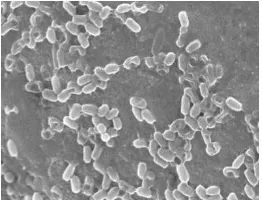
Further confirmation and complete microscopic analysis of the isolates was performed. SEM images of dried thin biofilm samples of all the three respective orthodontic brackets, that is, the positive control, the one treated with hexane solvent were obtained.
RESULTS AND DISCUSSION
Isolation of the strains
Bacterial strains were isolated Luria Bertani media. Two distinct strains were isolated and purified.
Characterization of the isolates
The isolates were found to be gram positive, short-rods, non-endospore forming bacteria. On performing biochemical tests, it was found to be IMVIC negative. It also showed negative and positive for catalase and oxidase, respectively.
Table1. Morphological and Biochemical characterization
Tests Results
Gram’s staining +
Endospore staining -
Indole Test -
Methyl Red -
Voger Proskauer -
Citrate Utilisation Test -
Catalase Test -
Oxidase Test +
Biofilm formation
The microscopic studies of the slides on which the Gram’s staining was performed indicated the presence of a meager number of Gram negative cocci cells. There was a predominance of Gram positive rod shaped cells, which was indicative of the fact that the microorganisms cultured was basically Gram positive bacilli strain[14].
Solvent extraction
Figure 1: Comparative analysis of biofilm formation on orthodontic brackets
Coating on orthodontic brackets
For soliciting the evidence as observed, SEM analysis was carried out of the thin dried film samples of all the three orthodontic brackets along with the positive control. Figure 2a shows the sample treated with hexane solvent. Figure 2b shows the sample treated with ethyl acetate solvent.
[image:4.595.371.502.316.420.2]
Figure 2a: SEM image of sample without bioactive compound Figure 2b: SEM image of sample with bioactive compound
SEM Analysis of biofilm
The SEM images justified the results observed, that the biofilm formation on the orthodontic bracket was maintained as the positive control indicated the presence of Bacilli (rods) cells. In comparison to the bioactive compounds extracted from both the solvents, that is, hexane and ethyl acetate used in this study, hexane showed a greater tendency for inhibition activity against the biofilm formed.
CONCLUSION
From this study we can conclude that the bioactive compounds from the hexane solvent extraction is more effective in its inhibitory activity against biofilm formation on orthodontic brackets as compared to the bioactive compound obtained from ethyl acetate solvent extraction.
Acknowledgments
The authors thank VIT lab technicians, Dr. Elumalai, Pondicherry University, for the SEM Analysis and Mr. Manoj, CMFRI for providing the seaweed sample and VIT management for financial support.
REFERENCES
[image:4.595.110.241.318.420.2][6] JW Costerton; KJ Cheng; GG Geesey; TI Ladd; JC Nickel; M Dasgupta; TJ Marrie. Annu. Rev. Microbiol.,1987, 41:435-464.
[7] JW Leung; YL Liu; T Desta; E Libby; JF Inciardi; K Lam. Gastrointest. Endosc.,1998, 48:250-257.
[8] PD Marsh. Dental plaque. In Lappin-Scott H.M. and Costerton J.W. (ed.), Microbial biofilms. Cambridge University Press, New York, N.Y., 1995, p. 282-300.
[9] KC Marshall. Mechanisms of bacterial adhesion at solid-water interfaces. In Savage D.C. and M. Fletcher (ed.), Bacterial adhesion: mechanisms and physiological significance. Plenum Press, New York, N.Y., 1985, p. 133-141. [10] A Bednarski; R Kranz; K Weston-Hafer; E Richards. Identifying Unknown Bacteria Using Biochemical and Molecular Methods. Washington University in Saint Louis, 2006.
[11] BE Christensen. J. Biotechnology., 1989, 10:181-202.
[12] Warkoyo, Elfis Anis Saati. The solvent effectiveness on extraction process of seaweed pigment.Makara Seri Teknologi. 2011, vol.15.
[13] MG Brading; J Jass; HM Lappin-Scott. Dynamics of bacterial biofilm formation, In Lappin-Scott H.M. and Costerton J.W. (ed.) Microbial biofilms. Cambridge University Press, New York, 1995, p. 46-63.
