NON-INVASIVE EARLY DIAGNOSIS OF DEEP VEIN THROMBOSIS
AHMAD NOOR ARIFF BIN ZAINAL ABIDIN
A project report submitted in partial fulfilment of the requirement for the award of the
Master of Electrical Engineering
Faculty of Electrical and Electronic Engineering Universiti Tun Hussein Onn Malaysia
ABSTRACT
ABSTRAK
Deep Vein Thrombosis (DVT) adalah pembentukan darah beku yang biasa berlaku di
bahagian bawah badan atau peha. Darah beku boleh menyebabkan saluran darah tersumbat dan boleh bergerak ke paru-paru dan menyebabkan arteri pulmonari tersumbat. Keadaan ini boleh menyebabkan Pulmonary Embolism (PE). DVT boleh didiagnosis menggunakan non-invasif teknik seperti ultrasonografi. Salah satu faktor yang menyebabkan Deep Vein Thrombosis adalah tingkah-laku injap vena. Oleh sebab itu, terdapat keperluaan untuk pergerakan injap vena dijejaki untuk menjalankan analisis berkaitan tingkah-lakunya bagi sistem pengesanan awal bagi
Deep Vein Thrombosis. Projek ini bermatlamat untuk mengesan pergerakan injap
TABLE OF CONTENT
STUDENT’S DECLARATION i
ACKNOWLEDGEMENT ii
ABSTRACT iii
ABSTRAK iv
TABLE OF CONTENT v
LIST OF TABLE ix
LIST OF FIGURES x
LIST OF SYMBOLS AND ABBREVIATIONS xii
Chapter 1 1
INTRODUCTION 1
1.1 Introduction 1
1.2 Problem Statement 3
1.3 Aim and Objectives 4
1.4 Scope of Project 5
1.5 Rationale and Significant 5
Chapter 2 7
LITERATURE REVIEW 7
2.1 Introduction 7
2.2 Deep Vein Thrombosis (DVT) Diagnosis 7
2.3 Venous Valve Cycle 8
2.4 Image Processing 11
2.5 Object Detection and Tracking 15
2.6 Summary 16
Chapter 3 17
METHODOLOGY 17
3.1 Introduction 17
3.2 Flowchart of algorithm 17
3.3 Ultrasound Image Acquisition 18
3.4 Region of Interest (ROI) Selection 19
3.5 Conversion from RGB image to Grayscale image 19
3.6 Image Enhancement 20
3.6.1 Contrast-limited Adaptive Histogram Equalization (CLAHE) 21
3.7 Invert Image 24
3.8 Conversion to Binary Image 25
3.9 Vein Valve Tracking 27
Chapter 4 32
RESULT AND DISCUSSION 32
4.1 Introduction 32
4.2 Result of Contrast Enhancement 32
4.3 Result of Noise Filtering 33
4.4 Result of Invert Image 35
4.5 Result for Conversion into Binary Image 36
4.6 Result Vein Valve Tracking 37
4.6.1 Result of Image Differencing Method 37
4.6.2 Result of tracking vein valve movement. 38
CONCLUSION 49
5.1 Introduction 49
5.2 Limitation 49
5.3 Recommendation 50
REFERENCES 52
LIST OF TABLE
LIST OF FIGURES
Figure 1.1: Pulmonary Embolism and Deep Vein Thrombosis 2
Figure 1.2: Example of incompetence of vein valve 3
Figure 2.1: The valve cycle 9
Figure 3.1: Flowchart for development of algorithm 18
Figure 3.2: Create Range of Interest from 1 frame of ultrasound images 19 Figure 3.3 : Conversion from RGB Image to Grayscale Image 20 Figure 3.4: Differences between original histogram and histogram after apply
CLAHE 22
Figure 3.5: Example for window 5x5 23
Figure 3.6: Differences between original image and image after apply Hybrid
Median Filter. 24
Figure 3.7: Image after apply complement method. 25
Figure 3.8: Image after thresholding process 27
Figure 4.3: Result image after apply Median Filter 34
Figure 4.4: Result image after apply Wiener Filter 35
Figure 4.5: Result after applied invert method. 36
Figure 4.6: Result after apply global threshold by using Otsu's Method. 37 Figure 4.7: Result of Image Differencing using imabsdiff function for Fr=2903 and
Fr=2904 38
Figure 4.8: Frame Difference vs Two Consecutive Close Vein Valve Frame
(Observation Method) 39
Figure 4.9: Condition of vein valve during open and close in single frame. The frame
captured from the input ultrasound video. 40
Figure 4.10: Distance between reference line and nearest point for both part (above and below reference line) for frames, Fr=2800 to Fr=3400 41 Figure 4.11: Sequence of distance pattern from frame, Fr=2800to Fr=3050 42 Figure 4.12: Unclear interval between peak to peak is observed 42 Figure 4.13: Sequence of distance pattern from frame, Fr=3150 to Fr=3350 43 Figure 4.14: Frame Difference vs Two Consecutive Close Vein Valve Frame
LIST OF SYMBOLS AND ABBREVIATIONS
DVT Deep Vein Thrombosis
PE Pulmonary Embolism
VTE Venous Thromboembolism
VDUS Venous Duplex Ultrasound
CDI Colour Doppler imaging
RF Radio Frequency
CLAHE Contrast-Limited Adaptive Histogram Equalization
SNR Signal to Noise Ratio
PSNR Peak Signal to Noise Ratio
MSE Mean Square Error
PSO Particle Swarm Optimization
CCA Common Carotid Artery
SRAD Speckle Reducing Anisotropic Diffusion OD&T Object detection and tracking
CHAPTER 1
INTRODUCTION
1.1 Introduction
Deep Vein Thrombosis (DVT) is a blood clot that forms in deep veins which commonly occur in lower limb or thigh. The blood clot blocks deep vein and can travel to lung and block pulmonary arteries. This condition will cause fatal Pulmonary Embolism (PE). Common symptoms of DVT are leg swelling or redness, thigh pain or unilateral calf [1].DVT, PE or combination is broader term that refers as Venous Thromboembolism (VTE) [2]. Figure 1.1 illustrates the concept of Pulmonary Embolism and Deep Vein Thrombosis.
Figure 1.1: Pulmonary Embolism and Deep Vein Thrombosis.[5]
Conventionally, DVT has been diagnosed by contrast venography. This method allows tremendous visualisation of the venous system and identification of both distal and proximal DVT but has limitations. The intravenous contrast usage may be unsafe for pregnancy patient, may lead to renal failure or known allergy, the complex and difficult procedure, expensive, needs professional explanation, and frequently uncomfortable for the patient. All those limitation has led to the exploration for cheaper, simpler, non-invasive tests for DVT [2].
Figure 1.2: Example of incompetence of vein valve [7]
Many researches have been done to study the diagnosis and treatment of patients with high risk of developing VTE. However, there is less research has been done that focus on early diagnosis of DVT especially on deep vein valve behaviour. Therefore, this project will focus on detection and tracking vein valve movement on deep vein ultrasound images (B-mode image) which will contribute to further research on early diagnosis of DVT.
1.2 Problem Statement
The efforts to prevent from VTE diseases are important as the impact of VTE disease severe to the patients. However, there is less research has been done that focus on early diagnosis of DVT. Therefore, there is a need to research the area on early diagnosis of DVT for prevention of DVT.
movement of vein valve need to be tracked to investigate the valve competency for early detection of Deep Vein Thrombosis.
Currently, the diagnosis of Deep Vein Thrombosis (DVT) uses ultrasonography method. The problem that always occurs during interpretation of ultrasound images are degradation of the fine details and edge definition due to speckle noises existence. This problem leads wrong diagnosis. Therefore, the excellent image processing method needs to propose to avoid this problem and help to improve the research.
1.3 Aim and Objectives
The aim of this project is to track the vein valve movement for early diagnosis of DVT. To achieve the aim of this project, the following objectives have been set up:
1. To develop the valve movement tracking algorithm based on image processing technique.
2. To validate the proposed algorithm via a series of experiments.
1.4 Scope of Project
The scopes of the project are:
1. To create an algorithm to detect and tracking popliteal vein valve movement on ultrasound image created by B-Mode images.
2. To use MATLAB® software for the suggested algorithm development.
1.5 Rationale and Significant
The impact of this project is to help the researcher to study the behaviour of venous valve of deep vein for early diagnosis of DVT. The project is intended to develop specific method and algorithm for tracking venous valve movement automatically.
The significant of the research is to study the best possibility method for image enhancement for ultrasound images and the tracking venous valve movement.
1.6 Project Outline
The thesis comprise in 5 chapters together including Chapter 1 is on Introduction, Chapter 2 is Literature Review, Chapter 3 is Methodology, Chapter 4 discuss result and discussion followed by last chapter which is Conclusion.
In Chapter 2, it reviews the previous works from thesis, journals, conference paper and experiments that related to the project. The literature review includes Deep Vein Thrombosis Diagnosis, Venous Valve Cycle, Image Processing and Object Detection and Tracking.
Chapter 3 represents the research methodology of the project. The step by step procedure used to run the project will be explained in details. It will also include the flowchart of processes involved in software development of entire project.
Chapter 4 will contain the simulation results and its respective analysis. The result will be discussed and explained with the aid of diagrams. The comparison of every finding will be explicated in detail.
CHAPTER 2
LITERATURE REVIEW
2.1 Introduction
This chapter present the concept of ultrasound imaging, noise occur on its images and the theory of venous valve closure cycle. Image processing for image enhancement, noise filtering, segmentation and object detection and tracking also has been discussed this chapter.
2.2 Deep Vein Thrombosis (DVT) Diagnosis
DVT can be diagnosed using non-invasive technique such as ultrasonography include assessment of venous condition, use grey-scale image to visual the blood clot in real-time, and recognise areas of venous thrombosis by using Doppler, colour flow and power Doppler imaging [4].
Basically, ultrasound imaging system uses sound waves with high frequency as imaging method. This ultrasound device is non-ionized system therefore it is quite popular for non-invasive diagnostic device. The device is capable to display structure and movement of the human’s internal organs including blood vessels. This imaging system consists of three basic types of data which are Radio Frequency (RF) signals, B-Mode images and envelope-detected signals. In medical application, there are many available ultrasound modes such as A mode, B-mode, colour Doppler and Continuous Doppler [9].
The ultrasound image contains noise such as speckle noise. This noise degrades the fine details and edge definition of ultrasound images [9]. Thus, it is difficult to detect small and low contrast lesions in body due to contrast resolution limit by the noise. All developed noise is due to air gap between ultrasound transducer and body during imaging process, beam forming process and stage of signal processing. Therefore, image enhancement and noise filtering process need to be applied for further analysis and diagnosis of the ultrasound images.
2.3 Venous Valve Cycle
A consistent pattern of flow event is identified in healthy femoral and great saphenous vein when the blood passes through the valve station. Valve cycle, the time period between two consecutive closures of the valve is physiological process of flow events and movement of the valve leaflets combination [10].
Figure 2.1: The valve cycle [10]
Table 2.1: The four phases of the venous valve cycle [10][11]
Phase Information
Opening phase Which the cusps move from the closed position toward the vein wall
Lasts on average 0.27 seconds
Equilibrium phase
Which the valves are no longer opening, but remain suspended open undergoing oscillation or fluttering in the blood flow Lasts on average 0.65seconds
Closing phase Which the valves move synchronously towards the centre of the vein
Lasts on average 0.41 seconds
2.4 Image Processing
[image:22.595.149.536.455.624.2]Basically, image enhancement is about image processing to get better and accurate image to correctly diagnose by doctors. The examples of image enhancement’s techniques are noise filtering, contrast stretching and histogram modification [12]. Noise filtering is used to clean the needless information from an image by using any filter type such as median filter or mean filter. Contrast stretching is technique can be used for image that has homogenous histogram. The technique expanses the narrow range to the entire of the existing dynamic range. Histogram is important to image processing as it provides the characteristic of image such as intensity and pixels. The modification of histogram can be used to change the image’s characteristics. One of the histogram modification techniques is histogram equalization which redistributes pixel values to create flat histogram. Contrast-Limited Adaptive Histogram Equalization (CLAHE) is one of histogram equalization [13].
The quality of filtered images can be evaluate quantitatively with Signal to Noise Ratio (SNR), Peak Signal to Noise Ratio (PSNR), Mean Square Error (MSE), Root Mean Square Error [15][16].
M i N j j i j i M i N j j i j i y x y x SNR 1 2 1 2 , 2 , 1 1 2 , 2 , 10 log .10 ---- (2.1)
MSE g PSNR max 2 10 log .
20 ---- (2.2)
M i N j j i j i y x MN MSE 1 2 1 2 , 2 , 1 ---- (2.3)
M i N j j i j i y x MN RMSE 1 2 1 2 , 2 , 1 ---- (2.4)Where M,N is size of tested image, x is filtered image, y is input image and g is maximum intensity of unfiltered image.
Figure 2.2 Diagram of neighbourhood pixel used in modified hybrid median filter. [9]
In Roomi [16], Particle Swarm Optimization (PSO) technique is introduced to reduce speckle noise in ultrasound image. The technique minimized the variance value in the sample uniform area of image by optimized the selection of weighting factors of neighbourhoods of each corrupted pixel. Then, the algorithm is tested and compared with others filter method such as Active filter, Lee filter, Frost filter, and Modified Hybrid median filter. From the results, the method is proves to remove the speckle noise without compromised the image’s information.
From the experimental results, the proposed technique is accomplished to segment and identify the CCA wall from the ultrasound images.
Figure 2.3: Contour extraction method. [17]
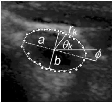
Figure 2.4: Ellipse model for transverse vessel area. The parameters for elliptical model are a, b, ɸ. Each contour point can be described in polar coordinates.[18]
2.5 Object Detection and Tracking
Object detection and tracking (OD&T) is one of the primary objectives for vision application system. It is used in many applications such as face detection, road traffic control and medical imaging. Karasulu[19] states, “object tracking in videos can be defined as the process of segmenting an object of interest from a sequence of video scenes”. In Mihaylova[20], OD&T techniques can be divided generally into two groups which are target object is behaved independently and not behaved independently. For vein valve, the movement is categorised as behaved independently because the movement of valve is only on their own path and not overlapped with each other path.
pixel intensity between current input image and background image. An effective method if the background image and the current input image are aligned [23]. In the frame differencing method, the moving objects existence is determined by calculating the difference between two consecutive images. If the object moves slowly, the differences not clearly identified as the method used previous frame as a reference image. Currently, there are some better methods of frame subtraction [24]. The detection of moving region based on optical flow method is use characteristics of flow vectors of moving objects over time in an image sequence. Even in the camera motion exists, this method can be used to detect independently moving objects. However, this method is required complex computational, very sensitive to noise, and without specialized hardware, this method cannot be applied to video streams in real time [25].
2.6 Summary
CHAPTER 3
METHODOLOGY
3.1 Introduction
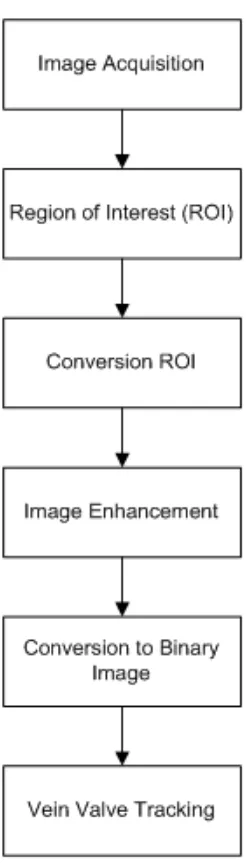
In this chapter, method that will use to track the vein valve is proposed. In this method, there are six stages involve which are Image Acquisition, Region of Interest Selection, Conversion of 3D image to 2D image, Image Enhancement, Conversion to Binary Image, Image Differencing and Vein Valve Tracking.
3.2 Flowchart of algorithm
Figure 3.1: Flowchart for development of algorithm
3.3 Ultrasound Image Acquisition
3.4 Region of Interest (ROI) Selection
[image:30.595.214.425.234.518.2]Movement of vein valve in deep vein ultrasound image are relatively small and there is difficulty to track in whole ultrasound images. Therefore, before further image analysis, region of interest (ROI) is selected.
Figure 3.2: Create Range of Interest from 1 frame of ultrasound images
3.5 Conversion from RGB image to Grayscale image
Figure 3.3 : Conversion from RGB Image to Grayscale Image
3.6 Image Enhancement
3.6.1 Contrast-limited Adaptive Histogram Equalization (CLAHE)
CLAHE is type of adaptive histogram equalization. The original image is divided into a contextual region (M x N). Each pixel of original image is in the centre of the contextual region. The original histogram is clipped and the clipped pixels are restructured to each gray level. The equation below describes the average number of pixels in each gray level:
gray Yp CR Xp CR aver N N N
N ---- (3.1)
Where,
aver
N is the average number of pixels;
gray
N is the number of the gray level in the contextual region;
Xp CR
N is the number of pixels in the x dimension of the contextual region;
Yp CR
Therefore, from equation 3.1, the NCLcan be calculated as below:
aver CLIP
CL N N
N
---- (3.2)
Where,
CL
N is actual clip-limit; Nclipis the maximum multiple of average pixels in each gray
[image:33.595.124.516.311.603.2]level of the contextual region.
3.6.2 Hybrid Median Filter
[image:34.595.249.389.250.378.2]Hybrid Median Filter (HMF) is nonlinear and adaptive filter. In moving window of 5x5 pixel neighbourhood, the median values of 45° neighbours creating an “X” form and median value of 90° neighbours creating an “+” form are compared with the central pixel to get median value. Then, the value will save as the new pixel value.
Figure 3.5: Example for window 5x5 [15].
Concept of the HMF algorithm:
1. Find the median MR for the marked R pixels and central pixels C in the window 5x5.
2. Find the median MD for the marked D pixels and central pixel C in the window 5x5.
3. Compute M where;
MR MDC
median
M , , ---- (3.3)
Figure 3.6: Differences between original image and image after apply Hybrid Median Filter.
3.7 Invert Image
REFERENCES
[1] P. Wells and D. Anderson, “The diagnosis and treatment of venous thromboembolism.,” Hematology Am. Soc. Hematol. Educ. Program, vol. 2013, pp. 457–63, Jan. 2013.
[2] S. Goodacre, F. Sampson, M. Stevenson, A. Wailoo, A. Sutton, S. Thomas, T. Locker, and A. Ryan, “Non-invasive diagnostic testing strategies for deep vein thrombosis.,” Health Technology Assessment, vol. 10, no. 15, 2006.
[3] S. Z. Goldhaber, “Pulmonary Embolism and Deep Vein Thrombosis,”
Circulation, vol. 106, no. 12, pp. 1436–1438, Sep. 2002.
[4] R. H. White, “The epidemiology of venous thromboembolism.,” Circulation, vol. 107, no. 23 Suppl 1, pp. I4–8, Jun. 2003.
[5] (2014, Mac 20). Pulmonary Embolism and Deep Vein Thrombosis / Venous
Thromboembolism [Online]. Available: http://www.medindia.net.
[6] R. Seshadri," Surgical Therapy for Deep Valve Incompetence" in Handbook
of Venous Disordes:Guidelines of the American Venous Forum,3rd ed., Hodder
Arnold, London, 2009.
[7] (2014, Mac 20). Interventional Radiology Clot-busting Treatment Prevents
Permanent Leg Damage [Online]. Available: http://www.sirweb.org.
[8] H. L. Gornik and A. M. Sharma, “Duplex ultrasound in the diagnosis of lower-extremity deep venous thrombosis.,” Circulation, vol. 129, no. 8, pp. 917–21, Feb. 2014.
[9] J. A. Noble and D. Boukerroui, “Ultrasound image segmentation: a survey.,”
IEEE Trans. Med. Imaging, vol. 25, no. 8, pp. 987–1010, Aug. 2006.
[10] F. Lurie, R. L. Kistner, B. Eklof & D. Kessler, “A new concept of the mechanism of venous valve closure and role of valves in circulation,” Phlebolymphology, vol. 13, no. 1, 2006.
[11] J. Zygmunt, O. Pichot & T. Dauplaise (2013). Practical Phlebology Venous
Ultrasound.CRC Press.
[13] S. M. Pizer, R. E. Johnston, J. P. Ericksen, B. C. Yankaskas, and K. E. Muller, “Contrast-Limited Adaptive Histogram Equalization: Speed and Effectiveness Stephen M. Pizer, R. Eugene Johnston, James,” 1990.
[14] Z. Xu, X. Liu, and X. Chen, “Fog Removal from Video Sequences Using Contrast Limited Adaptive Histogram Equalization,” 2009 Int. Conf. Comput.
Intell. Softw. Eng., pp. 1–4, Dec. 2009.
[15] R. Vanithamani, G. Umamaheswari, and M. Ezhilarasi, “Modified Hybrid Median Filter for Effective Speckle Reduction in Ultrasound Images 2 Model of Speckle Noise 3 Adaptive Speckle Filters.,” no. 1, pp. 166–171.
[16] S. M. M. Roomi and R. B. J. Rajee, “Speckle noise removal in ultrasound images using Particle Swarm Optimization technique,” 2011 Int. Conf. Recent
Trends Inf. Technol., pp. 926–931, Jun. 2011.
[17] M. Thangavel, M. Chandrasekaran, and M. Madheswaran, “Analysis of B-mode transverse ultrasound common carotid artery images using contour tracking by particle filtering technique,” 2012 Int. Conf. Devices, Circuits
Syst., pp. 470–473, Mar. 2012.
[18] J. Guerrero, S. E. Salcudean, J. a McEwen, B. a Masri, and S. Nicolaou, “Real-time vessel segmentation and tracking for ultrasound imaging applications.,” IEEE Trans. Med. Imaging, vol. 26, no. 8, pp. 1079–90, Aug. 2007.
[19] B. Karasulu, “Review And Evaluation Of Well-Known Methods For Moving Object Detection And Tracking In Videos", Journal Of Aeronautics And
Space Technologies, vol. 4, no. 4, pp. 11–22, 2010.
[20] L. Mihaylova, P. Brasnett, N. Canagarajah, and D. Bull, “Object Tracking by Particle Filtering Techniques in Video Sequences.”Advances and Challenges in Multisensor Data and Information. NATO Security Through Science
Series,pp.260-268, 2007.
[21] N. Singla, “Motion Detection Based on Frame Difference Method,” vol. 4, no. 15, pp. 1559–1565, 2014.
[22] S. Solehah, M. Radzi, and S. Nizam, “Extraction of Moving Objects Using Frame Differencing , Ghost and Shadow Removal,” pp. 229–234, 2014. [23] S. J. Mckenna, S. Jabri, Z. Duric, A. Rosenfeld & H. Wechsler, “Tracking
[24] D. P. Bertsekas (2012). Improved Temporal Difference Methods with Linear Function Approximation. Handbook of Learning and Approximate Dynamic Programming. Institute of Electrical and Electronics Engineers. pp. 231–255. [25] J. L. Barron, D. J Fleet & S.S. Beauchemin, “Performance of Optical Flow
![Figure 1.1: Pulmonary Embolism and Deep Vein Thrombosis.[5]](https://thumb-us.123doks.com/thumbv2/123dok_us/8766048.896482/13.595.225.412.104.361/figure-pulmonary-embolism-deep-vein-thrombosis.webp)
![Figure 1.2: Example of incompetence of vein valve [7]](https://thumb-us.123doks.com/thumbv2/123dok_us/8766048.896482/14.595.190.449.111.279/figure-example-of-incompetence-of-vein-valve.webp)
![Figure 2.1: The valve cycle [10]](https://thumb-us.123doks.com/thumbv2/123dok_us/8766048.896482/20.595.177.500.110.371/figure-the-valve-cycle.webp)
![Table 2.1: The four phases of the venous valve cycle [10][11]](https://thumb-us.123doks.com/thumbv2/123dok_us/8766048.896482/21.595.106.530.141.572/table-phases-venous-valve-cycle.webp)
![Figure 2.1 : Histogram modification by using CLAHE method [14].](https://thumb-us.123doks.com/thumbv2/123dok_us/8766048.896482/22.595.149.536.455.624/figure-histogram-modification-using-clahe-method.webp)
![Figure 2.3: Contour extraction method. [17]](https://thumb-us.123doks.com/thumbv2/123dok_us/8766048.896482/25.595.205.472.195.381/figure-contour-extraction-method.webp)