STEM CELLS AND REGENERATION RESEARCH ARTICLE
Notch signalling restricts inflammation and
serpine1
expression in
the dynamic endocardium of the regenerating zebrafish heart
Juliane Münch1,2, Dimitrios Grivas1,3, Álvaro González-Rajal1,4, Rebeca Torregrosa-Carrión1,3and JoséLuis de la Pompa1,3,*
ABSTRACT
The zebrafish heart regenerates after ventricular damage through a process involving inflammation, fibrotic tissue deposition/removal and myocardial regeneration. Using 3D whole-mount imaging, we reveal a highly dynamic endocardium during cardiac regeneration, including changes in cell morphology, behaviour and gene expression. These events lay the foundation for an initial expansion of the endocardium that matures to form a coherent endocardial structure within the injury site. We studied two important endocardial molecules, Serpine1 and Notch, which are implicated in different aspects of endocardial regeneration. Notch signalling regulates developmental gene expression and features of endocardial maturation. Also, Notch manipulation interferes with attenuation of the inflammatory response and cardiomyocyte proliferation and dedifferentiation. serpine1 is strongly expressed very early in the wound endocardium, with decreasing expression at later time points. serpine1 expression persists in Notch-abrogated hearts, via what appears to be a conserved mechanism. Functional inhibition studies show that Serpine1 controls endocardial maturation and proliferation and cardiomyocyte proliferation. Thus, we describe a highly dynamic endocardium in the regenerating zebrafish heart, with two key endocardial players, Serpine1 and Notch signalling, regulating crucial regenerative processes.
KEY WORDS: Heart regeneration, Endocardium, Myocardium, Signalling, Serpine1, Notch
INTRODUCTION
The adult mammalian heart fails to regenerate after cardiac injury owing to a permanent deposition of massive fibrotic tissue, together with the inability to replenish lost cardiac muscle (Conrad et al., 1995). The zebrafish exhibits a remarkable capacity for organ regeneration (Gemberling et al., 2013). Localised damage to the zebrafish heart using the cryoinjury model induces processes similar to those in the infarcted mammalian heart: cell death, inflammatory cell infiltration and scar deposition. In fish, this scar is dissolved and the injured tissue is replaced by new cardiomyocytes (González-Rosa et al., 2011; Schnabel et al., 2011; Chablais and Jazwinska,
2012b). Most studies have focused on how cardiomyocyte dedifferentiation and proliferation are achieved to regenerate cardiac muscle (Kikuchi et al., 2010; Fang et al., 2013; Aguirre et al., 2014; Kikuchi, 2015), but how different cardiac tissues interact to orchestrate heart regeneration is poorly understood. Ventricular resection rapidly activates the epicardium and endocardium, and endocardial retinoic acid is required for cardiomyocyte proliferation (Kikuchi et al., 2011). However, the behaviour and function of the endocardium after cryoinjury have not been investigated.
Endocardial Notch signalling regulates cardiomyocyte proliferation, differentiation and patterning in a non-cell-autonomous manner during mouse cardiac chamber development (Grego-Bessa et al., 2007; Luxán et al., 2013; D’Amato et al., 2016). Notch function in the damaged adult mouse heart is unclear; it has been suggested to control fibrotic and regenerative repair in a pressure-overload model (Nemir et al., 2014), but is unable to promote cardiomyocyte proliferation after myocardial infarction (Felician et al., 2014).
Here, we study the behaviour and morphology of the endocardium after cryoinjury, using three-dimensional (3D) confocal imaging to visualise the complete injured region. Our results revealed activated, proliferating and migrating endocardial cells at 3 days post cryoinjury (dpci), in contrast to the more mature, less proliferative, coherent endocardial structure in the injury site at 7 dpci. Furthermore, we describe the consequences of Notch manipulation on endocardial morphology, attenuation of the inflammatory response and cardiomyocyte proliferation. We also establish Serpine1 as an early endocardial injury-response factor, with regulatory roles in cellular maturation and proliferation. Our findings highlight the significance of endocardial signals regulating different processes during heart regeneration.
RESULTS
Endocardial cells reside and expand within the injured tissue after cryoinjury
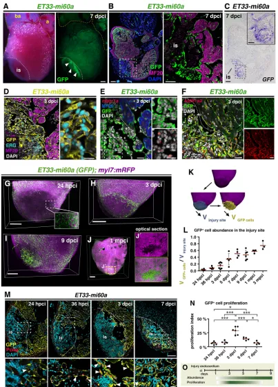
To study the endocardium during heart regeneration we used the endocardial enhancer trap line ET33-mi60a, in which GFP is inserted close to the promoter of the Notch signalling modulator lunatic fringe (lfng) (Poon et al., 2010). We used the cryoinjury model of heart regeneration that faithfully recapitulates mammalian ischemic injury (Chablais et al., 2011; González-Rosa et al., 2011; Schnabel et al., 2011). We detected GFP expression throughout the ventricle but also within the cryoinjury site ofET33-mi60ahearts (Fig. 1A,B; sample sizes for all figures are provided in Tables S1, S2). GFP+cells (GFP mRNA, Fig. 1C) expressed the endothelial
erythroblast transformation-specific transcription factor Erg (Fig. 1D, Fig. S1A) and the endocardial development gene nfatc1a (Fig. 1E, Fig. S1B) (Larson et al., 2004; Wong et al., 2012). This suggests that GFP+ cells in the wound endocardium Received 12 August 2016; Accepted 20 February 2017
1Intercellular Signalling in Cardiovascular Development and Disease Laboratory,
Centro Nacional de Investigaciones Cardiovasculares Carlos III (CNIC), Melchor Fernández Almagro 3, Madrid E-28029, Spain.2Institute of Biochemistry and
Biology, Potsdam University, Karl-Liebknecht-Straße 24-25, Potsdam D-14476, Germany.3CIBER CV, 28029 Madrid, Spain.4Cancer Division, Garvan Institute of
Medical Research, The Kinghorn Cancer Centre, 370 Victoria Street, Darlinghurst NSW 2010, Australia.
*Author for correspondence ( [email protected])
J.L.d., 0000-0001-6761-7265
DEVEL
O
Fig. 1. The endocardium expands at the injury site.(A)ET33-mi60atransgenic zebrafish heart, whole-mount views. Strong GFP expression is apparent in the cryoinjured region of the ventricle (arrowheads). (B)ET33-mi60aheart section. Immunohistochemistry (IHC) shows GFP+endocardial cells in a myosin
heavy chain (MF20)−area. (C)ET33-mi60aheart consecutive section to Fig. 1B showingGFPISH. (D-F) IHC, or IHC combined with FISH. GFP+cells express
Erg (D),nfatc1a(E) and Aldh1a2 (F). (B-F) Boxed areas are magnified on the right. (G-I) Volume rendering ofET33-mi60a;myl7:mRFPinjured ventricles. Endocardium, green; myocardium, magenta. (J,J′) Volume rendering (J) and amplified optical section from the centre (J′) of an injured ventricle. (K) Imaris-based volume quantification. The volume of the mRFP−region was determined (blue, Vinjury site) and used as a mask to label GFP+cells in this region (yellow) and
determine their volume (VGFP cells). (L) Scatter plot showing the relative volume occupied by GFP+cells in the injury site. Mean (red line)±s.d.; one-way
ANOVA and Newman-Keuls test (see Table S3). (M) IHC showing Pcna+GFP+endocardial nuclei (arrowheads). Boxed areas magnified beneath. (N) Scatter plot
showing percentage of Pcna+cells among GFP+cells within and adjacent (50μm) to the injury site. Mean±s.d.; one-way ANOVA and Newman-Keuls test,
*P<0.05, ***P<0.005 (see Table S4). (O) Schematic illustrating that endocardium proliferation precedes endocardial expansion at the injury site. is, injury site; a, atrium; ba, bulbus arterious. Dotted lines demarcate injured tissue. Scale bars: 200μm in A-C,G-J; 20μm in D-F; 100μm in M; 50μm in magnified views in B,C;
20μm in magnified views in D,E,M; 100μm in J.
DEVEL
O
region retain their endocardial/endothelial features after injury and are activated. These GFP+ wound endocardial cells expressed
Aldh1a2 protein (Fig. 1F), consistent with previous results (Kikuchi et al., 2011). Likewise, the endocardial reporter Tg(fli1a:GFP) (Lawson and Weinstein, 2002) presented GFP+cells in the injury
site (Fig. S1C).
We studied wound endocardium dynamics by high-resolution 3D confocal imaging. Optical clearing of ET33-mi60a;myl7:mRFP hearts by CUBIC (clear, unobstructed brain imaging cocktails and computational analysis; reagent 1; Susaki et al., 2014) allowed detection of endogenous fluorescence signals: GFP in the endocardium and membrane-bound RFP in cardiomyocytes (Rohr et al., 2008). At 24 hours post cryoinjury (hpci), strongly reduced mRFP fluorescence at the injury site relative to uninjured tissue indicated the absence of myocardial cells (Fig. 1G, Movie 1). Despite the overall reduced GFP fluorescence, we detected spared GFP+cells within the injury site (Fig. 1G, Movie 1). To estimate the
abundance of endocardial cells with respect to the injury site extension, we used Imaris software to 3D reconstruct confocal images and quantify the volume of the injury site, identified by the absence of mRFP+ cardiomyocytes (Fig. 1K; see Materials and
Methods). We also quantified the volume of the injury site occupied by GFP+cells (Fig. 1K). The relationship between these two values
provided an estimate of the relative volume occupied by GFP+
endocardial cells over the course of regeneration (Fig. 1K,L). GFP+
cells in an uninjured region, surrounding cardiomyocytes, occupied a volume of 46.1±2% (Fig. S1D). At 24 hpci, GFP+cells were rare
at the injury site, occupying 3.5±2.1% of the injured volume (Fig. 1G,L, Fig. S1D, Movie 1). By 36 hpci, the GFP+ volume
increased slightly but significantly (8.5±3.8%; Fig. 1L, Fig. S1E, Movie 2). At 3 dpci, most hearts exhibited a higher occupation by GFP+cells (15.5±8.9%; Fig. 1H,L, Movie 3). The proportion of
the injured volume occupied by GFP+ cells progressively
increased until 7 dpci (Fig. 1L, Fig. S1E, Movies 4, 5) and was stable at 9 dpci (47.1±13.7%; Fig. 1I,L, Movie 6). The relative volume occupied by GFP+ cells continued to increase up to 3
months post cryoinjury (mpci), albeit more gradually (57.6±4.4% at 1 mpci and 74.6±12.6% at 3 mpci; Fig. 1J,L, Fig. S1E, Movies 7, 8). This might be due to the decrease in the total size of the injury site over this period of time (Fig. S1F), when cardiomyocytes had already begun to replace lost cardiac muscle (Fig. 1J, 1 mpci; Fig. S1E, 3 mpci), consistent with published data (Chablais et al., 2011; Itou et al., 2012). Analysis of optical sections in 1 mpci hearts revealed that regions still lacking cardiomyocytes were filled with endocardial cells (Fig. 1J). Moreover, endocardial volume at 1 mpci and 3 mpci is beyond the value of an uninjured region of the heart (Fig. 1L, Fig. S1C), indicating that endocardial density is augmented at the injury site.
We next examined endocardial cell proliferation inET33-mi60a hearts. GFP+endocardial cells stained for proliferating cell nuclear
antigen (Pcna) were very rare in uninjured hearts (Fig. S1G; 0.45±0.15 cells/heart section). At 24 and 36 hpci, several GFP+endocardial cells
at the injury site were Pcna+(Fig. 1M,N). At 3 dpci, we observed a
massive proliferative response of GFP+cells adjacent to (Fig. 1M,
N) and throughout (Fig. S1H) the injury site. Endocardial proliferation decreased at 5 dpci (Fig. 1N, Fig. S1I) and was almost abolished at 7 dpci (Fig. 1M,N). This shows that the highest proliferation coincides with the initiation of endocardial expansion and suggests that endocardial cell proliferation at 3 and 5 dpci contributes to the endocardial expansion from 3 to 7 dpci (Fig. 1O). The observation that maximal endocardial proliferation (3 dpci) occurs before the peak of myocardial proliferation (7 dpci) (Kikuchi
et al., 2010; Sallin et al., 2014; Bednarek et al., 2015), and the presence of the endocardium at the injury site throughout regeneration (Kikuchi et al., 2010; Sallin et al., 2014; Bednarek et al., 2015), suggest that endocardial regeneration precedes myocardial regeneration.
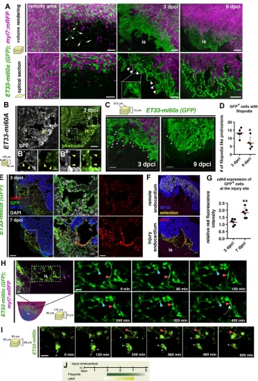
Wound endocardium is highly dynamic during regeneration We next analysed endocardial morphology at different time points after cryoinjury and conducted 3D imaging analysis at high magnification. In the undamaged region of the heart, a coherent network of endocardial cells with elongated morphology surrounded clusters of cardiomyocytes (Fig. 2A). Injury site endocardial cells at 24 hpci were similarly elongated (Fig. 2A). By contrast, we observed a dense and disorganised endocardial mesh at the injury site at 3 dpci (Fig. 2A). Endocardial cells appeared rounded, in clusters and displayed numerous filopodia-like protrusions (Fig. 2A, arrowheads). These co-stained with phalloidin (Fig. 2B, Fig. S2A), indicating an actin-rich cytoskeleton, one characteristic of filopodia (Mallavarapu and Mitchison, 1999; Mattila and Lappalainen, 2008). The high density of GFP+cells was maintained at 9 dpci; however, cells seemed more
orderly aligned than at 3 dpci, forming a coherent sheet (Fig. 2A). Moreover, filopodia-like protrusions were less abundant at 9 dpci than at 3 dpci (Fig. 2C,D).
We next examined expression of the endothelial cell-cell adhesion protein Cadherin 5 (Cdh5), which is involved in endocardial junction integrity (Mitchell et al., 2010). In situ hybridisation (ISH) showed strong cdh5 expression at the injury site, which was weaker throughout the ventricle and in the uninjured heart (Fig. S2B,C). We conducted fluorescent ISH (FISH) on ET33-mi60a heart sections and compared levels of cdh5 mRNA fluorescence of remote and wound endocardium (Fig. 2E,F). Relative cdh5expression at the injury site increased up to 1.156 (±0.089) at 3 dpci (Fig. 2G), and even more at 7 dpci (1.820±0.142), indicating that injury endocardium maturation coincides with increasing levels ofcdh5expression (Fig. 2G). Also, more mature injury endocardium at 7 dpci was strongly associated with collagen fibres at 7 dpci, but not at 3 dpci (Fig. S2D,E).
The presence of filopodia-like protrusions, a characteristic of migrating cells (Ridley, 2011), together with the observed endocardial expansion suggested endocardial cellular migration within the injury site. To test this, we cultured cryoinjured hearts and performed live imaging of the regenerating endocardium in ET33-mi60atransgenic fish at∼2-3 dpci. We observed that endocardial cells changed their location and that individual cells migrated short distances within the injury site (Fig. 2H,I, Movies 9, 10). We also detected endocardial cells sending out filopodia (Fig. 2I, Movie 10). Overall, we show by morphological, gene expression and live imaging analyses a dynamic post-injury endocardium (Fig. 2J), and distinguish an early activated, proliferative endocardium (3 dpci) from a more mature, organised, less-proliferative endocardial structure (7-9 dpci).
Notch pathway elements are expressed in the endocardium and are implicated in endocardial maturation and heart regeneration
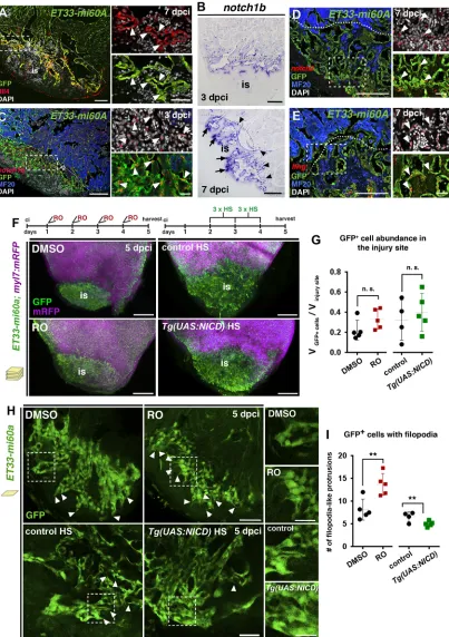
Next, we examined the involvement of Notch, a crucial endocardial cell signalling pathway during development and regeneration (Raya et al., 2003; Zhang et al., 2013; Luxán et al., 2016), in the cryoinjured heart. The ligand Delta-like 4 (Dll4) is expressed in GFP+wound endocardial cells inET33-mi60aandTg(fli1a:GFP)
hearts (Fig. 3A, Fig. S3A,E).notch1bexpression was initially low
DEVEL
O
Fig. 2. Characteristics of injury endocardium at different stages of regeneration.(A) Volume rendering and corresponding optical sections of part of the remote and injured region fromET33-mi60a;myl7:mRFPhearts. Remote region contains elongated and coherent GFP+cells. At 1 dpci individual GFP+cells are
seen at the injury site (is) (arrows). At 3 dpci, dense, clustered endocardial cells with filopodia-like protrusions (arrowheads) are observed. At 9 dpci the endocardial cells are more organised, aligned and mostly lack filopodia-like protrusions. (B-B″) Vibratome section ofET33-mi60aheart stained for GFP and with phalloidin (for F-actin). Filopodia-like protrusions of wound endocardial cells show phalloidin staining (arrowheads). (C) Volume rendering of part of the injured region fromET33-mi60a;myl7:mRFPhearts. (D) Quantification of filopodia-like protrusions from comparable 3D images. Mean±s.d.;t-test, *P<0.05. (E) FISH combined with IF showing highcdh5expression (red) in GFP+wound endocardium. Boxed areas magnified on the right. (F,G)cdh5(red) fluorescence intensity
measurements with ImageJ software: GFP+endocardium was selected (F, yellow) in the remote or wound region. Scatter plot (G) showing relative red
fluorescence intensity comparing values of both regions. Mean±s.d.;t-test, **P<0.01. (H,I) Confocal still pictures from time-lapse movies (Movies 9, 10) of wound endocardial cells in culturedET33-mi60a;myl7:mRFPhearts. The box in the schematic indicates the imaging region. Endocardial cells move (H,I, red arrowheads) and change their position (H, blue arrowhead). Dotted arrow (H) indicates the direction of cell migration. Endocardial cells present dynamic filopodia-like protrusions (I, asterisks). (J) Schematic showing wound endocardium characteristics. Filopodia-filopodia-like protrusions are more abundant at early (3 dpci) than at later (9 dpci) phases.cdh5expression increases when regeneration proceeds. Scale bars: 50μm in A-C; 100μm in E; 20μm H,I; 10μm in magnified views of B.
DEVEL
O
Fig. 3. Notch signalling elements are expressed in the endocardium and Notch signalling modulation affects injury endocardium maturation.(A) IHC showing Dll4 expression by GFP+wound endocardial cells (arrowheads). (B) ISH showingnotch1bexpression in endocardial cells at the injury site (arrows) and
lining injury-adjacent cardiomyocytes (arrowheads). (C-E) FISH fornotch1b(C),notch2(D) orlfng(E) combined with IHC showing transcripts in GFP+wound
endocardial cells (arrowheads). (F) Volume rendering of injuredET33-mi60a;myl7:mRFPventricles after DMSO or RO treatment and of heat shocked (HS)
Tg(UAS:NICD);ET33-mi60a;myl7:mRFPand control hearts. Treatment regimens are indicated at the top. (G) Scatter plot presenting relative wound GFP+cell
volume. Mean (dotted line)±s.d.;t-test. (H) Injury site optical sections showing the disorganisation and clustered appearance of endocardial cells and more filopodia-like protrusions (arrowheads) after RO treatment, and fewer filopodia-like protrusions after heat shock inTg(UAS:NICD);ET33-mi60a;myl7:mRFP
hearts. (I) Scatter plot showing the number of filopodia-like protrusions. For representative images used for quantification see Fig. S6A. Mean±s.d.;t-test
**P<0.05. Dotted lines (A,C,D,E) delineate the injury site (is). Boxed areas are magnified on the right. Scale bars: 100 µm in A-E; 200 µm in F; 50 µm in H; 20 µm in magnified views in A,C,D,E; 25 µm in magnified views in H.
DEVEL
O
(36 hpci, Fig. S3B), but strongnotch1b,notch2,notch3and lfng transcription was evident in endocardial cells lining injury-adjacent cardiomyocytes and within the injury site at 3 and 7 dpci (Fig. 3B, Fig. S3C,D). FISH combined with immunofluorescence (IF) confirmed notch1b,notch2 and lfngexpression by GFP+wound
endocardial cells inET33-mi60a (Fig. 3C-E) and Tg(fli1a:GFP) (Fig. S3F) transgenic fish.
To study the requirement of Notch for cryoinjured heart regeneration, we used the γ-secretase inhibitor RO492909 (RO), which effectively reduces Notch activity in the zebrafish embryo and adult fin (Munch et al., 2013), and in the retina (Conner et al., 2014). In the injured heart, RO treatment diminished Notch target gene transcription (Fig. S4A) and impaired heart regeneration, as indicated by the increased amount of fibrotic tissue at 30 dpci (Fig. S4B,C), similar to observations made in the ventricular resection model (Zhao et al., 2014).
We examined the impact of increased Notch activity on regeneration using the inducible modelTg(hsp70l:Gal4);Tg(UAS: myc-notch1a-intra)[abbreviated toTg(UAS:NICD)]. Heat shocks induced the expression of the Notch intracellular domain (NICD) (Scheer et al., 2001) and increased Notch target gene transcription in Tg(UAS:NICD) injured hearts (Fig. S4D). Differences in regeneration between control andTg(UAS:NICD)hearts were not evident until 33 dpci (Fig. S4E,F), presumably owing to heat shock-induced slowing of regeneration (Gemberling et al., 2013). At 90 dpci,Tg(UAS:NICD) hearts retained more fibrotic tissue than control fish (Fig. S4G,H), suggesting that long-term Notch overactivation impairs heart regeneration.
Next, we examined the requirement of Notch for wound endocardial cell expansion and maturation. In line with published results (Zhao et al., 2014), manipulation of Notch signalling did not interfere with endocardial activation (Kikuchi et al., 2011), as shown by unchanged expression ofaldh1a2(Fig. S5A,B), and did not alter endocardial cell proliferation at 3 dpci (Fig. S5C-E). In ET33-mi60atransgenic fish, GFP expression was unaffected by RO treatment or Notch pathway overactivation (Fig. S5G), allowing us to use this line for the following studies.
Notch signalling inhibition for 4 dpci did not significantly alter GFP+ wound endocardium expansion, which was similar in
Tg(UAS:NICD);ET33-mi60a and control hearts (Fig. 3F,G). Analysis of endocardial cell morphology in RO-treated hearts at 5 dpci revealed clustered endocardial cells at the injury site (Fig. 3H), similar to our observations at 3 dpci (Fig. 2A). DMSO-treated hearts, however, exhibited a more orderly aligned injury endocardium (Fig. 3H), indicating that endocardial maturation had occurred in control but not in RO-treated hearts. Quantification of filopodia-like protrusions as one characteristic of the early immature wound endocardium (Fig. 2C,D) revealed a higher abundance in RO-treated hearts (Fig. 3H,I, Fig. S6A). Moreover, overactivation of the Notch pathway inTg(UAS:NICD);ET33-mi60ahearts resulted in a reduced number of filopodia-like protrusions (Fig. 3H,I, Fig. S6A), suggesting a role for Notch in endocardial maturation. Additionally,cdh5transcript levels, analysed as before (Fig. 2F), did not significantly differ after Notch signalling manipulation (Fig. S6B-E). Thus, these results indicate that a precise level of Notch activation is crucial for heart regeneration after cryoinjury and that Notch is involved in regulating endocardial maturation.
Decreased Notch signalling affects endothelial, cardiovascular and wound healing processes
To study the molecular changes resulting from Notch abrogation in the cryoinjured heart, we extracted RNA from the injured region of
the ventricle after RO or DMSO treatment at 3 dpci (Fig. 4A). RNA-seq analysis identified 347 differentially expressed genes, 196 of which were upregulated and 151 downregulated (Fig. 4B, Table S6). Ingenuity-based gene ontology (GO) classification revealed that Notch signalling inhibition affected genes involved in various endothelial cell processes (Fig. 4C), suggesting a function for Notch in endocardial cells, since they are specialised endothelial cells and both cell types share structural and functional genes (Harris and Black, 2010). Furthermore, genes required in the cardiovascular system and for wound healing were dysregulated (Fig. 4C). GO assignments of differentially expressed genes to categories related to endothelial cells, the cardiovascular system and wound healing are indicated in Fig. S7A.
Notch inhibition dysregulated genes involved in the regulation of angiogenesis (vegfc, id1, efnb2a, egr1), endothelial integrity [cldn5b, heg (heg1)] and endothelial cell differentiation (klf2a, klf2b,aqp1a.1) (Fig. 4D). A subset of these (efnb2a,heg,klf2a, egr1,id1) are also endocardial genes (Mably et al., 2003; Grego-Bessa et al., 2007; Vermot et al., 2009; Zhao et al., 2011) and were expressed at the inner border of the injured heart (egr1,id1; Fig. S7B, C). Confirming the Notch signalling attenuation (Fig. S4A), RNA-seq detected the downregulation of efnb2a and bmp10, two Notch-dependent genes involved in chamber development (Grego-Bessa et al., 2007) (Fig. 4D). Three additional endocardial genes,heg,klf2a and klf2b, were upregulated upon Notch inhibition (Fig. 4D). heg regulates endothelial/endocardial integrity (Kleaveland et al., 2009) and growth of the zebrafish myocardium (Mably et al., 2003).klf2 gene endothelial expression is induced by shear forces and is related to a stretched, less migratory, differentiated endothelial phenotype (Dekker et al., 2002, 2006).klf2aandhegtranscripts were present in a subset of GFP+wound endocardial cells and also in the remote
region (Fig. 4E,F), the latter being consistent with previous reports after ventricular resection (Kikuchi et al., 2011).
qPCR analysis confirmed that Notch inhibition increased heg, klf2aandklf2bexpression (Fig. 4G) and that Notch overactivation attenuated heg expression (Fig. S7D). Moreover, heg and klf2a expression, which is normally restricted to non-chamber endocardium during cardiac development (DMSO, Fig. 4H) (Mably et al., 2003; Vermot et al., 2009), was expanded to the ventricular endocardium after Notch inhibition (RO, Fig. 4H), suggesting that Notch regulates these genes similarly during development and regeneration.
Notch signalling inhibition alters the expression of inflammatory genes and leads to increased inflammatory cell abundance
RNA-seq analysis revealed the involvement of Notch in cardiovascular and wound healing processes, which led us to study both more in detail. The expression of extracellular matrix (ECM)-degrading proteases [ctsba,ctssb.1 (ctss2.1), mmp9] and hyaluronidase 2 (hyal2), which degrades hyaluronan (Chowdhury et al., 2013), was increased in Notch-diminished hearts (Fig. 5A). Notch inhibition (Fig. 5B) also led to increased expression of inflammatory markers and regulators (ptgs2b,pde7a,sgpl1) (Smith et al., 2003; Ogryzko et al., 2014) and the pro-inflammatory endothelial genesarg2(Ryoo et al., 2008) and tnfrsf9a(CD137) (Olofsson et al., 2008; Teijeira et al., 2012) (Fig. 5B). This upregulation of inflammatory factors is consistent with the augmented protease gene expression because protease-induced low molecular weight ECM molecule fragments trigger pro-inflammatory signals and leukocyte recruitment (Frangogiannis,
2008; Dobaczewski et al., 2010).
DEVEL
O
In zebrafish, injured cardiac tissue is infiltrated by immune cells (Schnabel et al., 2011; Wang et al., 2011; Han et al., 2014) but little is known about the timely regulation of inflammatory signals in the cryoinjured heart. qPCR analysis revealed that inflammatory and ECM degradation genes were strongly upregulated at 36 hpci, with levels declining thereafter to reach near-baseline levels at 7 dpci (Fig. 5C,D). ISH revealedctssb.1,mmp9andtnfrsf9atranscription at the inner injury border after cryoinjury (Fig. S8A,B). We detected inflammatory cells, indicated byl-plastin(lcp1) ormpeg1expression (Herbomel et al., 1999; Ellett et al., 2011), mainly in the injury site and most of these macrophages were in close proximity to GFP+
endocardial cells in ET33-mi60a fish (Fig. 5E,F, Fig. S8C). We hypothesize that the early activated wound endocardium may be involved in the regulation of inflammatory cell recruitment, and investigated the function of Notch in this process.
High inflammatory gene expression (Fig. 5C,D) temporally coincided with the high number of l-plastin-expressing leukocytes at the inner injury border (Fig. S8D) and low Notch expression at 36 hpci (Fig. S3B). Consistent with the RNA-seq data (Fig. 5A,B), qPCR and ISH analyses revealed increased expression ofmmp9,ctssb.1andtnfrsf9a in regenerating hearts upon Notch inhibition at later stages (Fig. S8E-H). Moreover, an augmented number of wound endocardial cells expressed the pro-inflammatory gene tnfrsf9a in hearts after RO treatment, as compared with DMSO-treated hearts (Fig. 5G,H). Notch inhibition further caused elevated numbers of l-plastin+ and
mpeg1+ macrophages associated with wound endocardial cells
(Fig. 5I-K, Fig. S8I), and an increased abundance of l-plastin+
[image:7.612.113.507.58.466.2]macrophages could be detected until 30 dpci after long-term Notch inhibition (Fig. 5L). These observations suggest that Notch Fig. 4. Notch inhibition affects developmental and injury-related endocardial/endothelial genes.(A) RO injection and RNA extraction procedure from the ventricular apex (below the dotted line). (B) RNA-seq detected 347 genes differentially expressed in injured hearts after RO treatment (fold change >0.5,
P<0.05). (C) Ingenuity-based RNA-seq data analysis. The charts represent selected Ingenuity categories affected. (D) RNA-seq revealed differential cardiac expression of endothelial genes at 3 dpci in RO- or DMSO-treated fish. Yellow, downregulated; blue, upregulated. (E,F) FISH againstklf2a(E) orheg(F) combined with IHC. Mosaic expression of both genes was observed in wound and wound-adjacent GFP+endocardial cells (arrowheads). Boxed areas are
magnified on the right. (G) qPCR analysis ofheg,klf2aandklf2bexpression in injured hearts. Mean±s.d.;t-test, *P<0.05, **P<0.01, **P<0.005. (H) ISH in embryos at 3 days post fertilisation (dpf ). RO treatment (regime indicated at top) expanded ventricular expression ofhegandklf2a(arrowhead). Dotted lines delineate injury site. The number of embryos displaying the illustrated phenotype among the total number examined is indicated. Scale bars: 100 µm in E,F; 20 µm in magnified views in E,F.
DEVEL
O
Fig. 5. Notch signalling inhibition affects inflammatory gene expression and macrophage abundance.(A,B) RNA-seq analysis. Differential cardiac expression of genes related to ECM remodelling (A) and inflammation (B) in RO-treated fish at 3 dpci. (C,D) qPCR analysis of hearts with no cryoinjury (no ci) and at indicated time points following cryoinjury. Mean±s.d.;t-test, *P<0.05, **P<0.01, ***P<0.005. (E) FISH combined with IHC, showingl-plastin+cells (arrowheads)
in contact with GFP+cells.l-plastin+cell accumulations at the injury site locally coincide with high endocardial cell abundance (brackets). (F) The percentage of
mpeg1-expressing orl-plastin-expressing inflammatory cells that contact (green) or do not contact (blue) endocardial cells at the injury site or remote region. Inflammatory cells at the outer epicardial region were not considered. (G)tnfrsf9aFISH combined with IHC. RO-treated hearts (regime indicated at top) show more GFP+cells expressingtnfrsf9a. (H) Scatter plot showing percentage oftnfrsf9a+endocardial cells. Mean±s.d.;t-test, *P<0.05. (I)mpeg1FISH combined with IHC.
RO treatment (regime in G) increasedmpeg1+macrophage abundance associated with the GFP+endocardium. (J,K) Scatter plots of the number ofmpeg1+(J) or
l-plastin+(K) macrophages related to the area occupied by GFP+cells. Only macrophages contacting GFP+endocardial cells were considered. Mean±s.d.;t-test,
*P<0.05. (L) MF20 IHC andl-plastinISH on consecutive heart sections. RO treatment increased the numbers ofl-plastin+macrophages inside the MF20−injury
site (red arrowheads).l-plastin+macrophages in the outer region of the injury can be seen in both conditions (arrows). (M) Schematic showing time and intensity of
the inflammatory response and Notch signalling activation at the injury site. Dotted lines delineate the injury site (is). Scale bars: 100 µm, except 50 µm in
magnified views.
DEVEL
O
is required to restrict the inflammatory response in the injury site (Fig. 5M).
Notch signalling regulates cardiomyocyte proliferation and differentiation
We observed that endocardial activation and proliferation preceded myocardial regeneration (Fig. 1H-N). High-magnification 3D analysis showed that clusters of injury-adjacent cardiomyocytes were embedded in a dense endocardial network (Fig. 6A), and that myocardial cell protrusions, which are characteristic of migrating cardiomyocytes (Morikawa et al., 2015), were in close contact with endocardial cells (Fig. 6B). To investigate myocardial and endocardial interactions we analysed BrdU incorporation in Myocyte enhancer factor 2 (Mef2)+wound-adjacent cardiomyocytes
after Notch signalling manipulation at 7 dpci. Notch inhibition decreased cardiomyocyte proliferation (Fig. S9A,B), consistent with results obtained after ventricular resection (Zhao et al., 2014). Further, we observed that sustained Notch overactivation augmented cardiomyocyte proliferation (Fig. 6C,D), which differs from previous findings (Zhao et al., 2014). Analysis of Mef2+
cardiomyocyte density adjacent to the injury site at 7 dpci revealed higher cardiomyocyte numbers inTg(UAS:NICD)than in control hearts (Fig. 6E,F), suggesting that cardiomyocytes accumulate in this region after Notch overactivation.
ISH againsthand2andnkx2.5, hallmark transcription factors of dedifferentiated cardiomyocytes (Lepilina et al., 2006; Fig. S9C), indicated an expansion of dedifferentiated cardiomyocytes in Tg(UAS:NICD)hearts (Fig. 6G, Fig. S9D).
We next analysed myocardial genes that were differentially expressed in the RNA-seq, including the immediate early cardiac growth genesmycband fosab(Fig. 6H). RNA-seq and ISH data showed that Notch inhibition decreased mycb levels in injury-adjacent cardiomyocytes (Fig. 6I), similarly to fosab expression (Fig. 6J). Both genes were shown to be expressed in dedifferentiated, proliferating cardiomyocytes in fish (Aguirre et al., 2014; Beauchemin et al., 2015), suggesting that changes in mycbandfosabexpression might be associated with the reduction in cardiomyocyte proliferation in Notch-abrogated hearts. RNA-seq analysis indicated an upregulation of genes encoding sarcomere assembly and function proteins (Fig. 6H). During regeneration, dedifferentiating zebrafish and mouse cardiomyocytes disassemble the sarcomere for cell division to proceed (Jopling et al., 2010; Porrello et al., 2011), and high levels of sarcomeric proteins are characteristic of differentiated cardiomyocytes (O’Meara et al., 2015). Confirming the RNA-seq data, we detected fewer wound-adjacent cardiomyocytes showing decreased levels ofmyosin light chain kinase 3 (mylk3), which regulates sarcomere assembly (Seguchi et al., 2007), in hearts after RO treatment than in DMSO-treated hearts (Fig. 6K,L). Further, Notch overactivation decreased tcap expression in Tg(UAS:NICD) transgenic hearts (Fig. 6M). These results suggest that Notch signalling modulations interfere with sarcomeric gene expression and affect cardiomyocyte dedifferentiation.
The early endocardial geneserpine1is implicated in endocardial and myocardial proliferation
To identify a potential early endocardial factor, the downregulation of which may depend on Notch signalling at later stages (Fig. 4D), we focused on plasminogen activator inhibitor 1 (serpine1). Secreted Serpine1 is the main physiological inhibitor of urokinase plasminogen activator (uPA) and tissue plasminogen activator (tPA), and thus inhibits fibrinolysis (Declerck and Gils, 2013).
Moreover, Serpine1 regulates endothelial cell proliferation (Ploplis et al., 2004), apoptosis (Balsara and Ploplis, 2008; Abderrahmani et al., 2012) and migration (Isogai et al., 2001) and is upregulated in the injured neonatal heart (Darehzereshki et al., 2015).
serpine1 expression was absent in the uninjured adult heart (Fig. S10A) but strongly upregulated early after cryoinjury (36 hpci, 3 dpci, Fig. 7A) and decreased at later stages (Fig. 7A, Fig. S10B). FISH combined with antibody staining detected a high number ofserpine1-expressing GFP+wound and wound-adjacent
endocardial cells (Fig. 7C,D, Fig. S10C) in ET33-mi60a fish at 24 hpci and 36 hpci (36.7±6.6% and 35.5±6.8%, respectively). The percentage of serpine1-expressing endocardial cells dramatically decreased at 3 dpci (14.4±5.6%; Fig. 7C,D) and remained low at 7 dpci (4.7±0.9%; Fig. 7C,D). Notch inhibition increasedserpine1 expression levels (Fig. 7B, Fig. S10D) and indeed augmented the number of endocardial cells expressingserpine1(Fig. 7E,F), indicating thatserpine1downregulation in the injury site endocardium requires Notch signalling at later stages of regeneration.
To investigate if this regulation could be a common mechanism in endothelial/endocardial cells we treated porcine aortic valve endothelial cells (PAVECs) with RO for 48 h. SERPINE1 expression was increased in Notch-abrogated PAVECs (Fig. 7G) in parallel with a marked downregulation of Notch targets (not shown). Also, RNA-seq data obtained in various mouse mutants with disrupted endocardial Notch signalling at different time points of development (Luxán et al., 2013; D’Amato et al., 2016) showed significantly increased Serpine1 expression (Fig. 7G), indicating that, similar to the zebrafish situation (Fig. 7B,E-G), Serpine1 expression is upregulated in mouse after Notch abrogation.
To investigate the function of Serpine1 in the cryoinjured endocardium, we treated fish with the inhibitor Tiplaxtinin (PAI-039, abbreviated as TP) (Fig. 7H), which blocks Serpine1 protease activity (Gorlatova et al., 2007; Daniel et al., 2015). TP treatment did not interfere with fibrotic tissue deposition (Fig. S10E-G) or endocardial activation, as indicated by the expression ofaldh1a2 (Fig. S10H). However, we detected an augmented number of wound endocardial cells expressing Pcna after TP treatment for 3 dpci (Fig. 7H,I), suggesting that endocardial proliferation at 3 dpci is linked to serpine1 downregulation in endocardial cells. Also, relative levels ofcdh5in wound endocardial cells were significantly higher (1.4±0.1; Fig. 7J,K) in TP-treatedET33-mi60afish than in DMSO-treated controls (1.3±0.1; Fig. 7J,K) at 3 dpci, indicating that endocardial maturation had progressed further.
As Serpine1 is a secreted molecule, endocardial Serpine1 might also have non-cell-autonomous functions during heart regeneration. We examined the consequences of TP treatment on cardiomyocyte proliferation, and observed higher numbers of Pcna+
cardiomyocytes in TP-treated hearts (Fig. 7L,M), indicating that early Serpine1 abrogation augments cardiomyocyte proliferation. We next treated fish with TP or DMSO during the first days of regeneration (10 dpci), the time frame when we expectedserpine1 expression to be active in the endocardium. Examination of TP- or DMSO-treated injured hearts at 22 dpci did not reveal any difference in injury site size (Fig. 7N,O), suggesting that early Serpine1 inhibition does not result in accelerated regeneration or in interference with collagen deposition or degradation.
In summary, these results revealserpine1as an early endocardial injury-response gene, the downregulation of which at later stages depends on Notch signalling. Moreover, Serpine1 is involved in the regulation of endocardium proliferation and maturation, and non-cell-autonomously influences myocardial proliferation.
DEVEL
O
Fig. 6. Notch signalling regulates cardiomyocyte proliferation.(A)ET33-mi60a;myl7:mRFPventricle, volume rendering and optical section of a region of the inner injury border. Endocardial cells surround injury-adjacent cardiomyocytes (arrows) and precede into the wound. (B) IHC showing endocardial cells (GFP+) in
contact with myocardial protrusions (tropomyosin+; blue arrowheads). (C,D) IHC against BrdU and Mef2 (C). Heat shock regime is indicated at the top. Scatter plot
(D) of percentage of BrdU+wound-adjacent cardiomyocytes (Mef2+; arrowheads between dotted lines). Mean±s.d.;t-test,**P<0.01. (E,F) Mef2 IHC showing a
higher density of wound-adjacent cardiomyocytes (between the dotted lines) inTg(UAS:NICD)than in control hearts (E), as quantified in F. Mean±s.d.;t-test,
*P<0.05. (G) ISH showing higher numbers ofnkx2.5+wound-adjacent cardiomyocytes (between dotted lines) inTg(UAS:NICD)hearts. (H) RNA-seq data
showing differential myocardial gene expression in RO-treated hearts at 3 dpci. (I) ISH showingmycbexpression in injury-adjacent cardiomyocytes (arrowheads) in DMSO-treated but not in RO-treated hearts (asterisks). (J) qPCR levels offosabin the injured heart. Mean±s.d.;t-test, *P<0.05. LOF, loss of function. (K) MF20 IHC andmylk3ISH on heart sections. RO treatment decreased the numbers ofmylk3−cardiomyocytes adjacent to the injury site (arrowheads). (L) Scatter plot showing the percentage ofmylk3−wound-adjacent (50 µm) cardiomyocytes. Mean±s.d.;t-test,**P<0.01. (M) qPCR analysis ofmylk3andtcaplevels in the injured heart. Mean±s.d.;t-test, ***P<0.001. GOF, gain of function. Dotted lines delineate the injury site (is). Boxed areas are magnified in insets. Scale bars:
100 µm, except 25 µm in magnified views.
DEVEL
O
Fig. 7.serpine1is upregulated early upon cryoinjury and responds to Notch in endocardial/endothelial cells.(A) qPCR analysis ofserpine1in hearts with no cryoinjury (no ci) and at indicated time points following cryoinjury. Mean±s.d.;t-test, *P<0.05. (B)serpine1qPCR in the injured heart. Mean±s.d.;t-test, *P<0.05. (C)serpine1FISH plus IHC. At 36 hpci, numerous wound and wound-adjacent GFP+endocardial cells expressserpine1. At 3 and 7 dpci,serpine1
-expressing wound endocardial cells are less frequent. Boxed areas are magnified beneath. (D) Scatter plot of the percentage ofserpine1+cells among all wound
and wound-adjacent (50μm) GFP+cells. Mean±s.d.; one-way ANOVA and Newman-Keuls test, ***P<0.005 (see Table S5). (E)serpine1FISH combined with
IHC. RO-treated hearts (regime indicated at top) show more GPF+cells expressingserpine1(arrowheads). (F) Scatter plot indicating the percentage ofserpine1+
endocardial cells. Mean±s.d.;t-test, *P<0.05. (G) Log fold change (FC) ofserpine1showing increased expression as assessed by qPCR in RO-treated PAVECs, by RNA-seq in RO-treated zebrafish hearts and in various murine models of endocardial Notch disruption. (H) IF indicating Pcna+and GFP+cells after TP
treatment (regime indicated at top). (I) Scatter plot of the relative number of Pcna+cells among all wound and wound-adjacent GFP+cells. Mean±s.d.;t-test,
*P<0.05. (J) FISH combined with IHC showing that TP treatment increasescdh5mRNA levels in GFP+wound endocardium. (K) Scatter plot of relative red
fluorescence intensity, comparing values of remote and wound endocardium (see Fig. 2F). Mean±s.d.;t-test, **P<0.01. (L) IF revealed more Pcna+Mef2+
wound-adjacent cardiomyocytes (arrowheads) after TP treatment. (M) Scatter plot of the relative number of Pcna+cells among all wound-adjacent (100 µm) Mef2+
cardiomyocytes. Mean±s.d.;t-test, **P<0.01. (N) AFOG-stained hearts, treated with DMSO or TP (treatment regime indicated at top). (O) Scatter plot indicating injury site size (mean±s.d.). Dotted lines delineate injury site. Scale bars: 100 µm in C,E,H,J,L; 200 µm in N; 20 µm in magnified views.
DEVEL
O
DISCUSSION
Endocardial dynamics at the injury site
In this report we provide the first 3D image analysis of the whole injured region of the zebrafish heart, and describe the dynamics and possible functions of the injured endocardium. Previous studies have reported morphological changes and the activation of wound-adjacent endocardial cells following ventricular resection (Kikuchi et al., 2011). Our results, using the cryoinjury model, reveal a highly dynamic endocardium during the first days of regeneration, with changes in endocardial cell morphology, behaviour and gene expression occurring at distinct phases of regeneration (Fig. 8).
Signals controlling endocardial dynamics
We identified Serpine1 as an early injury-induced molecule in the damaged endocardium (Fig. 8). Despite its inhibitory role in fibrinolysis (Declerck and Gils, 2013), Serpine1 inhibition did not interfere with fibrotic tissue deposition or resolution. We describe one function for Serpine1 as a negative regulator of proliferation and ofcdh5 levels in wound endocardial cells (Fig. 8B). This could imply the involvement of Serpine1 in the maintenance of an initial activation state of the wound endocardium and is in line with its cell-autonomous role in endothelial cells, regulating proliferation or apoptosis (Bajou et al., 2008). Further, we identify Notch as an important player during wound endocardium maturation (Fig. 8), with similar functions in development and regeneration. This holds true for the augmented expression of the developmental genesheg andklf2aand the increased number of filopodia-presenting wound endocardial cells in Notch-inhibited hearts. During developmental angiogenesis, Notch blocks the migratory tip-cell fate of endothelial cells (Hellström et al., 2007; Lobov et al., 2007; Suchting et al.,
2007) and Notch inhibition interferes with blood vessel maturation (Ehling et al., 2013).
Implication of endocardial signals in the inflammatory response and cardiomyocyte proliferation
We observed that early activated endocardium coincides with a high abundance of inflammatory cells and inflammatory gene expression. This raises the hypothesis that endocardial signals may regulate regenerative processes, and that endocardial maturation might be crucial for cardiac regeneration to progress from the inflammatory to the reparative phase (Chablais and Jazwinska, 2012a). During inflammation, vascular endothelial cells mediate the recruitment, adherence and passage of inflammatory cells (Pober and Sessa, 2007). The endocardium might possess this role in the regenerating heart as it responds to inflammatory signals (Kikuchi et al., 2011) and expresses cytokines (Fang et al., 2013). We extend these studies, showing that inflammatory macrophages are associated with wound endocardium. Also, Notch signalling abrogation resulted in increased wound-related endothelial and inflammatory gene expression and an increased abundance of macrophages. This anti-inflammatory role of Notch might be direct, by regulating endothelial inflammatory genes such as tnfrsf9a (Olofsson et al., 2008; Teijeira et al., 2012), or might be linked to the appearance and maturation of the endocardium upon Notch inhibition.
[image:12.612.48.377.407.731.2]Cardiac injury induces the dedifferentiation and proliferation of existing cardiomyocytes, which peaks at 7 dpci (Kikuchi et al., 2010; Sallin et al., 2014; Bednarek et al., 2015), and endocardial signals are implicated in this process (Kikuchi et al., 2011; Zhao et al., 2014). Our results show that endocardial proliferation and
Fig. 8. Endocardial dynamics and signalling after cryoinjury.(A) The tip of the adult zebrafish ventricle in the uninjured situation (no ci), and at various time points after cryoinjury. Uninjured endocardium is depicted as a dark green monolayer and the myocardium in pink; the damaged tissue of the injury site (is) in purple and delineated by a dotted line, with the endocardium colonising the injury site in light green. (B) Injury endocardium is characterised by changes in morphology (filopodia, organisation), behaviour ( proliferation, migration) and gene expression (cdh5,serpine1) at different time points of regeneration. Alterations in endocardial signals (Notch, Serpine1) affect these characteristics (arrows, lines) and interfere with inflammation attenuation (blue) and cardiomyocyte proliferation (red).
DEVEL
O
regeneration precede the regenerating myocardium. In addition, we revealed a possible non-cell-autonomous effect of Serpine1 and Notch signalling on cardiomyocyte proliferation. Secreted Serpine1 might directly signal to cardiomyocytes, preventing proliferative signals or controlling the degradation of specific ECM components, which are crucial for myocardial proliferation and regeneration (Trinh and Stainier, 2004; Mercer et al., 2013; Wang et al., 2013). The inverse correlation between high endocardial serpine1 expression and high cardiomyocyte proliferation (Kikuchi et al., 2010; Sallin et al., 2014; Bednarek et al., 2015) (Fig. 8B) at different stages of heart regeneration supports the hypothesis thatserpine1 downregulation is one prerequisite for myocardial proliferation. Moreover, hearts with abrogated Notch signalling, where cardiomyocyte proliferation was decreased, also showed increased levels ofserpine1. These observations suggest a Notch-mediated downregulation ofserpine1to regulate cardiomyocyte proliferation. Serpine1 expression is also upregulated in endocardial cells in the cryoinjured mouse heart (Darehzereshki et al., 2015) and might be involved in an evolutionarily conserved mechanism of cardiac repair. The inhibitory relationship between Notch and Serpine1 might be part of this mechanism, as supported by our observation that abrogated Notch signalling caused increased SERPINE1 expression in endothelial cells (PAVECs) and in mouse embryos. Future studies should investigate the regulatory effect of Notch on serpine1, and whether Serpine1 inhibition affects dedifferentiation of cardiomyocytes. Precise regulation of Serpine1 is crucial, as elevated Serpine1 levels increase cardiac fibrosis upon myocardial infarction in mice (Takeshita et al., 2004), whereas Serpine1 deficiency leads to cardiac fibrosis (Moriwaki et al., 2004).
In our study, Notch overactivation led to increased BrdU incorporation by injury-adjacent cardiomyocytes (Fig. 8). This result appears to conflict with data from Zhao et al. (2014) showing decreased numbers of Pcna+ cardiomyocytes upon Notch overactivation. We
further observed the accumulation of dedifferentiated cardiomyocytes at the inner injury border, explaining why Notch overactivation impairs regeneration. We suggest two possible explanations. First, the dedifferentiated, proliferating state of cardiomyocytes might prevent their invasion into the injury. Second, cardiomyocytes might fail to enter the injury site due to altered ECM remodelling or endocardial organisation within the injury site. Our Notch inhibition data would support both possibilities, but this issue requires further investigation.
The results presented here reveal a highly dynamic endocardium after cryoinjury, with changes in cell behaviour, morphology and gene expression during regeneration. We identifiedserpine1as an early endocardial injury-response gene and Notch signalling as a player later during regeneration. The maturation of the endocardium and the control of inflammatory cell infiltration require Notch signalling, and our data suggest that the endocardium promotes myocardial regeneration by providing Serpine1-dependent negative and Notch-dependent positive proliferative signals (Fig. 8). These findings demonstrate the importance of the endocardium in the regulation of the injury response and in regeneration upon cardiac insult. Future analysis of the specific endocardial signals orchestrating inflammation, fibrotic tissue deposition and cardiomyocyte renewal could contribute to the development of therapeutic applications for cardiac diseases.
MATERIALS AND METHODS
Zebrafish husbandry
Animal studies were approved by the CNIC Animal Experimentation Ethics Committee and by the Community of Madrid (ref. PROEX 118/15). Animal
procedures conformed to EU Directive 2010/63EU and Recommendation 2007/526/EC regarding the protection of animals used for experimental and scientific purposes, enforced in Spanish law under Real Decreto 53/2013. Zebrafish were raised and maintained under standard conditions (Kimmel et al., 1995). Cryoinjury was performed as described (González-Rosa and Mercader, 2012). Details of transgenic lines used are provided in the supplementary Materials and Methods.
Treatments
Adult zebrafish were injected intraperitoneally with 30 µl RO4929097 (S1575, Selleckchem; 600 µM in PBS) or DMSO as a control. Treatment regimens of each experiment are indicated in the corresponding figure. Embryos were incubated in fishwater containing DMSO or RO (50 µM). 30 µl Tiplaxtinin (PAI-039, S7922, Selleckchem; 1500 µM) or DMSO in PBS was injected intraperitoneally following the treatment regime indicated in the figures. See the supplementary Materials and Methods for further details.
Histology
Hearts were fixed in 4% paraformaldehyde (PFA) overnight, dehydrated, embedded in paraffin and sectioned at 7 µm. Serial sections were distributed on several slides, such that each slide contained sections of several levels of the heart. One or two slides of each heart were used for Acid
Fuchsin-Orange G (AFOG) staining, immunohistochemistry (IHC) or in situ
hybridisation (ISH) to obtain stained sections randomly distributed throughout the heart for unbiased quantification (see below). Primers used to generate RNA probes are listed in Table S7. IHC was performed as described (González-Rosa et al., 2011). For IHC on vibratome sections, hearts were fixed in 4% PFA overnight, washed with PBST (0.1% Tween in PBS) and embedded in 4% low melting point agarose. Sections of 70 µm were obtained and kept in PBST. For IHC, sections were incubated in blocking solution (5% BSA, 5% goat serum, PBS) for 1 h and then in primary antibody overnight at 4°C. After extensive washes with PBST, heart sections were incubated overnight with secondary antibody and FITC-coupled phalloidin. Extensive washes and a 30 min incubation with DAPI followed. Sections were mounted in Fluoromount (Sigma) for imaging. For details of antibodies and imaging see the supplementary Materials and Methods.
Cell culture
Isolation of porcine aortic valve endothelial cells (PAVECs) was carried out as described (Gould et al., 2010). Cells at passage five were seeded on
gelatin-coated 6-well dishes (2.1×10⁵cells/well) and cultured in Dulbecco’s
Modified Eagle’s Medium (DMEM; Gibco) supplemented with fetal bovine
serum (FBS; 10%), penicillin-streptomycin (1%) and bovine brain extract (20 ng/ml). After 24 h incubation, cells near confluence were serum starved (with 0.5% FBS) and treated with 10 µM RO or vehicle (DMSO) for 12, 36 or 48 h. After the corresponding incubation periods, cells were washed twice with PBS and collected for RNA extraction. Each time point was conducted in triplicate.
Gene expression analysis
For quantitative PCR (qPCR) and RNA sequencing (RNA-seq), hearts were dissected at different time points and apical portions of the three ventricles were pooled and (Fig. 4A) used for RNA extraction using Direct-zol RNA MiniPrep (Zymo Research). cDNA was synthesized with the SuperScript III First Strand Kit (Invitrogen). For qRT-PCR, three hearts were pooled, and the data represented here are from four to six samples. Primer sequences are provided in Tables S8 and S9. See the supplementary Materials and Methods for further details. For RNA-seq analysis, we used three pools, each of three apexes from RO- or DMSO-treated fish. cDNA libraries were prepared with a TruSeq RNA Sample Preparation Kit v2 (Illumina) and were sequenced in a Genome Analyzer IIx Illumina sequencer using a 75 bp single-end elongation protocol. Sequenced reads were quality controlled and pre-processed using Cutadapt v1.6 to remove adaptor contaminants (Martin, 2011). The resulting reads were aligned and gene expression was
quantified with RSEM v1.2.3 (Li and Dewey, 2011), using the Zv9_75
DEVEL
O
zebrafish reference genome. Differentially expressed genes were defined as
those with altered expression levels with an adjustedP<0.05. RNA-seq data
were analysed using Ingenuity Pathway Analysis Software (QIAGEN Bioinformatics).
Quantification and statistical analysis
For details of quantification and statistical analysis, including 3D volume, filopodia-like protrusions, fibrotic tissue, IF or ISH staining, BrdU incorporation, macrophage numbers and cardiomyocyte density, see the supplementary Materials and Methods. Sample numbers for all data are listed in Tables S1 and S2.
Acknowledgements
We thank E. Dıaz for husbandry; B. Rios, V. Bou, R. Doohan, P. Mart́ ınez foŕ technical help; the CNIC Microscopy Unit for advice; F. Sánchez-Cabo for help with statistics; and S. Bartlett and K. McCreath for English editing. RNA-seq and bioinformatics analysis were performed by the CNIC Genomics and Bioinformatics Units. TheTg(hsp70l:Gal4)kca4;Tg(UAS:myc-Notch1a-intra)kca3line was from
N. Lawson and theET(krt4:EGFP)sqet33-mi60a
andET(krt4:EGFP)sqet33-1A
lines were from V. Korz. We are grateful to A. Oates, G. Weidinger, D. Goldman, S. Sumanas, J. Vermot and A. Kawakami for probes.
Competing interests
The authors declare no competing or financial interests.
Author contributions
J.M., D.G., A.G.-R., R.T.-C. performed experiments. J.M. and J.L.d.l.P. designed experiments, reviewed the data and wrote the manuscript. All authors reviewed the manuscript during its preparation.
Funding
This work was supported by grants SAF2013-45543-R, RD12/0042/0005 (RIC), RD12/0019/0003 (TERCEL) and CB16/11/00399 (CIBER CV) from the Spanish Ministry of Economy, Industry and Competitiveness [Ministerio de Economıa,́ Industria y Competitividad (MINECO)]; FP7-ITN 215761 (NotchIT) and PITN-GA-2011-289600 (CardioNeT) from the European Commission to J.L.d.l.P. J.M. held a PhD fellowship linked to grant FP7-ITN 215761 (NotchIT). The cost of this publication has been paid in part with Fonds Européen de Développement Régional (FEDER) funds. The CNIC is supported by MINECO and the Pro-CNIC Foundation, and is a‘Severo Ochoa’Center of Excellence (MINECO award SEV-2015-0505).
Data availability
RNA-seq data are deposited at NCBI Gene Expression Omnibus under accession number GSE68650.
Supplementary information
Supplementary information available online at
http://dev.biologists.org/lookup/doi/10.1242/dev.143362.supplemental
References
Abderrahmani, R., Francois, A., Buard, V., Tarlet, G., Blirando, K., Hneino, M., Vaurijoux, A., Benderitter, M., Sabourin, J.-C. and Milliat, F.(2012). PAI-1-dependent endothelial cell death determines severity of radiation-induced intestinal injury.PLoS ONE7, e35740.
Aguirre, A., Montserrat, N., Zacchigna, S., Nivet, E., Hishida, T., Krause, M. N., Kurian, L., Ocampo, A., Vazquez-Ferrer, E., Rodriguez-Esteban, C. et al.
(2014). In vivo activation of a conserved microRNA program induces mammalian heart regeneration.Cell Stem Cell15, 589-604.
Bajou, K., Peng, H., Laug, W. E., Maillard, C., Noel, A., Foidart, J. M., Martial, J. A. and DeClerck, Y. A.(2008). Plasminogen activator inhibitor-1 protects endothelial cells from FasL-mediated apoptosis.Cancer Cell14, 324-334.
Balsara, R. D. and Ploplis, V. A.(2008). Plasminogen activator inhibitor-1: the double-edged sword in apoptosis.Thromb. Haemostasis100, 1029-1036.
Beauchemin, M., Smith, A. and Yin, V. P.(2015). Dynamic microRNA-101a and Fosab expression controls zebrafish heart regeneration. Development 142, 4026-4037.
Bednarek, D., González-Rosa, J. M., Guzman-Martinez, G., Gutierrez-Gutierrez, O., Aguado, T., Sanchez-Ferrer, C., Marques, I. J., Galardi-Castilla, M., de Diego, I., Gomez, M. J. et al.(2015). Telomerase is essential for zebrafish heart regeneration.Cell Rep.12, 1691-1703.
Chablais, F. and Jazwinska, A.(2012a). The regenerative capacity of the zebrafish heart is dependent on TGFbeta signaling.Development139, 1921-1930.
Chablais, F. and Jazwinska, A.(2012b). Induction of myocardial infarction in adult zebrafish using cryoinjury.J. Vis. Exp.62, pii: 3666.
Chablais, F., Veit, J., Rainer, G. and Jaźwińska, A.(2011). The zebrafish heart regenerates after cryoinjury-induced myocardial infarction.BMC Dev. Biol.11, 21.
Chowdhury, B., Hemming, R., Hombach-Klonisch, S., Flamion, B. and Triggs-Raine, B.(2013). Murine hyaluronidase 2 deficiency results in extracellular hyaluronan accumulation and severe cardiopulmonary dysfunction.J. Biol. Chem.
288, 520-528.
Conner, C., Ackerman, K. M., Lahne, M., Hobgood, J. S. and Hyde, D. R.(2014). Repressing notch signaling and expressing TNFalpha are sufficient to mimic retinal regeneration by inducing Muller glial proliferation to generate committed progenitor cells.J. Neurosci.34, 14403-14419.
Conrad, C. H., Brooks, W. W., Hayes, J. A., Sen, S., Robinson, K. G. and Bing, O. H. L.(1995). Myocardial fibrosis and stiffness with hypertrophy and heart failure in the spontaneously hypertensive rat.Circulation91, 161-170.
D’Amato, G., Luxán, G., del Monte-Nieto, G., Martınez-Poveda, B., Torroja, C.,́ Walter, W., Bochter, M. S., Benedito, R., Cole, S., Martinez, F. et al.(2016). Sequential Notch activation regulates ventricular chamber development.Nat. Cell Biol.18, 7-20.
Daniel, A. E., Timmerman, I., Kovacevic, I., Hordijk, P. L., Adriaanse, L., Paatero, I., Belting, H.-G. and van Buul, J. D.(2015). Plasminogen activator inhibitor-1 controls vascular integrity by regulating VE-cadherin trafficking.PLoS ONE10, e0145684.
Darehzereshki, A., Rubin, N., Gamba, L., Kim, J., Fraser, J., Huang, Y., Billings, J., Mohammadzadeh, R., Wood, J., Warburton, D. et al.(2015). Differential regenerative capacity of neonatal mouse hearts after cryoinjury.Dev. Biol.399, 91-99.
Declerck, P. J. and Gils, A.(2013). Three decades of research on plasminogen activator inhibitor-1: a multifaceted serpin. Semin. Thromb. Hemost. 39, 356-364.
Dekker, R. J., van Soest, S., Fontijn, R. D., Salamanca, S., de Groot, P. G., VanBavel, E., Pannekoek, H. and Horrevoets, A. J.(2002). Prolonged fluid shear stress induces a distinct set of endothelial cell genes, most specifically lung Kruppel-like factor (KLF2).Blood100, 1689-1698.
Dekker, R. J., Boon, R. A., Rondaij, M. G., Kragt, A., Volger, O. L., Elderkamp, Y. W., Meijers, J. C., Voorberg, J., Pannekoek, H. and Horrevoets, A. J.(2006). KLF2 provokes a gene expression pattern that establishes functional quiescent differentiation of the endothelium.Blood107, 4354-4363.
Dobaczewski, M., Gonzalez-Quesada, C. and Frangogiannis, N. G.(2010). The extracellular matrix as a modulator of the inflammatory and reparative response following myocardial infarction.J. Mol. Cell. Cardiol.48, 504-511.
Ehling, M., Adams, S., Benedito, R. and Adams, R. H.(2013). Notch controls retinal blood vessel maturation and quiescence.Development140, 3051-3061.
Ellett, F., Pase, L., Hayman, J. W., Andrianopoulos, A. and Lieschke, G. J.
(2011). mpeg1 promoter transgenes direct macrophage-lineage expression in zebrafish.Blood117, e49-e56.
Fang, Y., Gupta, V., Karra, R., Holdway, J. E., Kikuchi, K. and Poss, K. D.(2013). Translational profiling of cardiomyocytes identifies an early Jak1/Stat3 injury response required for zebrafish heart regeneration.Proc. Natl. Acad. Sci. USA
110, 13416-13421.
Felician, G., Collesi, C., Lusic, M., Martinelli, V., Ferro, M. D., Zentilin, L., Zacchigna, S. and Giacca, M. (2014). Epigenetic modification at Notch responsive promoters blunts efficacy of inducing notch pathway reactivation after myocardial infarction.Circ. Res.115, 636-649.
Frangogiannis, N. G.(2008). The immune system and cardiac repair.Pharmacol. Res.58, 88-111.
Gemberling, M., Bailey, T. J., Hyde, D. R. and Poss, K. D.(2013). The zebrafish as a model for complex tissue regeneration.Trends Genet.29, 611-620.
González-Rosa, J. M. and Mercader, N.(2012). Cryoinjury as a myocardial infarction model for the study of cardiac regeneration in the zebrafish.Nat. Protoc.
7, 782-788.
González-Rosa, J. M., Martin, V., Peralta, M., Torres, M. and Mercader, N.(2011). Extensive scar formation and regression during heart regeneration after cryoinjury in zebrafish.Development138, 1663-1674.
Gorlatova, N. V., Cale, J. M., Elokdah, H., Li, D., Fan, K., Warnock, M., Crandall, D. L. and Lawrence, D. A.(2007). Mechanism of inactivation of plasminogen activator inhibitor-1 by a small molecule inhibitor.J. Biol. Chem.
282, 9288-9296.
Gould, R. A. and Butcher, J. T.(2010). Isolation of valvular endothelial cells.J. Vis. Exp.46, 2158.
Grego-Bessa, J., Luna-Zurita, L., del Monte, G., Bolos, V., Melgar, P., Arandilla, A., Garratt, A. N., Zang, H., Mukouyama, Y.-S., Chen, H. et al.
(2007). Notch signaling is essential for ventricular chamber development.Dev. Cell12, 415-429.
Han, P., Zhou, X.-H., Chang, N., Xiao, C.-L., Yan, S., Ren, H., Yang, X.-Z., Zhang, M.-L., Wu, Q., Tang, B. et al. (2014). Hydrogen peroxide primes heart regeneration with a derepression mechanism.Cell Res.24, 1091-1107.
Harris, I. S. and Black, B. L.(2010). Development of the endocardium.Pediatr. Cardiol.31, 391-399.
Hellström, M., Phng, L.-K., Hofmann, J. J., Wallgard, E., Coultas, L., Lindblom, P., Alva, J., Nilsson, A.-K., Karlsson, L., Gaiano, N. et al.(2007). Dll4 signalling