IJPSR (2015), Vol. 6, Issue 11 (Research Article)
Received on 30 April, 2015; received in revised form, 14 June, 2015; accepted, 10 September, 2015; published 01 November, 2015
KARANJIN AMELIORATE DSS INDUCED COLITIS IN C57BL/6 MICE
Praful P Patel 1* and Naitikumar D Trivedi 2
Department of Pharmaceutical Sciences 1, JJT University, Jhunjunu, Rajasthan, India. Department of Pharmacology 2, A.R., College of Pharmacy, Anand, Gujarat, India.
ABSTRACT: Background: Karanjin a furanoflavanoid from Pongamia pinnata
(L.) Seeds, has gastroprotective, anti-arthritic properties rendering it a natural drug having prophylactic and therapeutic property. But, the effect of Karanjin on colitis till not known. Objectives: To evaluate the beneficial effect of karanjin for the treatment of experimental colitis. Method: Colitis were induced in the C57BL6 mice by oral administration of 2.5% solution of DSS in drinking water for 7 days. Karanjin (>98% pure) was administered in two different concentrations 100 and 200 mg/kg and 5-ASA (100 mg/kg) as reference for 7 consecutive days to the DSS induced colitic mice. On 8 day mice were sacrificed and degree of inflammation was assessed by Disease Activity Index (DAI), histology and biochemical estimation of myeloperoxidase (MPO), Nitric oxide (NO), malondialdehyde (MDA), Catalase (CAT), Superoxide dismutase (SOD), and reduced glutathione (GSH) level were measured. Result: Karanjin significantly and dose dependently ameliorate the macroscopic damage, histological changes such as cellular infiltration, tissue necrosis, mucosal and submucosal damage, reduce the activity of MPO. Depressed MDA and NO level and help in restoring the level of CAT, SOD and GSH to normal when compare to the experimental colitic group. Discussion: we demonstrated for the first time that karanjin proposed marked protective effect on experimental colitis through its anti-oxidation and immunomodulation which ultimately reduce the production of inflammatory mediator produced by the immune cells. Conclusion:
Result of the present study indicates that karanjin has potential to cure colitis induced by administration of DSS.
INTRODUCTION: Ulcerative colitis (UC) and Crohn’s disease (CD) collectively called as inflammatory bowel disease (IBD) are serious and major complications of colon. These two diseases have some common characteristics but some genetic and clinical differences exist are - UC affects the mucosal lining of the rectum and colon while CD affects whole intestinal wall and may extend to whole gastrointestinal tract.
QUICK RESPONSE CODE
DOI:
10.13040/IJPSR.0975-8232.6(11).4866-74
Article can be accessed online on:
www.ijpsr.com
DOI link: http://dx.doi.org/10.13040/IJPSR.0975-8232.6(11).4866-74
At present drugs used for the treatment of IBD are aminosalicylates, corticosteroids, immune modulatory drugs, antibiotics, anti-TNF-α antibody. 1 but, unfortunately these drugs have serious side effects, which will limit their use. DSS administration induces colitis having signs of diarrhea, wasting, rectal prolapse and rectal bleeding, histopathologically it produce colonic edema, granuloma formation, loss of normal architecture of the colon and colon shortening. 2
Immunologically DSS induces T-helper-1(Th-1) response by increasing production of IL-12, IL-1, TNF-α, and INF-γ which is similar to the inflammatory course observed in Crohn’s disease. 3
C57BL/6 mice develops chronic acute DSS colitis as compare to BALB/c mice because after DSS
Keywords:
Pongamia pinnata,
Karanjin, Furanoflavonoid, DSS Induced Colitis
Correspondence to Author: Mr. Praful Prakash Patel
Department of Pharmaceutical Sciences, JJT University, Jhunjunu, Rajasthan, India.
removal BALB/c mice recovered fastly from colitic conditions while even after DSS removal C57BL/6 mice develops chronic colitis and require 6-7 weeks for the recovery 4 so, in present study Balb/c mice were used.
Pongamia pinnata (L.) Pierre (Fabaceae; synonym
Pongamia glabra Vent.), is popularly known as “Karanja” in Hindi, “Pungam” in Siddha. It is widely grown forest tree. Seeds of Pongamia pinnata contains 33–36% oil, which is known as Karanja oil and is traditionally used for treating various disease conditions such as rheumatic pain, flatulence, diarrhea and cough. 5 Leaves of
Pongamia pinnata reported to have
anti-inflammatory 6, Seeds have antioxidant 7, Anti-ulcer 8, anthelmintic 9 activity. Karanjin from
Pongamia pinnata seeds has gastroprotective activity by inhibiting H+, K+- ATPase and oxidation 10 of gastric tissues. Owing to its various ethno-pharmacological properties, there is no report available in the literature on the screening of anti-colitis property of karanjin which is a major constituent of Karanja seed oil.
To the best of our knowledge, activity of karanjin against the DSS induced colitis was never been investigated. Therefore, the present study was planned, we explored effects of Karanjin in the treatment of DSS induced colitis and to investigate its effect on enzyme activities in colon tissue which is associated with the intestinal inflammation.
MATERIALS AND METHODS:
Chemicals: Dextran sodium sulphate (MW; 36,000–50,000; Catalogue No. 160110) were purchased from MP Biochemical (Aurora, OH), 5-ASA was obtained as a gift from Zydus Cadila (Ahemdabad, India). Hydroxylamine,5-5-dithio bis-(2-nitrobenzoic acid) (DTNB), thiobarbituric
acid & other chemicals for oxidative parameter measurement were purchased from sigma, India. All other chemicals were of analytical grade.The test drugs karanjin were isolated from Karanja oil.
Animals:
Female C57BL6 mice (17-25 g) were obtained from the animal house facility of National Institute of Nutrition, Hyderabad (Andhra Pradesh). Animals were housed in cages at room temperature (25 °C) with 12:12-h light-dark cycles. The animals were feed on standard pellets and water supplied ad libitum. Animals were acclimatised to the environmental conditions for 10 days before the start of the treatment and were randomized to the respective groups using body weight. The study was approved by the institutional animal ethics committee (IAEC/09/2013-14) which follows the CPCSEA guideline.
Induction of colitis:
Mice were randomly divided into five groups, each containing 6 mice (n=6). The first group (Vehicle control) received normal drinking water. Second group (DSS control) received 2.5% (w/v) DSS (MW; 36,000–50,000) in their drinking water ad libitum for 7 days for induction of colonic inflammation & treated with drinking water. The Third group designated as standard were treated with 100 mg/kg of 5-ASA and 2.5% DSS. The Fourth group (Karanjin low dose) were treated with Karanjin (100 mg/kg/p.o./day) and 2.5% DSS. The fifth group (Karanjin high dose) were treated with Karanjin (200 mg/kg/p.o./day).
Mice of each group were monitored carefully every day to confirm that they consumed an approximately equal volume of DSS-containing water 11.
Score Weight loss Stool Consistency Occult/gross bleeding
0 - Normal (well-formed pellets) Normal
1 1-5%
2 6-10% Loose
(pasty stools that do not stick to the anus)
Occult bleeding
3 11-15%
4 > 15% Diarrhea
(liquid stools that stick to the anus)
Gross bleeding
Evaluation Parameters:- Assessment of colitis severity:
Severity of colitis was assessed daily using a DAI based on the scoring system of 12 DAI was determined on the basis of scores for body weight loss, stool consistency, and rectal bleeding. Scoring was performed according to the criteria described in the table.
Measurement of body weight and colon length: In each experiment, the body weight was monitored every day. Loss of body weight was calculated as the difference between the initial and final weight.
13
on the end of the experiment, mice were killed and the colons were separated from the proximal rectum, close to its passage under the pelvisternum.
The colon length and weight was measured between ileocecal junction and the proximal rectum by using Vernier caliper (Mitutoyo, Japan) colon edema was calculated by weight/length ratio. The spleen were also obtained and their weight is measured. 14
Histology:
To assess the severity of colitis, disease activity index (DAI) score and weight gain were monitored daily. The animals were scarified on 8th day, colons were immediately removed and fixed in 10% buffered formalin, paraffin-embedded, sectioned, and stained with haematoxylin and eosin 15 All tissue sections were examined with a Trinocular microscope (S-500) fitted with CMOS camera and analysed with Biowizad 3.5 image analysis software (Japan), in a blinded manner by a skilled pathologist as described by Funakoshi 16 et al.
2012. Briefly, Grading index was as follows: inflammation severity (0: none, 1:mild, 2: moderate, 3: severe), inflammation extent (0:none; 1:mucosa, 2: mucosa and submucosa, 3: transmural), crypt damage (0: none, 1: basal one-third damaged, 2: basal two-one-thirds damaged, 3: only surface epithelium intact, 4: entire crypt and epithelium lost), and the percentage involvement in the ulcer or erosion (1:< 1%, 2: 1%–15%, 3: 16%– 30%, 4: 31%–45%, 5: 46%–100%). The sum of the first 3 scores (inflammation severity, inflammation extent, and crypt damage) was multiplied by the score of the percentage involvement.
Biochemical Estimations:
The colonic tissues were removed, homogenized and used for the determination of Nitric oxide (NO), Myeloperoxidase (MPO), lipid peroxidation, levels of reduced glutathione (GSH), Superoxide dismutase (SOD), and Catalase.
Procedure for myeloperoxidase (MPO)
determination:
Quickly excised colon wash with cold sodium phosphate buffer (pH: 7.4). 200 mg of tissue homogenized in 2 ml of potassium phosphate-buffered saline (0.1 Mol/l) (10% w/v) and centrifuged at 4◦C for 20 minutes at 12000 rpm. Pellets are resuspend in 2ml 0.05M potassium phosphate buffer (pH 7.4) containing 0.5% (w/v) hexadecyl trimethylammonium bromide (0.1g/20 ml phosphate buffer) homogenate was frozen and thawed three times, centrifuged at 15000 rpm for 15 min at 4 °C. The supernatant obtained was used for estimation of MPO. 17 100 µl phosphate buffer (pH 7.4) containing 0.167 mg/ml O-dianisidine dihydrochloride was added to 100 µl of supernatant and 10 µl 0.05 % H2O2.
Change in optical density was measured at 460 nm. MPO activity was expressed as the amount of an enzyme necessary for the degradation of 1 μmol H2O2/min/100 mg tissue at 250C. 18
Procedure for nitric oxide determination (NO): NO level is estimated as nitrite and nitrate by the Griess reaction after reduction of nitrate to nitrite by vanadium trichloride according to the method described by Miranda 19 et al., (2001). 0.1ml supernatant of tissue homogenate is mixed with 0.1 ml of vanadium (III) chloride. Then, 50 µl of sulfanilamide (2% w/v) solution and 50 µl of N-(1-naphthyl) ethylene diamine dihydrochloride (0.1% w/v) was added respectively, and incubated at 370C for 30 min. Optical density was measured at 540 nm against a blank. The concentrations were determined using a standard curve of sodium nitrate and the results were expressed as nmol/g wet tissue.
Estimation of SOD, GSH, CAT, MDA:
estimation of MDA 24, activity of endogenous antioxidant enzymes such as SOD 20, CAT 23 and GSH 22.
Statistical analysis:
Data were expressed as means ± SEM. Statistical analysis was performed using graph pad prism software. The statistical significance of the data was evaluated by one-way analysis of variance (ANOVA) followed by Dunnett’s t- test. A value of P<0.05 was considered statistically significant.
RESULT:
Colon edema, colon length and histological grading.
Colon weight/length ratio was consider as an indicator of disease associated intestinal wall thickening and intensity of colonic inflammation. The increase in colonic edema was observed after DSS administration (0.148±0.01) as compared to vehicle control (0.09 ±0.005, P<0.001). The 5-ASA at 100 mg/kg (0.10±0.006, P<0.05) and karanjin 200 mg/kg (0.116±0.002, P<0.05) significantly inhibited the colonic edema expressed as Colon weight/length ratio Table. 1.
TABLE 1: PARAMETERS ASSESSED IN DSS INDUCED COLITIS.
Parameter vehicle
control
DSS control 5-ASA 100 mg/kg
Karanjin 100 mg/kg
Karanjin 200 mg/kg
Colon length (cm) 10.38±0.46 7.96 ±0.44 10.28 ±0.19 *** 9.16 ±0.18 ** 9.78 ±0.18 *** Colon weight (g) 0.925 ±0.064 1.172 ±0.047 1.045 ±0.069 1.150±0.045 1.53 ±0.021
Colon Edema 0.09 ±0.005 0.148 ±0.01 0.10 ±0.006 * 0.125 ±0.005 0.116±0.002* HDS 0 13.4 ±0.42 3.32 ±0.43 *** 8.35 ±0.35 * 7.02 ±0.51 ** Spleen weight (mg) 143.8 ±9.93 190.3 ±6.66 164.8 ±6.69 * 172 ±7.38 160.3 ±4.24 * Where- MDS- Macroscopic Damage Score, HDS- Histological Damage Score
The values are expressed as mean ± SEM (n=6) significance was determined by one-way ANOVA followed by post-hoc dunnet’s test. *
p<0.05, **p < 0.01, ***p < 0.001 versus DSS-treated control group.
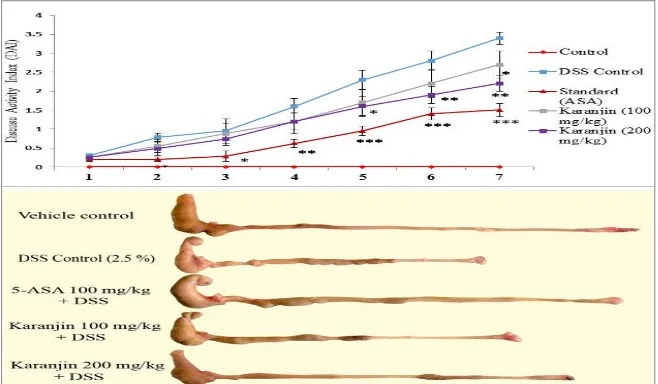
The colons of vehicle control mice showed no inflammation, whereas the DSS control group exhibited significant hyperemia, erosion, ulceration at 7 days after induction of colitis. Karanjin treatments reduced the size of necrosis and ulceration as well as reduce the Disease Activity Index (DAI) (Fig. 1). The oral administration of karanjin reduced shortening of colon length in a dose-dependent manner as compared with DSS control group; the mice with DSS-induced colitis
versus 5-ASA (100mg/kg)-treated mice with DSS-induced colitis (7.96 ± 0.45 cm vs. 10.28 ± 0.19 cm, P < 0.01), mice with DSS-induced colitis versus Karanjin (100 mg/kg)-treated mice with DSS induced colitis (7.96 ± 0.45 cm vs. 9.16 ± 0.23 cm, P < 0.05), and mice with DSS-induced colitis versus Karanjin (200mg/kg)-treated mice with DSS-induced colitis (7.96±0.45cm vs. 9.78±0.18cm, P<0.01)(Fig. 1).
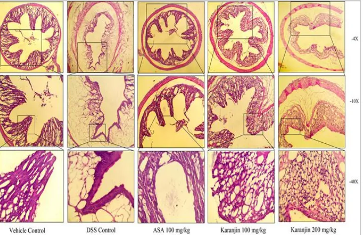
[image:4.612.142.471.533.725.2]Histological examination:
DSS induced colitis shows the dense infiltration of mononuclear inflammatory cells, irregular epithelial structure, and persistent deposits of collagen, edema. Treatment with 5-ASA (100 mg/kg) shows intact epithelial cell, mild edema, inflammation and necrosis Treatment of mice with
[image:5.612.126.492.156.393.2]low dose of Karanjin (100 mg/kg) is able to restore these pathologic conditions such as necrosis, inflammation but shows mild epithelial infiltration, and slight edema. High dose of karanjin (200 mg/kg) significantly ameliorated experimental colitis and histological injury and restore the normal architecture (Fig.2).
FIG. 2: REPRESENTATIVE LIGHT MICROGRAPH OF VEHICLE CONTROL C57BL/6 MICE COLON SHOWING NORMAL ARCHITECTURE OF COLON, DSS-CONTROL GROUP COLON SHOWING DESTRUCTION OF EPITHELIAL ARCHITECTURE WITH LOSS OF CRYPTS AND EPITHELIAL INTEGRITY, SUBMUCOSAL EDEMA, AND INTENSE INFLAMMATORY CELLULAR INFILTRATION, 5-ASA (100 MG/KG/DAY) SHOWING INTACT EPITHELIAL CELL, NO EDEMA, INFLAMMATION AND NECROSIS, KARANJIN 100 MG/KG + DSS TREATED RAT COLON SHOWING MILD CONGESTION IN EPITHELIAL CELLS, AND KARANJIN 200 MG/KG + DSS SHOWING MARKED RESTORATION OF
NORMAL PHYSIOLOGIC ARCHITECTURE.
Effect of karanjin on the MPO activity, NO level:
MPO level in colonic tissue reflects the degree of neutrophils infiltration. Thus colonic inflammation was measured by determining MPO in colonic tissues. MPO level (Table 2) was significantly
[image:5.612.45.568.585.725.2]increase in DSS (61.42±5.65) control group as compared to vehicle (17.10±6.43) control group. Karanjin 100 mg/kg (49.41±6.03), Karanjin 200 mg/kg (37.52±6.56, P˂0.01) and 5-ASA 100 mg/kg (30.70±6.98, P˂0.001) produces decrease in the activity of MPO as compare to DSS control group.
TABLE 2: BIOCHEMICAL ESTIMATION CARRIED OUT IN DSS INDUCED COLITIS.
Groups Vehicle control DSS control 5-ASA 100
mg/kg
Karanjin 100mg/kg
Karanjin 200 mg/kg Myeloperoxidase (MPO)
(U/100 mg protein)
17.10±6.43 61.42±5.65 30.70±6.98 ***
49.41±6.03 37.52±6.56 ** Nitric oxide (NO)
(nmol/g tissue)
15.82± 4.11 48.21± 6.23 24.85±6.45 ***
33.17± 6.37 * 28.93± 5.11 ** Catalase (CAT)
(U/mg protein)
42.43 ± 5.85 26.41± 4.72 40.1± 5.59 **
30.1± 5.35 37.08± 4.6 * Lipid peroxidation (LPO)
(nmol/g tissue)
31.95± 5.65 54.10± 6.43 33.20± 5.06 **
44.79± 3.68 36.10±6.74 * Superoxide Dismutase (SOD)
(U/mg protein)
12.67± 2.16 6.57± 1.31 11.21± 3.2 **
6.5± 1.83 9.78± 1.98 * Reduced Glutathione (GSH) (µg/g tissue) 3.37 ±0.35 1.10±0.20 2.66± 0.42 *** 1.5± 0.50 1.9±0.14 *
The values are expressed as mean ± SEM (n=6). Significance was determined by One-Way ANOVA followed by Dunnet’s test.
Increased production of nitric oxide in intestinal mucosa observed in active colitis and in excess NO act as pro-inflammatory mediator. 25
Therefore, we investigated the effect of karanjin on NO production in mice with DSS induced colitis. NO level (Table 2) was significantly increase in DSS (48.21±6.23) control group as compared to vehicle (15.82±4.11) control group. Oral administration of karanjin (200 mg/kg) significantly (28.93±5.11, P˂0.01) dose dependently inhibited NO production in mice.
Effect of karanjin on lipid peroxidation:
The DSS- control group showed a significant (P<0.001) increase in levels of TBARS in the colon from 31.955.65 to 54.106.43when compared to vehicle control group (Table 2). Karanjin dose dependently (100 and 200 mg/kg), decreased the levels of TBARS in the colon when compared to DSS-control rats. However, significance could be achieved only with 200 mg/kg dose of Karanjin (36.10 6.74,P<0.05).
Effect of karanjin on antioxidant enzymes: DSS administration led to a significant (P < 0.001) decrease in activities of catalase from 42.435.85 to 26.414.72 U/mg protein, SOD from 12.672.16 to 6.571.31U/mg protein and GSH level from 3.370.35 to 1.100.20µg/g tissue as compared to the vehicle control group, indicating that elevated oxidative stress is involved in the DSS mediated colitis in mice (Table 2). Karanjin dose dependently (100 and 200 mg/kg, oral) counteracted the deleterious effect of DSS by increasing the level of these antioxidants. A significant increase in the levels of catalase (37.084.6), GSH (1.90.14) and SOD (9.781.98) (P<0.05) were observed with 200 mg/kg Karanjin dose.
In the present study, mice with DSS-induced colitis exhibited splenic enlargement. The effect of 5-ASA at inhibiting spleen enlargement (190 ± 6.7 mg vs. 165 ± 6.7 mg, P < 0.05) was same as that of Karanjin (100 mg/kg) with no significant difference versus mice with DSS-induced colitis (190 ± 6.7 mg vs. 172 ± 7.4 mg). The Karanjin (200 mg/kg) inhibited splenic enlargement with significant difference versus mice with DSS-induced colitis
(190 ± 6.7 mg vs. 160 ± 4.2 mg, P < 0.05) (Table 1).
DISCUSSION: In present research was
particularly focused on the effect of Karanjin on DSS-induced colitis in mice, this model was widely used to screen potential drugs for treatment of colitis because of its similarity to human IBD particularly with Crohn’s disease. Karanjin and the reference drug 5-ASA were orally administrated to the C57BL/6 mice with colitis, then body weight change, stool consistency score was recorded every day.
Colon length is inversely proportional to the severity of colitis. Karanjin significantly attenuated shortening of colon and colon edema in a dose dependent manner. Administration of Karanjin on DSS-induced colitic mice could attenuate the body weight loss, Karanjin decreased stool consistency score, but not significantly, bleeding is completely stopped in treated groups. 5-ASA shows significant decrease in stool consistency score and bleeding.
Activated neutrophils produce reactive oxygen and nitrogen species which induces oxidative stress, and plays a significant role in the pathogenesis of IBD. 26 Inflammaogenic reactive oxygen metabolites includes hydrogen peroxide, superoxide radicals, hydroxyl radicals and nitric oxide in mucosal cells Induces inflammatory and immune responses which could directly or indirectly cause damage to the intestinal epithelial cells, disrupt the integrity of the intestinal mucosal barrier initiate an inflammatory signalling cascade and leads to severe impairment in experimental colitis. 27, 3 MDA is a product derived from the colonic oxidative stress. Increase in MDA activity causes cross linking between proteins and nucleic acid and cell toxicity. Studies demonstrate that toxic colitic injury, increase MDA level in rats/mice and treatment of such colitic injury decreases the MDA level in the colon.
Two important Enzymes, present in the cell to combat with the superoxide radicals and hydrogen peroxide they are SOD which convert superoxide radical into hydrogen peroxide and molecular oxygen (O2), and CAT which convert hydrogen
cellular level of these enzymes cause increases the superoxide and hydrogen peroxide which cause injury to cells, this cellular injury contributes to the inflammation. SOD reduces peroxidation reactions, adhesion molecule expression in the inflamed colon and thus, inhibit intestinal endothelial activation, which indirectly reduces the infiltration of neutrophils into the inflamed colon. 28
Therefore; recruitment of neutrophils seems to be influenced by antioxidants. On treatment with Karanjin significantly increase the level of CAT and SOD as compare to the disease control group in a dose dependent manner when estimated at the end of the experiment. Glutathione (GSH), the most abundant cytosolic low molecular weight thiol synthesized by glutathione peroxidase from two molecules of ATP. The glutathione peroxidase provides protection against oxidative damage and detoxification of reactive chemicals by forming GSH, whichreacts with free radicals and form reduced glutathione which is nontoxic to host tissues. The decreased GSH level has been observed in a number of chronic and acute inflammations. 29 Karanjin significantly prevent the decrease in glutathione level.
Neutrophils infiltration is one of the most prominent histopathological features in the inflamed colonic mucosa in IBD. The MPO is an enzyme exist in neutrophils, which catalyses the formation of potent cytotoxic oxidants such as hypochlorous acid (HOCl) from chloride and hydrogen peroxide (H2O2) and N-chloramine, MPO
activity reflects the degree of neutrophil infiltration
30
in intestinal inflammatory process and the reduction of MPO activity can be interpreted as a manifestation of the anticolitic effect of the test drug. Our research demonstrated that karanjin inhibit the increase of MPO activity in a dose dependent manner in DSS colitis.
NO is a potent inflammatory mediator because of its strong reactivity with superoxide leading to tissue damage by the production of peroxynitrite (ONOO-) 31 and the level of NO was determined by estimation of nitrite level. Treatment with Karanjin showed significant inhibition in NO production probably by suppressing TNF-α production.
The spleen is an essential organ of the immune system. The spleen plays important role in modulation of the immune system by clearance of circulating apoptotic cells, differentiation and activation of T- cells and B- cells. 32 It holds a reservoir of blood. It is a centre of activity in the reticuloendothelial system. 33 Spleen may be enlarged because of increase in the number of apoptotic cells in response to the destruction of epithelial barrier which cause the entry of foreign material or intestinal bacteria into circulation. The relation between spleen enlargement and colitis is not yet studied.
In our study, we observed significant decrease in the spleen enlargement in 5-ASA and karanjin (200 mg/kg) treated groups. This response due to immunomodulation by karanjin.
DSS induced colitis histologically shows mucosal and submucosal lymphocyte, macrophages, polymorphonuclear leukocyte, tissue mast cell infiltration, destruction of epithelial architecture with loss of epithelial integrity, edema, segmental ulcer, and granuloma. Focal confluent necrosis of muscle fiber with fibroblastic proliferation and phagocytosis along with extravasations of red blood cells.34 In our study, we successfully produced DSS induced colitis in C57BL/6 mice. Microscopic examination of karanjin (100 mg/kg) low dose treated animal shows decrease in monocyte, lymphocyte infiltration, necrosis and epithelial destruction. High dose of karanjin (200 mg/kg) significantly ameliorated inflammatory infiltration, edema, necrosis, epithelial destruction, colon shortening and ulceration in DSS induced colitis and restores the normal architecture (Fig.2). This is the first study which demonstrates the curative effect of karanjin on DSS induced colitis.
CONCLUSION: Karanjin isolated from Pongamia pinnata seed oil prevented progression of DSS induced colitis in mice probably by inhibiting inflammatory cell recruitment and production of oxidant. Thus, these findings suggest that karanjin may be a promising agent for treatment of intestinal inflammation.
ACKNOWLEDGEMENTS: Authors are thankful
CONFLICT OF INTEREST: The authors have declared that there is no conflict of interest.
REFERENCES:
1. Ardizzone S, Porro GB. Inflammatory bowel disease: new insights into pathogenesis and treatment. Journal of Internal Medicine 2002; 252:475–496.
2. Rumi G, Tsubouchi R, Nishio H, Kato S, Mozsik G, Takeuchia K. Dual Role Of Endogenous Nitric Oxide In Development Of Dextran Sulfate Sodium-Induced Colitis In Rats, Journal Of Physiology And Pharmacology 2004; 55 (4), 823-836.
3. Egger B, Bajaj EM, Macdonald TT, Inglin R, Eysselein VE, Buchler MW. Characterisation of acute murine dextran sodium sulphate colitis: cytokine profile and dose dependency. Digestion 2000; 62(4):240-248.
4. Melgar S, Karisson A, Michaelsson E.Acute colitis induced by dextran sulfate sodium progresses to chronicity in C57BL/6 but not in BALB/c mice: correlation between symptoms and inflammation. Am J Physiol Gastrointest Liver Physiol.2005; 288: G1328–G1338.
5. Khare CP. Pongamia pinnata. Indian medicinal plants. Springer2007; 511-512.
6. Srinivasan K, Muruganandan S, Lal J, Chandra S, Tandan SK, Ravi PV. Evaluation of anti-inflammatory activity of
Pongamia pinnata leaves in rats. Journal of
Ethnopharmacology 2001; 78: 151 – 157.
7. Vadivel V, Biesalski HK. Contribution of phenolic compounds to the antioxidant potential and type II diabetes related enzyme inhibition properties of Pongamia pinnata L. Pierre seeds. Process Biochemistry 2011; 46: 1973– 1980.
8. Goel RK, Prabha T, Dorababu M, Goel S, Agrawal PK, Singh A, Joshi VK. Effect of methanolic extract of
Pongamia pinnata Linn seed on gastro-duodenal
ulceration and mucosal offensive and defensive factor in rats. India Journal of Experimental Biology 2009; 47: 649-659.
9. Nirmal SA, Malwadkar G, Laware RB. Anthelmintic activity of Pongamia glabra. Songklanakarin. J. Sci. Technol 2007; 29 (3): 755-757.
10. Dharmesh SM, Vismaya, Belagihally SM, Rajashekhar S, Jayaram VB, Kanya S, Thirumakudalu C. Gastroprotective properties of karanjin from karanja (Pongamia pinnata) seeds; role as antioxidant and H+, K+ -ATPase inhibitor. Evidence-Based Complementary and Alternative Medicine 2011; 1-10.
11. Beloqui A, Coco R, Alhouayek M.et al. Budesonide-loaded nanostructured lipid carriers reduce inflammation in murine DSS-induced colitis. International Journal of
Pharmaceuteutics. 2013.
http://dx.doi.org/10.1016/j.ijpharm.2013.05.017.
12. Yao J, Wang JY, Liu L, Li YX, Xun AY. et al. Anti-oxidant Effects of Resveratrol on Mice with DSS-induced Ulcerative Colitis. Archives of Medical Research 2010; 41, 288-294.
13. Park JW, Bu Y, BaeJ. et al. Protective effects of Ulmus macrocarpa on experimental colitis mice models. Orient Pharm Exp Med, 2011; 11:107–112.
14. Cho E, Shin J, Young SN.et al., Anti-inflammatory effects of methanol extract of Patrinia scabiosaefolia in mice with ulcerative colitis. Journal of Ethnopharmacology, 2011; 136: 428–435.
15. Zhao ZJ, Xiang JY, Liu L.et al. Parthenolide, an inhibitor of the nuclear factor-κB pathway, ameliorates dextran
sulfate sodium-induced colitis in mice. International Immunopharmacology 2012; 12: 169–174.
16. Funakoshi T, Yamashita K, IchikawaN.et al. A novel NF-κB inhibitor, dehydroxymethylepoxyquinomicin, ameliorates inflammatory colonic injury in mice. Journal of Crohn's and Colitis 2012; 6: 215–225.
17. Francischi JN, Junior CMQ, Pacheco CMF. et al., Myeloperoxidase content is a marker of systemic inflammation in a chronic condition: the example given by the periodontal disease in rats. Mediators of Inflammation2009; 1-7.
18. Jawhara S, Thuru X, Standaert-Vitse A, Jouault T, Mordon S, Sendid B, Desreumaux, P, Poulain D. Colonization of mice by candida albicans is promoted by chemically induced colitis and augments inflammatory responses through galectin-3. The Journal of Infectious Diseases 2008; 197: 972–980.
19. Miranda KM, Michael GE, David AW. A rapid, simple spectrophotometric method for simultaneous detection of nitrate and nitrite. Nitric Oxide: Biology and Chemistry2001; 5(1): 62–71.
20. Chopra K, Sharma S, Kulkarni SK. Curcumin, the active principle of turmeric (curcuma longa), ameliorates diabetic nephropathy in rats. Clinical and Experimental Pharmacology and Physiology 2006; 33: 940–945. 21. Kono, Yasuhisa. Generation of superoxide radical during
autoxidation of hydroxylamine and an assay for superoxide dismutase. Archives of Biochemistry and Biophysics 1978; 186(1): 189-195.
22. Ellman GL. Tissue sulfhydryl groups. Arch Biochem Biophys 1959; 82: 70-77.
23. Yi Li, Schellhorn HE. Rapid kinetic microassay for catalase activity. Journal of Biomolecular Techniques 2007; 18(4): 185–187.
24. Ohkawa H, Ohishi N, Yagi K. Assay for lipid peroxidation in animal tissues by thiobarbituric acid reaction. Anal Biochemim 1979; 95: 351-358.
25. Kolios G, Valatas V, Ward SG. Nitric oxide in inflammatory bowel disease: a universal messenger in an unsolved puzzle. Immunology 2004; 113: 427–437. 26. Matthew G, Jason H, Laura G, Pavlick KP, Stephenlaroux
F, Fuseler J, Wolf R. Role of reactive metabolites of oxygen and nitrogen in inflammatory bowel disease. Free Radical Biology and Medicine 2002; 33(3): 311–322. 27. Vetuschi A, Latella G, Sferra R, Caprilli R, Gaudio E.
Increased Proliferation and Apoptosis of Colonic Epithelial Cells in Dextran Sulfate Sodium-Induced Colitis in Rats. Digestive Diseases and Sciences 2002; 47(7): 1447-1457.
28. Segui J, Gironella M, Sans M, Granell S, Gil F, Gimeno M, Coronel P, Pique JM, Panes J. Superoxide dismutase ameliorates TNBS-induced colitis by reducing oxidative stress, adhesion molecule expression, and leukocyte recruitment into the inflamed intestine. Journal of Leukocyte Biology2004; 76: 537–544.
29. Townsend DM, Tew KD, Tapiero H. The importance of glutathione in human disease. Biomedicine and Pharmacotherapy 2003; 57: 145–155.
30. Bradley PP, Priebat DA, Christensen RD, Rothstein G. Measurement of cutaneous inflammation: estimation of neutrophil content with an enzyme marker. The journal of investigative dermatology 1982; 78:206-209.
31. Beckman JS, Willem HK. Nitric oxide, superoxide and peroxynitrite: the good, the bad, and ugly. American journal of physiology 1996; 271(40): C1424-C1437. 32. Tarantino G, Scalera A, Finelli C. Liver-spleen axis:
metabolism. World J Gastroenterol. 2013; 19(23): 3534-3542.
33. Patil MVK, Kandhare AD, Bhise SD. Anti-inflammatory effect of Daucus carota root on experimental colitis in rat. International journal of pharmacy and pharmaceutical sciences2012; 4(1): 337-343.
34. Coskun ZK, Kerem M, Gurbuz N, Omeroglu S, Pasaoglu H, Demirtas C, et al. The study of biochemical and histopathological effect of spirulina in rats with TNBS- induced colitis. Bratisl Lek Listy, 2011; 112(5): 235-243.
All © 2013 are reserved by International Journal of Pharmaceutical Sciences and Research. This Journal licensed under a Creative Commons Attribution-NonCommercial-ShareAlike 3.0 Unported License. This article can be downloaded to ANDROID OS based mobile. Scan QR Code using Code/Bar Scanner from your mobile. (Scanners are available on Google Playstore)
How to cite this article: