ISSN Online: 2162-2019 ISSN Print: 2162-2000
Tarsal Tunnel Syndrome—A New Way to
Diagnose an Old Problem
Conor O’Brien1, Rob Byrden2
1Neurophysiology Department, Sports Surgery Clinic, Santry, Dublin, Ireland 2Department of Engineering, University College, Dublin, Ireland
Abstract
Tarsal Tunnel Syndrome [TTS] is the most common lower limb focal neuro-pathy but it has a poor pick up rate in most Electrodiagnostic (EXD) Labora-tories. There is no gold standard for assessing TTS. The tibial nerve has a complex branching system with 4 main branches and 9 different patterns of division. This study evaluated potential TTS with a similar and extensive as-sessment of the tibial nerve. The protocol involved 2 tibial motor studies to the Adductor Hallucis Longus (AH) and Adductor Digiti Quinti (ADQ) mus-cles, assessing amplitudes and distal latencies; medial plantar, lateral plantar and calcaneal sensory studies assessing amplitudes and distal latencies. A needle EMG to the tibial innervated AH and ADQ muscles was also per-formed. This protocol evaluated 12 different parameters which significantly increased the diagnostic yield. TTS has a low pick up rate using current stan-dard assessment methods accounting for between 0.5% and 0.6% of positive cases referred to electrodiagnostic laboratories. This study had a pick up rate of 3.3% with 40 positive cases identified out of a population of 1210 patients referred to an electrodiagnostic laboratory in a calendar year. A combination of positive findings was observed. There were on average 4.3 positive parame-ters. The calcaneal sensory study and the needle EMG to the distal AH and ADQ muscles were the most sensitive tests. These 3 tests are not routinely performed in most labs. Of the 40 cases of TTS over 80% had a history of ei-ther prior injury or surgery to affected lower limb. This study suggests that this 12 parameter assessment will increase diagnostic sensitivity.
Keywords
Tarsal Tunnel Syndrome, Electrodiagnostic, Tibial Nerve, Focal Neuropathy, Adductor Hallucis Longus, Adductor Digiti Quinti
1. Introduction
Tarsal Tunnel Syndrome is a compression neuropathy involving the tibial nerve How to cite this paper: O’Brien, C. and
Byrden, R. (2017) Tarsal Tunnel Syn-drome—A New Way to Diagnose an Old Problem. World Journal of Neuroscience, 7, 172-180.
https://doi.org/10.4236/wjns.2017.71012
Received: January 4, 2017 Accepted: February 6, 2017 Published: February 9, 2017
Copyright © 2017 by authors and Scientific Research Publishing Inc. This work is licensed under the Creative Commons Attribution International License (CC BY 4.0).
http://creativecommons.org/licenses/by/4.0/
or its branches as they pass through the tarsal tunnel under the flexor retinacu-lum. Tarsal tunnel syndrome is the entrapment of the posterior tibial nerve or one of its branches. This entrapment typically occurs within or distal to the tar-sal canal. This results in pain and/or sensory disturbance on the medial aspect of the ankle or on the plantar aspect of the foot. Tarsal Tunnel Syndrome is not recognized as readily as its counterpart in the wrist, Carpal Tunnel Syndrome, although the clinical presentation of painful burning sensation in the medial border of the foot and into the great toe in the case of the tibial nerve entrap-ment is analogous to the sensory alteration in the thumb, index, long and ring finger in cases of median nerve entrapment at the wrist. Tarsal tunnel syndrome frequently involves sensory changes in the heel and the lateral part of the sole of the foot as well as the remaining toes. In addition, it may lead to weakness of the intrinsic muscles of the foot. This syndrome often goes unrecognized or mis-diagnosed as plantar fasciitis, sciatic neuropathy or an S1 radiculopathy, partic-ularly in the athletic population.
It is also associated with weakness in the muscles supplied by the motor ele-ment of the tibial nerve in particular the adductor hallucis longus and the ab-ductor digiti quinti.
The first reports of the posterior tibial nerve entrapment were in 1960 when Coppel and Thompson described the condition [1]. In 1962 Keck and Lam in-dependently used the term Tarsal Tunnel Syndrome [2] [3].
The initial descriptions of tarsal tunnel syndrome described the entrapment of the posterior tibial nerve in the fibro-osseous tunnel behind the medial malleo-lus. This condition was considered to be rare. In the 1980’s the identification of the lateral plantar nerve and its branches by Baxter and colleagues confirmed that the nerve could be entrapped at other sites [4]. In these individuals where the lateral plantar nerve or the calcaneal branch of the lateral plantar nerve is af-fected, patients can present with heel pain. This is frequently misdiagnosed as plantar fasciitis, the calcaneal branch of the nerve supplying the heel directly.
Tarsal tunnel syndrome is the most common entrapment neuropathy in the foot and ankle, with patients typically presenting with burning pain in the sole of the foot with a worsening of symptoms with prolonged standing or walking.
The results of surgical treatment for tarsal tunnel syndrome have been subop-timal. This is attributed to a poor understanding of the detailed anatomy of the “tarsal tunnel” and potential sites of nerve compression.
There is a slight female predominance in some studies; the range of ages is wide reporting from the age of 14 to 80 years. The condition is frequently asso-ciated with a previous injury to the lower limb or ankle, in a similar way to its near relation the carpal tunnel which can frequently present following a wrist or scaphoid fracture. The condition is common in non-athletes who sustain inju-ries; however Baxter noted the condition to be quite prevalent in long distance runners [4] where it is well recognized.
1.1. Anatomy
supply of L4, L5, S1, S2 and S3.
[image:3.595.246.500.371.638.2]The nerve enters the leg between the two heads of the gastrocnemius muscle and the nerve lies deep to the soleus muscle in the deep posterior compartment of the leg. The nerve can be entrapped at this level in cases of a posteromedial compartment syndrome. In the lower leg the nerve travels between the flexor di-gitorum longus and the flexor hallucis longus. It then travels behind the medial malleolus through the proximal tarsal tunnel where it divides into its terminal branches (Figure 1), the medial plantar nerve, the lateral plantar nerve and the calcaneal nerve. In 93% of the cases the bifurcation occurs within 2cms of an imaginary line drawn between the middle of the medial malleolus and the mid calcaneus. The calcaneal branches have more variable anatomy. Most individuals (79%) have a single calcaneal nerve usually rising from the posterior tibial nerve but sometimes arising from the lateral plantar nerve. About 21% have multiple calcaneal branches originating from the posterior tibial nerve or the lateral plan-tar nerve or the medial planplan-tar nerve or from a combination of these. The calca-neal branch travels over the adductor hallucis muscle and supplies sensation of the medial heel pad hence the confusion with cases of plantar fasciitis. The medial calcaneal nerve or terminal nerves penetrate the flexor retinaculum and innervates the skin over the medial and posterior heel.
The first branch of the lateral plantar nerve travels between the deep fascia of the abductor hallucis and the medial fascia of the quadratus plantae and then continues on to the deep digitorum brevis muscle. It usually has several branches one of which supplies the abductor digiti quinti [ADQ]. This first branch which supplies the ADQ is called the inferior calcaneal nerve, and is often described as the “Baxter Nerve”, branches separately from the main tibial nerve in 46% of feet [4]. The lateral plantar nerve typically provides a sensory branch to the medial calcaneal tuberosity and motor branch to the flexor digitorum brevis. It then provides a sensory branch to the lateral heel and a motor branch to the abductor digiti quinti muscle. This anatomy is important when making an accurate neu-rophysiological diagnosis.
There are three well-defined, tough fascial septae in the sole of the foot. In ad-dition to the flexor retinaculum and the abductor hallucis, two of these septae represented potential sites of compression of the posterior tibial nerve and its branches. The medial plantar nerve may be entrapped under the medial septum. In a significant number of cases the medial plantar nerve does not traverse neath the septum in comparison to the lateral plantar nerve which traverses be-neath the medial septum in all specimens. The Baxter nerve to abductor digiti minimi may also be trapped under the medial and intermediate septum [4].
Various anomalies have been reported including a direct origination of all branches from the medial and lateral plantar nerves and from the posterior tibial nerve [4]. In N paper the bifurcation of the tibial nerve into the medial and lat-eral plantar nerves mostly occurred within the flexor retinaculum and the medial calcaneal branch showed many anatomical variations, the neurovascular bundle was separated from other tendon sheaths, and enclosed in its own tunnel [5]. Hence accurate needle EMG is a cornerstone is diagnosis of this condition as simply relying on the single nerve conduction studies will fails to identify an en-trapment due to an anomalous anatomy.
The medial plantar nerve provides sensation to the median half of the foot and the middle 31
2 digits, the nerve provides motor branches to the abductor hallucis, flexor digitorum brevis, flexor hallucis brevis as well as the first Lumbrical.
1.2. Aetiology
The tibial nerve can be entrapped anywhere along it course, the most common location is distal to the ankle. Entrapment above the ankle has been reported and can occur in the popliteal fossa or in association with a Baker’s cyst or in the posteromedial compartment in cases of compartment syndrome. External com-pression through the tarsal tunnel causes would include ganglion cysts, lipomas, varicosities, as well as tumours.
spondy-litis, acromegaly and talocalcaneal coalition. There is also an association of tarsal tunnel syndrome in cases of Diabetes Mellitus. Rheumatoid arthritis can also be associated with this condition due to the proliferative synovitis. Direct blunt trauma to the nerve and traction injuries to the nerve as the result of trauma or a valgus heel are also well reported.
The association of tarsal tunnel syndrome and athletic activity was reported by Baxter & Thigpen [3] who described a biomechanical base for the entrapment of the first branch of the lateral plantar nerve in the athletic population. The as-sociation of posterior tibial nerve entrapment and athletic pursuit was first iden-tified by Rask when he described it as Jogger’s Foot in 1978 [5].
2. Methods
An audit was made of a one year period when 1210 patients were referred to a clinical neurophysiologist for a electro diagnostic evaluation of suspected peri-pheral nerve pathology. The population of patients was tertiary referrals from hospital based consultants, who were specialists in Orthopaedic Surgery, Rheu-matology, Musculoskeletal Medicine and General Medicine. The age range was 20 - 83 years, with a female to male bias of 53% to 47%. Of these 65% were re-ferred for assessment of suspected Carpal Tunnel Syndrome or Cervical Radicu-lopathy. 10% were referred for assessment of Lumbar RadicuRadicu-lopathy. Brachial plexopathy accounted for 8% of referrals. Suspected pudendal neuropathy ac-counted for 3% of referrals. The remaining 14% were referred for a variety of suspected polyneuropathies and focal neuropathies. Of these 59 patients [4.8%] were referred for assessment of possible Tarsal Tunnel Syndrome. This discrete subset of patients was assessed for both radiculopathy, peripheral neuropathy and had a full assessment of the function of the tibial nerve. Patients with Di-abetes Mellitus were excluded. Patients with Rheumatoid arthritis were included The tibial nerve assessment involved nerve conduction studies to the follow-ing nerves assessfollow-ing amplitudes and distal latencies (10 parameters)
Tibial motor study to the adductor halluces muscle Tibial motor study to the adductor digiti quinti muscle Medial plantar sensory study
Lateral plantar sensory study Calcaneal sensory study
A needle EMG to the tibial innervated Adductor Halluis muscle (AH) and Adductor Digiti Quinti muscle (ADQ) were also performed This EMG assess-ment analysed rest activity, insertional activity and recruitassess-ment pattern. The re-sults were scored as either denervated or normal (2 parameter) The rere-sults were scored as either denervated or normal (2 parameter). All results were compared to standard laboratory values. In all 12 parameters were evaluated in the TTS as-sessment.
3. Results
(68%) showing evidence of the abnormality. 18 of the cohort of 59 showed no evidence of TTS, of these 5 showed no abnormality and the remaining 14 showed evidence of a combination of radiculopathy, peripheral neuropathy or focal neuropathies.
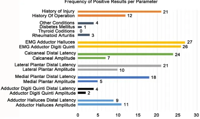
Of the 40 TTS cases there was on average 4.3 positive test parameters present in each of the cases (Figure 2).
[image:6.595.208.537.447.739.2]37.5% tested positive for at least 6 of the 12 electro-diagnostic parameters (Table 1).
67.5% of the Tarsal Tunnel Syndrome group had history of a previous injury or operation in the lower limb (Table 2).
30% had history of a previous operation alone. 52.5% had a prior injury to the lower limb. 6 individuals reported both a prior injury and ankle surgery.
The 12 parameters assessment of TTS resulted in a diagnostic pick up rate of 3.3% with 40 positive cases out of 1210 patient referrals in a calendar year.
The calcaneal sensory study and the needle EMG tp the AH and ADQ muscles proved to be the most sensitive tests. The calcaneal sensory study distal latency being prolonged in 60% of cases. The needle EMG to the AH muscle showed denervation in 65% of cases. The needle EMG to the ADQ muscle showed de-nervation in 67.5% of cases (Table 2).
Needle EMG to the AH and the ADQ and the calcaneal sensory study are not routinely preformed in cases of suspected TTS. Their addition to the range of electro-diagnostic tests contributed to the increased detection rates shown in this study.
Table 1. Positive test parameters in cases of tarsal tunnel syndrome.
Test
Number: Test Parameter:
% Positive Results:
Number of positive results:
1 Tibial motor study to Adductor
Halluces muscle: Amplitude of Response 27.5% 11
2 Tibial motor study to Adductor
Halluces muscle: Distal Latency 22.5% 9
3 Digiti Quinti muscle: Amplitude of response Tibial motor study to the Adductor 5% 2
4 Digiti Quinti muscle: Distal Latency Tibial motor study to the Adductor 10% 4
5 Medial Plantar: Amplitude of Response 12.5% 5 6 Medial Plantar: Distal Latency 45% 18 7 Lateral Plantar: Amplitude of Response 25% 10 8 Lateral Plantar: Distal Latency 52.5% 21 9 Calcaneal: Amplitude of Response 17.5% 7
10 Calcaneal: Distal Latency 60% 24
11 Needle EMG to tibial innervated
Adductor Halluces 65% 26
Figure 2. Frequency of positive results per parameter in cases of TTS.
Table 2. Prior history of injury and or surgery in cases of TTS.
% of total: Total Number: History of Previous Injury: 52.5% 21 History of previous Operation: 30.0% 12 History of injury or Operation: 67.5% 27
4. Discussion
A patient with tarsal tunnel syndrome often describes vague discomfort and pain, specifically the pain posterior to the medial malleolus and radiating to the arch of the foot. There is usually sensory alteration in the sole of the foot in both the medial and lateral aspects. Commonly there is heel pain due to the involve-ment of the calcaneal branch. Frequently individuals will also describe weakness in the foot biomechanics both in abductor and adduction. Patients are often asymptomatic in the morning after taking the first two steps. Symptoms usually worsen with increased activity and towards the end of the day after long periods of walking or running. Also standing in the same place for long periods of time can also aggravate the problem. The pain and burning in the feet usually persist for half an hour or more after the patient has gone off his feet.
peri-pheral neuropathy, all of which are associated with an increased incidence of TTS [1]. A history of a previous trauma to the foot or ankle should also be sought, as the current study confirms the association of previous injury and sur-gery in cases of TTS.
Electro-diagnostic testing should be extensive and consistent and follow a similar pattern in every individual. This is to take into account the significant variability in the anatomy of the posterior tibial nerve. A limited examination, as has been recommended by some authors [2], is restricted to tibial motor, and medial and lateral plantar sensory studies. This limited approach will reduce the diagnostic yield. The current study confirms that the most sensitive EXD tests for TTS were the needle EMG to the AH and ADQ muscles and the calcaneal sensory study. These elements are not routinely performed in the EXD assess-ment of suspected cases of TTS.
This study recommends a 12-parameter test protocol: 3 sensory nerve con-duction studies, 2 motor nerve concon-duction studies and a 2-muscle needle EMG assessment. This methodology is a detailed interrogation of the tibial nerve at the Tarsal Tunnel and is cognizant of the significant variability in the branching pattern of the nerve at this anatomical site, which therefore requires a detailed EXD examination. The approach resulted in a significant increase in the detec-tion rate of TTS, annualized at 3.3% which is a 5-fold increase on reported di-agnostic rates [6].
This study recommends a 12-parameter test protocol, 3 sensory nerve con-duction studies, 2 motor nerve concon-duction studies and a 2 muscle needle EMG assessment. This methodology resulted in a significant increase in the detection rate of TTS, annualized at 3.3% which is a 5-fold increase on reported diagnostic rates [6].
A literature review of the of Tarsal Tunnel evaluation techniques [7] con-firmed a significant lack of standardization in methodology , and little regard for the variability and complex nature of the anatomy of the tibial nerve in sus-pected cases. This extensive review of 317 articles and studies on the topic con-firmed that that needle EMG to the Adductor Halluces and the Adductor Digiti Quinti was never used in the assessment of TTS. Calcaneal nerve conduction sensory studies were similarly not employed in evaluating this condition. The current study has highlighted that these 3 tests proved to be the most sensitive tests in this study for evaluation of TTS. Non standardization of assessment and failure to use available techniques may account for the poor detection rate of this the most common lower limb focal neuropathy.
This study suggests that a heightened index of suspicion in cases of foot sen-sory alteration where there is a prior history of lower limb trauma coupled with this 12 parameter assessment may improve detection rate for TTS whose true incidence may be under reported.
References
[2] Keck, C. (1962) The Tarsal Tunnel Syndrome. The Journal of Bone & Joint Surgery, 44, 180-184.https://doi.org/10.2106/00004623-196244010-00015
[3] Lam, S.J. (1962) A Tarsal Tunnel Syndrome. Lancet, 2, 1354-1355. https://doi.org/10.1016/S0140-6736(62)91024-3
[4] Baxter, D.E. and Thigpen, M. (1984) Heel Pain Operative Results. Foot Ankle, 5, 16- 25.https://doi.org/10.1177/107110078400500103
[5] Baxter, D.E. and Pferrer, G.B. (1992) Treatment of Chronic Heel Pain by Surgical Release of the First Branch of the Lateral Plantar Nerve. Clinical Orthopaedics and Related Research, 279, 229-236.https://doi.org/10.1097/00003086-199206000-00029 [6] Saeed, M.A. (2004) Tarsal Tunnel Syndrome. Course C: Painful Foot and Ankle
AAEM 51ST Annual Scientific Meeting, Savannah, Georgia, November 2004, 17-26.
[7] Patel, A.T., Gaines, K., Malamut, R., Park, T., Del Toro, D. and Holland, N. (2005) Usefulness of Electrodiagnostic Techniques in the Evaluation of Suspected Tarsal Tunnel Syndrome: An Evidence Based Review. Muscle and Nerve, 32, 236-240. https://doi.org/10.1002/mus.20393
Submit or recommend next manuscript to SCIRP and we will provide best service for you:
Accepting pre-submission inquiries through Email, Facebook, LinkedIn, Twitter, etc. A wide selection of journals (inclusive of 9 subjects, more than 200 journals)
Providing 24-hour high-quality service User-friendly online submission system Fair and swift peer-review system
Efficient typesetting and proofreading procedure
Display of the result of downloads and visits, as well as the number of cited articles Maximum dissemination of your research work