REVIEW
Proliferation, survival and metabolism: the role of PI3K/AKT/
mTOR signalling in pluripotency and cell fate determination
Jason S. L. Yu and Wei Cui*ABSTRACT
Phosphatidylinositide 3 kinases (PI3Ks) and their downstream mediators AKT and mammalian target of rapamycin (mTOR) constitute the core components of the PI3K/AKT/mTOR signalling cascade, regulating cell proliferation, survival and metabolism. Although these functions are well-defined in the context of tumorigenesis, recent studies–in particular those using pluripotent stem cells – have highlighted the importance of this pathway to development and cellular differentiation. Here, we review the recent in vitroandin vivoevidence for the role PI3K/AKT/mTOR signalling plays in the control of pluripotency and differentiation, with a particular focus on the molecular mechanisms underlying these functions.
KEY WORDS: PI3K, AKT, mTOR, Pluripotency, Proliferation and differentiation, Metabolism
Introduction
During development, cellular differentiation is a crucial process by which all major tissues of the adult organism are formed. This is governed by the spatial and temporal activation of signalling pathways, classically demonstrated through gain- and loss-of-function studies performed in developmental models, such as zebrafish, Xenopus and mice. Genetic manipulation of signalling components induces defects in either specific tissues or developmental stages, thus implying that distinct pathways specify particular lineages. However, it is increasingly evident that this is a gross oversimplification, given that signalling pathways do not operate in isolation, but participate in a myriad of crosstalk interactions that cumulatively determine cell fate. Therefore, even pathways that do not appear to specify a lineage independently may act to augment or inhibit pathways that are essential for lineage specification. However, the inherent complexity and redundant nature of signalling makes the molecular dissection of such crosstalk events difficult.
PI3K/AKT/mTOR signalling contributes to a variety of processes that are critical in mediating many aspects of cellular function, including nutrient uptake, anabolic reactions, cell growth and survival. The core components of the pathway, namely phosphatidylinositide 3-kinases (PI3Ks), AKT and mammalian target of rapamycin (mTOR, also known as mechanistic TOR), have been shown to be frequently hyperactivated in the majority of cancers and have therefore been the focus of many studies in this field (Vanhaesebroeck et al., 2010; Thorpe et al., 2015). By contrast, the importance of this pathway in development is understated, even though loss any of the core components often results in embryonic lethality (Bi et al., 1999; Peng et al., 2003; Murakami et al., 2004).
The embryonic lethality that results from disruption of PI3K/ AKT/mTOR signalling hinders the interrogation of this pathway during differentiation through traditional developmental models. Derivation of pluripotent stem cells (PSCs), including embryonic stem cells (ESCs) and induced PSCs (iPSCs), has mitigated this problem to some extent. These cells can undergo self-renewal indefinitely, and can also give rise to all the cells of the adult organism, providing researchers with a unique cell-based model to interrogate cellular differentiation. This, coupled with increasingly defined culture conditions and better tools for genetic manipulation, has allowed researchers to overcome the technically challenging interrogation of the molecular basis for crosstalk interactions. As a result, recent studies have begun to delineate the contribution of PI3K/AKT/mTOR signalling in preserving the ability of PSCs to self-renew and differentiate (Paling et al., 2004; Zhou et al., 2009; Singh et al., 2012; Yu et al., 2015), and it has become apparent that this signalling pathway is crucial for embryonic development and is indispensable for the self-renewal of PSCs. In this Review, we discuss the functions of this pathway during embryonic development and in cell fate determination of PSCs, with a particular focus on the putative molecular mechanisms underlying these functions.
PI3K/AKT/mTOR signalling
PI3Ks are a unique family of intracellular lipid kinases that phosphorylate the 3′-hydroxyl group of the inositol ring of phosphatidylinositides (PtdIns) and are divided into three classes. The most studied is class I PI3K (Fig. 1), which stimulates the conversion of membrane-bound phosphatidylinositol-(4,5)-bisphosphate [PtdIns(4,5)P2; PIP2] to phosphatidylinositol-(3,4,5)-trisphosphate [PtdIns(3,4,5)P3; PIP3] (Vanhaesebroeck et al., 2010; Thorpe et al., 2015). PIP3 acts as a secondary messenger, facilitating the recruitment and activation of kinases that possess the pleckstrin homology (PH) domain, such as PI3K-dependent kinase-1 (PDK1). The signalling duration of PIP3 is subject to regulation by phosphatase and tensin homolog (PTEN), which acts to oppose PI3K activity. The serine/threonine kinase AKT, also known as protein kinase B (PKB), possesses a PH domain, and is recruited to the plasma membrane along with PDK1. Phosphorylation of amino acid residues T308 and S473 by PDK1 and mTORC2, respectively, is essential for full AKT activation. Following activation, AKT can phosphorylate many target proteins, most notably glycogen synthase kinase 3 (GSK3), tuberous sclerosis 2 (TSC2), caspase 9 and PRAS40 (AKT1S1), which explains its relatively wide spectrum of downstream effects in promoting cell proliferation, differentiation, apoptosis, angiogenesis and metabolism.
mTOR is an evolutionarily conserved serine/threonine protein kinase that belongs to the PI3K-related kinase family (PIKK). The accessibility of the mTOR active site is governed in part by mTOR-associated proteins that form two distinct complexes: mTOR complex 1 (mTORC1) and 2 (mTORC2). These complexes differ in their composition, mode of activation and rapamycin sensitivity
Institute of Reproductive and Developmental Biology, Department of Surgery and Cancer, Imperial College London, Du Cane Road, London W12 0NN, UK.
*Author for correspondence (wei.cui@imperial.ac.uk)
J.S.L.Y., 0000-0001-5203-3603; W.C., 0000-0003-2019-380X
DEVEL
O
(Yang et al., 2013; Liu et al., 2015). Although mTOR in both complexes interacts with DEPTOR and mLST8, the other core components are distinct between them. Instead of having RAPTOR (RPTOR) and PRAS40 in mTORC1, mTORC2 contains RICTOR (rapamycin-insensitive companion of mTOR) and mSIN1 (mammalian stress-activated protein kinase interacting protein 1; MAPKAP1) (Fig. 1). Activation of mTORC1 is regulated by multiple intracellular and extracellular cues, such as growth factors, nutrients, energy status and stress, in which the RAG and RHEB (RAS homolog enriched in brain) guanosine triphosphatases (GTPase) as well as TSC1/2 play essential and distinct roles (Box 1). Upon activation, mTORC1 regulates many cellular functions, such as cell growth, protein synthesis and autophagy via S6 kinase (S6K; RPS6K), eukaryotic translation initiation factor 4E-binding protein 1 (4E-BP1; EIF4EBP1) and ULK1, respectively (Laplante and Sabatini, 2012). In contrast to mTORC1, mTORC2 activation, stabilisation and function requires RICTOR and mSIN1, although the precise molecular mechanism by which this occurs remains unclear (Laplante and Sabatini, 2012). The downstream effectors of mTORC2 identified thus far are mainly members of the AGC kinase family, including AKT, protein kinase C (PKC) and serum and glucocorticoid induced kinases (SGKs), which modulate cytoskeleton organisation, cell survival, lipid homeostasis and metabolism (Laplante and Sabatini, 2012).
Involvement of PI3K/AKT/mTOR in early embryonic development
Given the complex interplay between PI3K, AKT, mTORC1 and mTORC2, it is challenging to identify the physiological functions and contributions of each factor within this signalling pathway,
particularly in the context of embryonic development. Nonetheless, genetic manipulation of key components by way of gene knockout has ascertained their importance in driving organismal growth and tissue formation, and has highlighted the essential role these signalling components play during embryonic development (summarised in Table 1).
Critical and divergent roles of PI3K isoforms
Although the various isoforms of class I PI3Ks share conserved domain structures and lipid substrates, thus generating identical PIP3 upon activation, knockout studies in mice have revealed that they have distinct roles in embryonic development. Mice with dysfunctional p110α (PIK3CA) die around embryonic day (E) 10.5 (Bi et al., 1999; Foukas et al., 2006), whereas mice lacking p110β (PIK3CB) die much earlier at E3.5 (Bi et al., 2002). Interestingly, mice with inactive (but not absent) p110βdevelop normally (Ciraolo et al., 2008), suggesting that the p110β, unlike p110α, has a kinase-independent function that is indispensable for early embryonic development. These results also suggest non-redundant roles for ubiquitously expressed p110α and p110β, which might reflect differences in their activation mechanisms. Whereas p110αis activated by phosphorylated tyrosine receptors, their adaptors and by RAS, p110βis directly activated by CDC42 and the Rho small G protein RAC1 (Fritsch et al., 2013). Mice carrying defects in either p110δ(PIK3CD) or p110γ(PIK3CG) show no defect in their overall development but demonstrate impaired lymphocyte production (Clayton et al., 2002; Sasaki et al., 2000), which is consistent with the enriched expression of these isoforms in leukocytes. Deficiency of all Class IA p85 regulatory subunits in mice leads to perinatal death, but elimination of p85α (PIK3R1) alone produces viable mice (Fruman et al., 2000; Terauchi et al., 1999; Ueki et al., 2002), suggesting that there are certain redundant functions among P
P
PIP2 PIP3
P
P PP P
S473 PI3K
T308
TSC2 TSC1 P
RHEB
PKB/AKTP P
PIP2
P P
PTEN
mTOR mLST8
RAPTOR DEPTOR PRAS40
mTORC1
4E-BP1 P
ULK1P S6K1/2
P
S6
P
EIF4E
T
mTOR mLST8
RICTOR mTORC2
mSIN1
DEPTOR
SGK1P PKCP GSK3
P
PDK1
[image:2.612.50.300.60.259.2]Cell membrane RTK
Fig. 1. Schematic of the PI3K/AKT/mTOR signalling cascade and its main downstream effectors.Upon growth factor stimulation (yellow diamond), active PI3K phosphorylates phosphatidylinositol (4,5)-bisphosphate (PIP2) to phosphatidylinositol (3,4,5)-triphosphate (PIP3), which then recruits AKT and permits phosphorylation of T308 and S473 through PDK1 and mTORC2, respectively. Activated AKT inhibits GSK3 and TSC2 via phosphorylation, inactive TSC1/2 is unable to bind RAS homolog enriched in brain (RHEB), which subsequently enables its activation of mTORC1 at the surface of lysosome, initiating its effect on many downstream proteins, including S6K, 4E-BP1 and ULK1. Active mTORC2 can regulate several AGC kinases, such as SGK1 and PKC, in addition to AKT.‘P’in a green circle indicates activating phosphorylation, in a red circle inhibitory phosphorylation and in a brown circle phosphorylation that is not induced by this signalling cascade.
Box 1. Regulation of mTOR complexes mTORC1
Activation of mTORC1 is regulated by multiple intracellular and extracellular cues, involving both RAG and RHEB (Ras homolog enriched in brain) guanosine triphosphatases (GTPases). RAG GTPase exists as a heterodimer of RAGA/B (RRAGA/B) with RAGC/D (RRAGC/D) and is anchored to the lysosome by the Ragulator complex (Bar-Peled et al., 2012). RAG GTPase is activated by the presence of nutrients (e.g. amino acids) through the combined action of the amino acid transporter SLC38A9 (Rebsamen et al., 2015; Wang et al., 2015) and the Ragulator complex as a guanine nucleotide exchange factor (GEF), along with other proteins, which recruit mTORC1 to the lysosome surface where it encounters RHEB. The activation of RHEB is negatively controlled by tuberous sclerosis 1 and 2 (TSC1/2). Notably, many upstream signalling inputs regulate mTORC1 via phosphorylation of TSC1/2. For example, growth factor-induced PI3K/AKT inactivates TSC2 by phosphorylation of S939/T1462, resulting in the activation of RHEB and therefore mTORC1. Conversely, energy deficiency-activated adenosine monophosphate protein kinase (AMPK; PRKA) triggers the phosphorylation of TSC2-T1227/S1387, enhancing TSC1/2 activity and inhibiting mTORC1. Recently, arginine has been shown to activate mTORC1 via a RAG GTPase-independent mechanism (Carroll et al., 2016).
mTORC2
In contrast to mTORC1, the factors and regulative processes driving mTORC2 activation remain relatively understudied. So far, only PI3K stimulation has been shown to promote mTORC2 activation in association with the mSIN1 PH domain and the ribosome (Liu et al., 2015; Zinzalla et al., 2011). The exact molecular details await further elucidation.
DEVEL
O
regulatory subunits of class I PI3Ks. In contrast to class I PI3Ks, the primary product of class II and III PI3Ks is PtdIns(3)P, indicating that they may have different roles in embryonic development. Taken together, these findings suggest that the majority of PI3K isoforms have distinct, non-compensating and indispensable roles in early embryonic development.
Different but redundant roles of AKT isoforms
In contrast to the highly specific roles attributed to PI3K isoforms, the three AKT isoforms appear to have an assorted but clearly redundant role in early embryonic development (Table 1). Germline deletion of any of the isoforms alone does not lead to clear embryonic lethality despite the various physiological and pathological defects that these mice exhibit (Chen et al., 2001; Cho et al., 2001; Tschopp et al., 2005). However, deletion ofAkt1 together withAkt2orAkt3leads to embryonic death, with theAkt1/ Akt3double-knockout mice exhibiting more severe phenotype and earlier lethality (Peng et al., 2003; Yang et al., 2005). By contrast, Akt2/Akt3 double-knockout mice are viable with only mild phenotypes (Dummler et al., 2006). Thus, even though AKT1 can sufficiently sustain full development by itself and appears to play a more important role during development than the other isoforms owing to its higher and broader expression pattern, all isoforms are required for proper development and function.
[image:3.612.46.561.69.447.2]Distinct roles of mTORC1 and mTORC2 in embryonic development Given the key roles that mTOR complexes play in AKT activation, protein translation and nucleotide biosynthesis, it is perhaps unsurprising to find that knockout mice of various components of the complexes lead to embryonic lethality (Rennebeck et al., 1998; Murakami et al., 2004; Guertin et al., 2006; Shiota et al., 2006; Jacinto et al., 2006; Goorden et al., 2011). However, upon closer inspection, mTORC1 and mTORC2 may have different functions during developmental progression, and indeed their deletion arrests the embryos at distinct developmental stages (Table 1). Mice with a lack of mTOR or the mTORC1-specific protein RAPTOR display similar phenotypes of embryonic lethality at E5.5-6.5, which is due to defective proliferation of the inner cell mass (ICM) and trophectoderm (Murakami et al., 2004; Guertin et al., 2006). By contrast, mice deficient in the mTORC2-specific proteins RICTOR or mSIN1 progress through development until E10.5, which is considerably later than that of mTOR- and RAPTOR-null mutants (Shiota et al., 2006; Guertin et al., 2006; Jacinto et al., 2006). Defects in cardiovascular function may account for the lethality in these mice as knockout ofRictorin the developing cardiovascular system alone leads to the similar phenotype (Zhao et al., 2014). Interestingly, knockout ofMlst8, which encodes a core component of both complexes, produces a phenotype that exhibits vascular defects in the yolk sac and embryonic lethality at E10.5, Table 1. Summary of knockout mice with germline deletion of key PI3K/AKT/mTOR signalling genes
Genetic manipulation Reported phenotype References
PI3K Class I
p110α−/− Embryonic lethality around E9.5-10.5 (Bi et al., 1999)
p110αD933A/D933A Growth retardation at E9 and death at E10-11 (Foukas et al., 2006)
p110β−/− Embryonic lethality at∼E3.5 (Bi et al., 2002)
p110βK805R/K805R Develop normally but small with metabolic defect; male infertility (Ciraolo et al., 2008)
p110δ−/− Develop normally but with defects in lymphocyte development (Ciraolo et al., 2008)
p110γ−/− Develop normally but with impaired thymocyte development and T-cell activation (Sasaki et al., 2000) p85a-p55a-p50a−/−* Perinatal lethality; extensive hepatocyte necrosis (Fruman et al., 2000)
p85a−/− Hypoglycaemia; increased insulin sensitivity (Terauchi et al., 1999)
p85b−/− Hypoinsulinemia; hypoglycemia; improved insulin sensitivity (Ueki et al., 2002) Class II
Pi3kc2a(Pik3c2a)−/− Growth retardation from E8.5 onwards; death at E10.5-11.5 (Yoshioka et al., 2012)
Pi3kc2atruncation Viable but smaller with less fat; late-onset kidney failure (Harris et al., 2011)
Pi3kc2b−/− Develop normally with no obvious phenotype (Harada et al., 2005)
Class III
Vps34-del Embryonic lethality at E7.5-8.5, with normal epiblast but abnormal visceral endoderm cells (Zhou et al., 2011) PTEN
Pten−/− Embryonic lethality at∼E8 (Di Cristofano et al., 1998)
AKT
Akt1−/− Mild growth retardation; accelerated apoptosis in thymus and testes (Chen et al., 2001)
Akt2−/− Insulin resistance phenotype (Cho et al., 2001)
Akt3−/− Smaller brain size (Tschopp et al., 2005)
Akt1−/−andAkt2−/− 50% smaller; immediate post-natal death (Peng et al., 2003) Akt1−/−andAkt3−/− Embryonic lethality at E10.5-11.5, possibly due to placenta vasculature defects (Yang et al., 2005)
Akt2−/−andAkt3−/− Combined phenotype ofAkt2−/−andAkt3−/− (Dummler et al., 2006) TSC/RHEB
Tsc1−/− Embryonic lethality at E10.5-11.5 with failed closure of neural tube (Kobayashi et al., 2001) Tsc2−/− Embryonic lethality at E9.5-12.5 with defective neural tube or liver development (Rennebeck et al., 1998)
Rheb−/− Embryonic lethality at E10.5-12.5 with defective cardiovascular development (Goorden et al., 2011) mTORC
Mtor−/− Embryonic lethality at E5.5-6.5; proliferation defect of ICM and trophoblast giant cells (Murakami et al., 2004) Rptor−/− Embryonic lethality at E5.5-6.5; implanted blastocysts fail to expand (Guertin et al., 2006)
Mlst8−/− Embryonic lethality at E10.5; severe vasculature defects (Guertin et al., 2006)
Rictor−/− Embryonic lethal at E10.5-11.5; severe vasculature defects (Guertin et al., 2006; Shiota et al., 2006)
Sin1−/− Embryonic lethality at E10.5-11.5 (Jacinto et al., 2006)
*Pi3kr1gene.
DEVEL
O
phenocopying RICTOR deficiency but dissimilar to that of mTOR or RAPTOR deficiency (Guertin et al., 2006). Together, these studies suggest that a deficiency in mTORC1 activity is largely responsible for proliferative defects that lead to early developmental arrest, whereas mTORC2 deficiency induces arrest at mid-gestation due to defective cardiovascular development, and that, although mLST8 is involved in the assembly of both mTORC1 and mTORC2 complexes, it is only required for mTORC2 activity during embryonic development. Furthermore, it is worth noting that mTORC2 does not appear to affect overall cytoskeleton organisation during mouse development as previously reported in yeast (Guertin et al., 2006; Shiota et al., 2006; Jacinto et al., 2006). In contrast to the lethal phenotypes that result from the deletion of key components of the mTOR complexes, there are no severe developmental abnormalities associated with the deficiency of effectors downstream of mTOR complexes, such as S6K1 (RPS6KB1), S6K2 (RPS6KB2), ULK1, 4E-BP1/2 and SGK1, with the exception of perinatal death in S6k1/2double-knockout mice due to heart failure (Pende et al., 2004; Kundu et al., 2008; Le Bacquer et al., 2007; Wulff et al., 2002). This is most likely to be due to the inherent redundancies in the regulation of downstream processes, whereby although each factor plays a crucial role, none is indispensable in inducing the phenotypic outcome. Interestingly, germline deficiency of any of the three mTORC1 upstream regulators TSC1, TSC2 and RHEB result in embryonic lethality around E11, which is much later than mTORC1 knockout mice (Rennebeck et al., 1998; Kobayashi et al., 2001; Goorden et al., 2011). This suggests that mTORC1 could be activated by other signalling pathways in addition to the TSC1/2-RHEB cascade and that TSC1/2 as well as RHEB may also have mTORC1-independent developmental functions (Neuman and Henske, 2011).
PI3K/AKT/mTOR signalling in pluripotent stem cells
Although in vivo studies have been informative in terms of establishing a role for PI3K/AKT/mTOR signalling in the regulation of early embryonic development, the inherent complexity and multiple layers of redundancy present a formidable barrier to the elucidation of the molecular details involved. ESCs isolated from pre-implantation embryos possess the developmental competency of the early embryo and their differentiation recapitulates to a certain extent early embryonic development. Thus, ESCs serve as an excellent basis for interrogating the molecular contribution of this signalling pathway to development (Zhu and Huangfu, 2013).
PI3K/AKT/mTOR are essential for maintaining pluripotency
Despite the considerable differences between human (hESCs) and mouse (mESCs) ESCs in terms of the signalling requirements for their culture and propagation, studies using kinase inhibitors and genetic approaches have shown that both types of ESCs require active PI3K/AKT signalling to maintain their undifferentiated properties. Treatment of mESCs with either a general PI3K or p110β-specific inhibitor compromises their pluripotency, and deficiency of PI3K genes also results in loss of pluripotency in mESCs (Paling et al., 2004; Kingham and Welham, 2009). Similarly, PI3K inhibition in hESCs results in the downregulation of pluripotency markers concomitant with the upregulation of lineage-specific genes, which together strongly suggest the loss of pluripotency (Armstrong et al., 2006; McLean et al., 2007; Singh et al., 2012). Given that active cell proliferation is essential for ESC self-renewal and that PI3K plays a key role in regulation of cell proliferation, this implicit requirement for PI3K in ESC self-renewal is probably expected. In fact, knockout ofPtenin both mESCs and
hESCs duly augments the proliferation and survival of these cells (Alva et al., 2011). However, PI3K deficiency appears to have no obvious effect on ESC survival or proliferation (Paling et al., 2004), suggesting that PI3K signalling is not limited to driving the growth and survival of ESCs, but also in inherently preserving their pluripotent properties.
Interestingly, despite the fact that PI3K signalling is active in both mESCs and hESCs, it is unclear whether this activation is through canonical induction via IGF1/insulin or whether it is simply a resultant effect from the activation of other pathways (Fig. 2). PI3K signalling in mESCs was found to be activated by an ESC-specific RAS protein (ERAS) via direct interaction with its p110 subunit (Takahashi et al., 2003). However, deficiency of ERAS does not affect pluripotency of mESCs, indicating that the PI3K-medicated effect on pluripotency is not associated with ERAS. By utilising chemically defined culture conditions, it was proposed that leukaemia inhibitory factor (LIF), a key player in maintenance of mESC self-renewal, may activate PI3K signalling through phosphorylation of its receptor JAK (Niwa et al., 2009; Paling et al., 2004), yet it remains unclear how LIF induces both JAK-STAT3 and JAK-PI3K pathways. Similarly, activation of PI3K in hESCs may also occur through non-canonical means. The presence of basic fibroblast growth factor (bFGF) in hESC cultures is thought to stimulate PI3K signalling in both fibroblast-conditioned medium or chemically defined culture conditions by activating EGFR and IGF1R (Zoumaro-Djayoon et al., 2011) Given that heregulin (neuregulin 1) and IGF1 can act in the absence of FGF to maintain hESC pluripotency, FGF-mediated PI3K induction appears to be the more crucial effect in preserving hESC properties opposed to stimulation of the FGF/RAS/MAPK axis (Singh et al., 2012). However, an important caveat to such a conclusion is that both
Insulin/IGF
IR/IGF1R
PI3K
MEK/ERK GSK3
LIF
JAK
STAT3
TCF3 bFGF
FGFR
MEK/ERK ELA
AKT
Pluripotency
β-catenin
[image:4.612.317.562.416.616.2]hESCs mESCs
Fig. 2. PI3K/AKT regulation of mESC and hESC pluripotency.PI3K/AKT can be activated by insulin and IGF1 in both mESCs and hESCs, which maintains pluripotency through inhibition of MEK/ERK signalling. In mESCs, PI3K/AKT may also be activated by the LIF/JAK pathway to inhibit GSK3 signalling in addition to inhibition of MEK/ERK. Inhibition of GSK3 attenuates theβ -catenin-mediated TCF3 repression of pluripotent genes, thus promoting pluripotency. In hESCs, PI3K is activated by an endogenous peptide ELABELA (ELA) in addition to insulin/IGF1. This may have a positive effect on GSK3 through the inhibition of MEK/ERK, thereby inhibiting nuclear translocation ofβ-catenin and enhancing pluripotency (see text for details). Green arrows represent activation, red arrows represent inhibition. Dashed arrows represent non-canonical
induction by other pathways. IR, insulin receptor.
DEVEL
O
heregulin and IGF1 can also conversely activate RAS/MAPK signalling, making it difficult to distinguish the independent effects of either pathway on pluripotency. Nonetheless, a recent report showed that under similar defined culture conditions, insulin but not bFGF, is able to activate PI3K signalling (Ho et al., 2015), suggesting that the magnitude of FGF-mediated PI3K activation is insufficient for maintaining hESC pluripotency. Intriguingly, the same group also reported the identification of an endogenous peptide ELABELA (ELA; APELA) that acts to induce PI3K/AKT activity via an alternative, unknown receptor that is distinctly different from canonical induction via insulin (Fig. 2). Introduction of exogenous ELA alone not only sufficiently activates PI3K to the degree required for preserving pluripotency, but also primes the cells for mesendoderm differentiation, further underscoring the importance of this pathway in the regulation of ESC identity.
The mechanisms by which PI3K regulates pluripotency remain somewhat elusive. In mESCs, increasing evidence suggests that PI3K promotes the retention of ESC properties mainly through the inhibition of two downstream pathways: the mitogen-activated protein kinases/extracellular signal-regulated kinase (MAPK/ERK) pathway and the GSK3 signalling pathway. This is consistent with the finding that PI3K signalling is dispensable in naive mESC 2i culture conditions in which both MAPK/ERK and GSK3 pathways are suppressed (Hishida et al., 2015). Active MAPK/ERK signalling is a key requirement for mESCs to undergo differentiation (Ying et al., 2008); thus, inhibition of MAPK/ERK signalling by LIF-induced PI3K may contribute to the maintenance of pluripotency even though the exact mechanisms by which PI3K inhibits MAPK/ERK are unclear (Paling et al., 2004). With respect to the inhibition of GSK3 signalling, several reports have shown that PI3K signalling inhibits GSK3α/βactivity by phosphorylating the S21/S9 residue via AKT activation in mESCs, which leads to the upregulation of pluripotent transcription factors TBX3 and NANOG that subsequently promote pluripotency in these cells (Paling et al., 2004; Niwa et al., 2009; Wray et al., 2011). However, the exact underlying mechanisms remain ambiguous, particularly regarding the involvement of β-catenin. Wellham and colleagues have shown that inactivation of GSK3 by inhibition of PI3K has no clear effect onβ-catenin phosphorylation, indicating that the effect of PI3K signalling on TBX3 and NANOG in mESCs could be through β-catenin-independent mechanisms (Paling et al., 2004). However, a more recent study using chemically defined culture media combined with genetic approaches has revealed that enhancement of NANOG expression by GSK3 inhibition results from the alleviation of TCF3 repression, which is largely mediated through β-catenin, although independent of its transcriptional activity and even though β-catenin is not essential for the maintenance of pluripotency (Wray et al., 2011).
In hESCs, the mechanisms by which PI3K may regulate pluripotency are even more ambiguous. Similar to mESCs, active PI3K signalling inhibits the MAPK/ERK pathway under chemically defined culture conditions. However, unlike mESCs, active PI3K signalling in hESCs appears to activate GSK3 activity through inhibition of MAPK/ERK, which leads to suppression ofβ-catenin (Singh et al., 2012). In support of this notion, undifferentiated hESCs contain mainly inactiveβ-catenin, and activation ofβ-catenin promotes hESC differentiation, probably throughβ-catenin-mediated transcription of differentiation genes (Davidson et al., 2012). However, little is known about the expression and function of TCF3 and other TCF/LEF proteins in hESCs as well as what mechanism accounts for the differential regulation of GSK3 phosphorylation in mESCs and hESCs by PI3K/AKT.
Nonetheless, the role of PI3K signalling remains largely preserved between species and is required to maintain both self-renewal and pluripotency of ESCs.
In agreement with the positive role PI3K plays in maintaining ESC pluripotency, ectopic expression of constitutively active AKT is sufficient to preserve the pluripotency of mouse and primate ESCs in the absence of feeder cells (Watanabe et al., 2006). By contrast, rapamycin-mediated inhibition of mTORC1 disrupts the pluripotency of hESCs (Zhou et al., 2009). The latter study also demonstrated that this effect is independent of the growth-inhibiting properties of rapamycin, given that treatment with a cyclin-dependent kinase (CDK) inhibitor failed to reproduce the same effect. However, long-term treatment of cells with rapamycin can also result in inhibition of mTORC2 (Sarbassov et al., 2006), and it is therefore unclear whether this phenomenon is purely an mTORC1-dependent effect. Nonetheless, knockdown of either the mTORC1 upstream suppressor TSC2 or the downstream target folliculin prevents mESC lineage commitment (Betschinger et al., 2013), further supporting a role for mTORC1 signalling in maintaining pluripotency. Taken together, PI3K/AKT/mTOR signalling is therefore indispensable for modulation of ESC self-renewal and pluripotency through multiple regulatory mechanisms.
The function of PI3K/AKT/mTOR in regulating glycolytic metabolism An increasing number of studies have demonstrated that cellular metabolism also plays an essential role in regulation of pluripotency, specifically through modulation of the epigenetic landscape (Zhou et al., 2012; Sperber et al., 2015; Zhang et al., 2016). In primed human PSCs, metabolism typically proceeds via the glycolytic pathway, which rapidly switches to oxidative phosphorylation upon differentiation (Zhou et al., 2012; Moussaieff et al., 2015). Compared with differentiated cells, which utilise oxidative phosphorylation as the primary metabolic pathway, these PSCs possess largely underdeveloped and immature mitochondria that are few in number, thus maintaining high rates of glycolysis even under aerobic culture conditions, although a recent report found that these cells can nonetheless utilise oxidative phosphorylation for energy generation through glutamine metabolism (Prigione et al., 2010; Varum et al., 2011; Tohyama et al., 2016). This preference for glycolytic metabolism is reminiscent of the Warburg effect in many highly proliferative cancer cells, typified by an increased conversion of pyruvate to lactate. However, a recent study showed that although glycolytic ESCs produce lactate via lactic fermentation, it is the acetyl-CoA by-product that functions to maintain pluripotency via modulation of histone acetylation (Moussaieff et al., 2015). Epigenetic modification by the cellular metabolome has also been shown to regulate the transition from naive to primed PSC states. Naive human PSCs accumulate high levels of nicotinamide N-methyltransferase, which acts to prevent their progression through pluripotency by relieving repressive methylation of WNT via the reduction of S-adenosyl methionine (SAM), a key precursor required by histone methyltransferases (Sperber et al., 2015). Equally, murine naive-to-primed PSC conversion is regulated by the RNA-binding proteins LIN28A and LIN28B, which act to downregulate mitochondrial metabolism and potentiate glycolysis (Zhang et al., 2016). Thus, naive PSCs, primed PSCs and differentiated cells possess unique metabolic signatures that ultimately potentiate their fate.
Signalling pathways play an important role in regulating cellular metabolism; PI3K/AKT/mTOR signalling is a master regulator of aerobic glycolytic metabolism (Ward and Thompson, 2012). The
DEVEL
O
active PI3K/AKT pathway increases glucose uptake by upregulating the expression of cell surface glucose transporters and by stimulating several glycolytic enzymes, such as hexokinase (Ward and Thompson, 2012). In addition, AKT has been found to promote activation of ATP citrate lyase (ACL; ACLY), which enhances the conversion of citrate to acetyl-CoA bothin vitroand in vivo (Lee et al., 2014). The role of mTOR complexes in the regulation of metabolism is more complex. On the one hand, mTORC1 upregulates the expression of hypoxia-inducible factor 1 (HIF1), which promotes glycolysis by diverting pyruvate into lactate (Kim et al., 2006; Cheng et al., 2014) and is further enhanced by mTORC2-mediated activation of AKT via S473 phosphorylation. On the other hand, mTORC1 positively regulates mitochondrial function and oxidative metabolism by selectively promoting translation of nucleus-encoded mitochondria-related mRNAs (Morita et al., 2013).
In addition to a direct role in regulating glycolytic metabolism, both mTORC1 signalling and nutrients are subject to reciprocal regulation by metabolic substrates. mTORC1 activation is induced by amino acids, particularly leucine, glutamine and arginine through both RAG GTPase-dependent and -independent mechanisms (Jewell et al., 2015; Carroll et al., 2016), and also activates feedback mechanisms that further increase nutrient uptake to fuel anabolic reactions. Conversely, upon starvation, mTORC1 activity is reduced, alleviating the repression of ULK1/2. Active ULK1/2 is then able to phosphorylate key glycolytic enzymes and initiate autophagy in a bid to sustain glycolysis, maintaining cellular energy and redox homeostasis (Li et al., 2016) (Box 2).
Compelling evidence has shown that mTORC1 negatively regulates autophagy via inhibition of ULK1/ATG1 and other
autophagy-associated proteins, which are essential to embryonic development (Mizushima and Levine, 2010; Ktistakis and Tooze, 2016). Under culture conditions permissive for the propagation of primed ESCs and where there is an abundance of nutrients, autophagy occurs at a basal level and therefore its deficiency has no detrimental effect in both mESCs and hESCs (Mizushima et al., 2001; Cho et al., 2014). However, stress conditions such as starvation act to reduce mTORC1 signalling, leading to the activation of autophagy, which is required to support ESC homeostasis via the expression of pluripotent genes (Mizushima et al., 2001). Deficiency of autophagy in ESCs under such stress conditions leads to apoptosis or detrimental accumulation of pluripotency-associated proteins that compromise the pluripotent state (Mizushima et al., 2001; Cho et al., 2014). Expanding on the crucial role of mTORC1 in nutrient sensing, several amino acid pathways have been reported to be essential for the maintenance of PSC properties (Wang et al., 2009; Carroll et al., 2016; Shiraki et al., 2014; Carey et al., 2015). Pluripotent mESCs are dependent on threonine catabolism by threonine dehydrogenase (TDH) for the synthesis of SAM, which in turn regulates histone methylations that drive ESC self-renewal and pluripotency (Wang et al., 2009; Shyh-Chang et al., 2013). This process appears to be dependent on PI3K/ AKT/mTOR signalling, as inhibition of this signalling attenuates the effect (Ryu and Han, 2011). However, TDH is inactive in hESCs owing to genetic mutations in human; thus, it is unclear how hESCs, particularly the fast growing naive hESCs, circumvent TDH deficiency. Nonetheless, methionine and arginine have been demonstrated to be essential for regulating hESC self-renewal, epigenetic landscape and gene expression, possibly in a PI3K/AKT/ mTOR-dependent manner (Shiraki et al., 2014; Carroll et al., 2016). Therefore, this signalling cascade also has crucial functions in maintaining PSC properties through the modulation of cellular metabolism.
The role of PI3K/AKT/mTOR in somatic cell reprogramming
Differentiated somatic cells can be reprogrammed back to an embryonic-like pluripotent state, either by nuclear transfer, cell fusion or the delivery of certain factors, including transcription factors and small molecule inhibitors (Wilmut et al., 1997; Cowan et al., 2005; Takahashi and Yamanaka, 2006; Zhu et al., 2010), which requires large-scale changes in epigenetic configuration. Although transcription factors are the key initiators of reprogramming, extracellular signalling pathways are also important for the stabilisation of the pluripotent state, as they can alter the expression of transcription factors, remodel cellular metabolism and modify the epigenetic landscape following the reprogramming event. This is particularly evident in the observation that small molecule inhibitors can directly substitute for traditional reprogramming factors during the event (Zhu et al., 2010). Although the role of PI3K/AKT/mTOR in this process has not been fully uncovered, several reports have demonstrated that PI3K/ AKT signalling promotes iPSC generation through inhibition of GSK3βand FOXO1, while concurrently enhancing the glycolytic metabolism (Silva et al., 2008; Yu et al., 2014; Zhu et al., 2011). This is further supported by experimental evidence indicating that inhibition of AKT hinders somatic cell reprogramming, which is alleviated by the inhibition of GSK3 (Tang et al., 2014).
In stark contrast, mTOR signalling has mainly been shown to play a negative role in reprogramming, yet the underlying mechanisms are largely unclear. Given that PSCs generally have fewer mitochondria than somatic cells, it is rational to propose that mTORC1-regulated autophagy might function to aid the clearance Box 2. Autophagy
Autophagy is an evolutionarily conserved catabolic‘self-eating’process by which cytoplasmic components, including macromolecules and organelles, are degraded through a lysosome-dependent mechanism. This process is crucial for maintaining cellular homeostasis, and plays an important role in development, immune defence and tumour suppression. Several forms of autophagy have been described (Mizushima and Levine, 2010), of which macroautophagy has been the most extensively studied and will hereafter simply be referred to as autophagy.
Canonical autophagy
Autophagy begins with the stepwise enclosure of cytoplasmic materials by isolation membranes called phagophores to form double membrane-bound vesicles named autophagosomes. Canonical autophagy requires a set of autophagy-related (ATG) proteins that function in a hierarchical order. This stepwise process includes initiation of the autophagosomal membrane, which involves the ULK1-ATG13 complex that is suppressed by mTORC1; nucleation, which depends on the formation of a complex containing VPS34 (PIK3C3; the class III PI3K), beclin 1 and ATG14L; and elongation and closure of autophagosomes, which requires the ATG12–ATG5-ATG7 ubiquitin-like conjugation systems that convert soluble light chain 3 (LC3-I) to the autophagic-vesicle-associated LC3-II. The completed autophagosomes then fuse with the lysosomes, which enables the degradation of proteins, lipids and organelles engulfed inside.
Non-canonical autophagy
Non-canonical autophagy is an alternative process by which autophagy occurs in the absence of key factors, circumventing the hierarchical, stepwise induction observed during the canonical process (Ktistakis and Tooze, 2016). For example, an ULK1-mediated ATG5/ATG7-independent non-canonical autophagy has been shown to be involved in the elimination of mitochondria in embryonic reticulocytes for erythrocyte differentiation (Honda et al., 2014).
DEVEL
O
and remodelling of these organelles for successful reprogramming. Indeed, Wang et al. reported that in the early stages of reprogramming, mTORC1 activity is inhibited, which is accompanied by an increase in ATG5-dependent canonical autophagy (Box 2). This increased autophagy is crucial for
successful reprogramming as ATG5-deficient mouse embryonic fibroblasts (MEFs) fail to be reprogrammed in this study (Wang et al., 2013). However, this finding was opposed by later reports showing that ATG5-deficient MEFs can be reprogrammed to PSCs, and that canonical autophagy is not required for and may even inhibit reprogramming (Wu et al., 2015; Ma et al., 2015). Instead, somatic mitochondria are cleared by ATG5-independent, non-canonical autophagy in a manner similar to that during differentiation of embryonic reticulocytes to erythrocytes (Ma and Blenis, 2009; Honda et al., 2014). Both PI3K and mTOR signalling are crucial regulators of this alternative autophagy pathway, as the process requires both PtdIns(3)P, a product of class II and III PI3Ks, and ULK1, an mTORC1 inhibitory target required for the formation of autosomes, further supporting both positive and negative roles of PI3K and mTOR, respectively.
A recent study that investigated the effect of signalling pathways in end-stage reprogramming cells revealed that PI3K/AKT/mTOR is absolutely required for the generation and maintenance of iPSCs (Zunder et al., 2015). This is consistent with the role of this pathway in maintaining ESC pluripotency. Therefore, PI3K/AKT/mTOR signalling may have dynamic and differential roles during somatic cell reprogramming in which PI3K/AKT signalling generally has a positive role, whereas mTOR signalling appears to be inhibited initially for the clearance of mitochondria, then subsequently required for the establishment new gene expression profile and metabolic activities.
PI3K/AKT/mTOR signalling in cell fate determination
Although the role that PI3K/AKT/mTOR signalling plays in the maintaining pluripotency is well established, its function in regulating the exit from pluripotency is less defined. The rapid loss of glycolysis and the establishment of oxidative phosphorylation as the primary metabolic pathway is a characteristic of early PSC differentiation, with the mitochondria serving as the central organelles in which this takes place (Moussaieff et al., 2015). Given that PI3K/AKT/mTOR signalling is vital in regulating cellular metabolism, it is conceivable that this signalling is also important for controlling the exit of PSCs from pluripotency (Betschinger et al., 2013).
Emerging evidence suggests that PI3K/AKT/mTOR signalling may also play an additional role in mediating lineage specification upon the exit of PSCs from the pluripotent state. It has been shown that PI3K/AKT/mTOR signalling, particularly through mTOR, is crucial for the establishment of proper neural architecturein vivo, and in the regulation of neural specification and function. Genetic ablation of mTORC2 components RICTOR or mLST8 revealed a significant loss of telencephalic cells, either by diminished proliferative potential or increased apoptosis (Guertin et al., 2006). Mutation of TSC2, an upstream regulator of mTORC1, results in embryonic lethality in the Eker rat due to a grossly TGF /activinβ
PI3K
PKB/AKT
P P
mTORC1
mTORC2 GSK3
MEK/ERK
SMAD2/3 β-cateninP
P P
/
β βacac F /ββ/a
PI3K
PKB/AKT
P P
mTORC1
mTORC2 GSK3
MEK/ERK
SMAD2/3 β-cateninP
P
PI3K
PKB/AKT
P P
mTORC1
mTORC2 GSK3
MEK/ERK
SMAD2/3 β-catenin
P
P P Insulin/IGF FGF/EGF
Pluripotency Self-renewal
TGF /activin
Insulin/IGF FGF/EGF
Pluripotency
Neuroectoderm
Neurodifferentiation
Insulin/IGF FGF/EGF
Mesendoderm
Mesendoderm
Mesendoderm differentiation
ββ
[image:7.612.57.285.60.658.2]TGF /activinβ
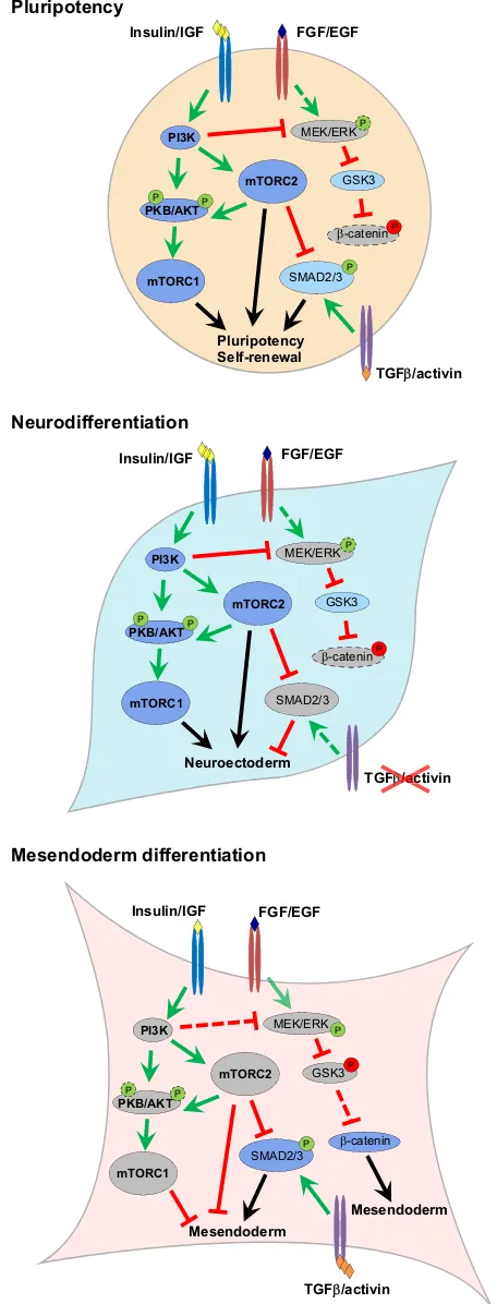
Fig. 3. Regulation of hPSC differentiation by PI3K/AKT/mTOR signalling.
Under culture conditions conducive for both naive and primed hPSC propagation, high levels of PI3K/AKT/mTOR signalling acts to maintain pluripotency while preventing aberrant differentiation (top). Under conditions permissive to differentiation, PI3K/AKT/mTOR can act to drive
neurodifferentiation via the suppression of mesoderm/endoderm-specifying pathways (middle). Specification of mesendoderm fates requires the alleviation of PI3K/AKT/mTOR-mediated inhibition of activin/SMAD2/3 and WNT/
β-catenin pathways (bottom). Dark blue indicates fully active pathways, light blue indicates components with partial activity, grey indicates inactive components. Dashed arrows indicate lower levels of stimulation/inhibition.‘P’in a green or red circle indicates activating or inhibitory phoshorylation, respectively. Dashed bubbles indicate reduced phosphorylation levels or protein expression.
DEVEL
O
expanded forebrain (Guertin et al., 2006; Rennebeck et al., 1998). This is pathologically manifested in humans as tuberous sclerosis syndrome, whereby loss or defective TSC2 function induces the formation of brain hamartomas primarily consisting of giant cell astrocytes, cortical tubers and subependymal nodules (Curatolo et al., 1991). Although hyperactivation of mTORC1 may well explain the tumour formation aspect of this syndrome due to hyperplasic tissue growth, the mixed cell-type phenotype and loss of local tissue architecture suggests further roles for mTORC1 in the regulation of neural cell identity, particularly with regard to the acquisition of neuronal functions. This is further supported by the fact that PI3K/AKT/mTOR signalling, or more precisely mTOR activity, is required for neuronal differentiation following the formation of the neural tube, as treatment with the mTOR inhibitor rapamycin reduces the expression of early neuronal marker genes in the chick (Fishwick et al., 2010). However, as with the formation of the telencephalon, this is not due to defects in cell proliferation, as stimulation of PI3K signalling does not result in increased neuron production in neural tube explants.
Consistent with the notion that sustained PI3K/AKT/mTOR signalling promotes neural fates, inhibition of this pathway in PSCs enhances the specification of mesendoderm over neuroectoderm (McLean et al., 2007; Zhou et al., 2009; Yu et al., 2015). Indeed, whereas insulin itself has the capacity to redirect the differentiation of cardiac mesoderm and endoderm to neuroectoderm in differentiating hESCs (Freund et al., 2008), efficient differentiation of hESCs to mesendoderm and definitive endoderm requires transient suppression of PI3K/AKT/mTOR signalling in addition to activin (McLean et al., 2007; Zhou et al., 2009; Yu et al., 2015). Several underlying mechanisms have been suggested for this negative effect of PI3K/AKT/mTOR upon mesendoderm/endoderm differentiation. PI3K/AKT signalling has been shown to suppress MAPK/ERK activity, acting to inhibit the WNT/β-catenin signalling required for activin-induced mesendoderm/endoderm differentiation (Singh et al., 2012; Sumi et al., 2008). Inhibition of PI3K/AKT therefore releases this inhibition, promoting endoderm differentiation. Additionally, mTORC2 activity directly inhibits activin-stimulated SMAD2/3 activities by increasing the ubiquitin-mediated degradation of SMAD2/3 via modulation of the phosphorylation status of the SMAD2/3 linker region (Yu et al., 2015). Therefore, PI3K/AKT/ mTOR signalling appears to facilitate neural differentiation and impede mesendoderm/endoderm formation once PSCs exit pluripotency (Fig. 3). However, it is unclear whether mTOR complexes play any part in actively driving the commitment of PSCs towards the neural fate or, alternatively, simply act to inhibit other pathways that drive mesendoderm commitment. Nevertheless, these studies highlight a prominent role for PI3K/AKT/mTOR signalling during lineage specification and the determination of cell fate.
Outlook
The PI3K/AKT/mTOR pathway is a major regulator of PSC properties, not only governing their proliferation and survival, but also modulating the gene expression, epigenetic configuration and metabolic activities of these cells. This, in turn, regulates both the maintenance of pluripotency and the acquisition of specialised functions during differentiation. Although it is remarkable that we can now describe the involvement of nearly all of the components of this pathway in fine mechanistic detail within the context of PSC fate determination, there are nonetheless a number of outstanding issues that need to be addressed. Chief of these is the fact that
no signalling pathway operates in isolation, which necessitates further investigation into how this pathway interacts with other signalling mediators or genetic factors to induce the large-scale changes associated with differentiation and/or reprogramming. Establishment and application of chemically defined culture conditions would greatly facilitate such investigations and reduce non-associated signalling interference. The advent of clustered regularly interspaced short palindromic repeats/CRISPR-associated protein 9 (CRISPR/Cas9) technologies and the development of guide RNA sub-libraries that not only allow simultaneous knockout of but also precise mutations in multiple signalling components are major tools that will enable us to develop a more complete molecular description of these processes. Equally, recent advancements in the ability to observe human development in vitro for extended periods beyond implantation (Deglincerti et al., 2016; Shahbazi et al., 2016) and single-cell gene expression profiling (Petropoulos et al., 2016; Yan et al., 2013) represent exciting avenues through which we may further improve defined culture conditions and explore the species-specific impact of PI3K/ AKT/mTOR signalling in hPSCs via the modulation of nutrition availability and other metabolic cues. When coupled with increasingly specific and efficient gene editing technologies, it is with utmost certainty that new roles for PI3K/AKT/mTOR will be uncovered that will transform our understanding of development, ageing, tumorigenesis and disease.
Acknowledgements
We apologise to those authors whose papers could not be cited owing to space constraints.
Competing interests
The authors declare no competing or financial interests.
Funding
This work was supported by UK Medical Research Council; and Genesis Research Trust.
References
Alva, J. A., Lee, G. E., Escobar, E. E. and Pyle, A. D.(2011). Phosphatase and
tensin homolog regulates the pluripotent state and lineage fate choice in human embryonic stem cells.Stem Cells29, 1952-1962.
Armstrong, L., Hughes, O., Yung, S., Hyslop, L., Stewart, R., Wappler, I., Peters, H., Walter, T., Stojkovic, P., Evans, J. et al.(2006). The role of PI3K/AKT, MAPK/ ERK and NFkappabeta signalling in the maintenance of human embryonic stem cell pluripotency and viability highlighted by transcriptional profiling and functional analysis.Hum. Mol. Genet.15, 1894-1913.
Bar-Peled, L., Schweitzer, L. D., Zoncu, R. and Sabatini, D. M.(2012). Ragulator
is a GEF for the rag GTPases that signal amino acid levels to mTORC1.Cell150, 1196-1208.
Betschinger, J., Nichols, J., Dietmann, S., Corrin, P. D., Paddison, P. J. and Smith, A.(2013). Exit from pluripotency is gated by intracellular redistribution of the bHLH transcription factor Tfe3.Cell153, 335-347.
Bi, L., Okabe, I., Bernard, D. J., Wynshaw-Boris, A. and Nussbaum, R. L.(1999).
Proliferative defect and embryonic lethality in mice homozygous for a deletion in the p110alpha subunit of phosphoinositide 3-kinase. J. Biol. Chem. 274, 10963-10968.
Bi, L., Okabe, I., Bernard, D. J. and Nussbaum, R. L.(2002). Early embryonic
lethality in mice deficient in the p110beta catalytic subunit of PI 3-kinase.Mamm. Genome13, 169-172.
Carey, B. W., Finley, L. W. S., Cross, J. R., Allis, C. D. and Thompson, C. B.
(2015). Intracellular alpha-ketoglutarate maintains the pluripotency of embryonic stem cells.Nature518, 413-416.
Carroll, B., Maetzel, D., Maddocks, O. D. K., Otten, G., Ratcliff, M., Smith, G. R., Dunlop, E. A., Passos, J. F., Davies, O. R., Jaenisch, R. et al.(2016). Control of TSC2-Rheb signaling axis by arginine regulates mTORC1 activity. eLife5, e11058.
Chen, W. S., Xu, P.-Z., Gottlob, K., Chen, M.-L., Sokol, K., Shiyanova, T.,
Roninson, I., Weng, W., Suzuki, R., Tobe, K. et al.(2001). Growth retardation
and increased apoptosis in mice with homozygous disruption of the Akt1 gene.
Genes Dev.15, 2203-2208.
DEVEL
O
Cheng, S.-C., Quintin, J., Cramer, R. A., Shepardson, K. M., Saeed, S., Kumar, V., Giamarellos-Bourboulis, E. J., Martens, J. H. A., Rao, N. A.,
Aghajanirefah, A. et al. (2014). mTOR- and HIF-1alpha-mediated aerobic
glycolysis as metabolic basis for trained immunity.Science345, 1250684.
Cho, H., Mu, J., Kim, J. K., Thorvaldsen, J. L., Chu, Q., Crenshaw, E. B., III,
Kaestner, K. H., Bartolomei, M. S., Shulman, G. I. and Birnbaum, M. J.(2001).
Insulin resistance and a diabetes mellitus-like syndrome in mice lacking the protein kinase Akt2 (PKB beta).Science292, 1728-1731.
Cho, Y.-H., Han, K.-M., Kim, D., Lee, J., Lee, S.-H., Choi, K.-W., Kim, J. and Han,
Y.-M. (2014). Autophagy regulates homeostasis of pluripotency-associated
proteins in hESCs.Stem Cells32, 424-435.
Ciraolo, E., Iezzi, M., Marone, R., Marengo, S., Curcio, C., Costa, C., Azzolino,
O., Gonella, C., Rubinetto, C., Wu, H. et al.(2008). Phosphoinositide 3-kinase
p110beta activity: key role in metabolism and mammary gland cancer but not development.Sci. Signal.1, ra3.
Clayton, E., Bardi, G., Bell, S. E., Chantry, D., Downes, C. P., Gray, A.,
Humphries, L. A., Rawlings, D., Reynolds, H., Vigorito, E. et al.(2002). A
crucial role for the p110delta subunit of phosphatidylinositol 3-kinase in B cell development and activation.J. Exp. Med.196, 753-763.
Cowan, C. A., Atienza, J., Melton, D. A. and Eggan, K. (2005). Nuclear
reprogramming of somatic cells after fusion with human embryonic stem cells.
Science309, 1369-1373.
Curatolo, P., Cusmai, R., Cortesi, F., Chiron, C., Jambaque, I. and Dulac, O.
(1991). Neuropsychiatric aspects of tuberous sclerosis.Ann. N. Y. Acad. Sci.615, 8-16.
Davidson, K. C., Adams, A. M., Goodson, J. M., McDonald, C. E., Potter, J. C.,
Berndt, J. D., Biechele, T. L., Taylor, R. J. and Moon, R. T.(2012).
Wnt/beta-catenin signaling promotes differentiation, not self-renewal, of human embryonic stem cells and is repressed by Oct4.Proc. Natl. Acad. Sci. USA109, 4485-4490.
Deglincerti, A., Croft, G. F., Pietila, L. N., Zernicka-Goetz, M., Siggia, E. D. and Brivanlou, A. H.(2016). Self-organization of the in vitro attached human embryo.
Nature533, 251-254.
Di Cristofano, A., Pesce, B., Cordon-Cardo, C. and Pandolfi, P. P.(1998). Pten is essential for embryonic development and tumour suppression.Nat. Genet.19, 348-355.
Dummler, B., Tschopp, O., Hynx, D., Yang, Z.-Z., Dirnhofer, S. and Hemmings,
B. A.(2006). Life with a single isoform of Akt: mice lacking Akt2 and Akt3 are
viable but display impaired glucose homeostasis and growth deficiencies.Mol. Cell. Biol.26, 8042-8051.
Fishwick, K. J., Li, R. A., Halley, P., Deng, P. and Storey, K. G.(2010). Initiation of neuronal differentiation requires PI3-kinase/TOR signalling in the vertebrate neural tube.Dev. Biol.338, 215-225.
Foukas, L. C., Claret, M., Pearce, W., Okkenhaug, K., Meek, S., Peskett, E.,
Sancho, S., Smith, A. J., Withers, D. J. and Vanhaesebroeck, B.(2006).
Critical role for the p110alpha phosphoinositide-3-OH kinase in growth and metabolic regulation.Nature441, 366-370.
Freund, C., Ward-van Oostwaard, D., Monshouwer-Kloots, J., van den Brink, S., van Rooijen, M., Xu, X., Zweigerdt, R., Mummery, C. and Passier, R.
(2008). Insulin redirects differentiation from cardiogenic mesoderm and endoderm to neuroectoderm in differentiating human embryonic stem cells.Stem Cells26, 724-733.
Fritsch, R., de Krijger, I., Fritsch, K., George, R., Reason, B., Kumar, M. S.,
Diefenbacher, M., Stamp, G. and Downward, J.(2013). RAS and RHO Families
of GTPases Directly Regulate Distinct Phosphoinositide 3-Kinase Isoforms.Cell 153, 1050-1063.
Fruman, D. A., Mauvais-Jarvis, F., Pollard, D. A., Yballe, C. M., Brazil, D.,
Bronson, R. T., Kahn, C. R. and Cantley, L. C.(2000). Hypoglycaemia, liver
necrosis and perinatal death in mice lacking all isoforms of phosphoinositide 3-kinase p85 alpha.Nat. Genet.26, 379-382.
Goorden, S. M. I., Hoogeveen-Westerveld, M., Cheng, C., van Woerden, G. M.,
Mozaffari, M., Post, L., Duckers, H. J., Nellist, M. and Elgersma, Y.(2011).
Rheb is essential for murine development.Mol. Cell. Biol.31, 1672-1678.
Guertin, D. A., Stevens, D. M., Thoreen, C. C., Burds, A. A., Kalaany, N. Y., Moffat, J., Brown, M., Fitzgerald, K. J. and Sabatini, D. M.(2006). Ablation in mice of the mTORC components raptor, rictor, or mLST8 reveals that mTORC2 is required for signaling to Akt-FOXO and PKCalpha, but not S6K1.Dev. Cell11, 859-871.
Harada, K., Truong, A. B., Cai, T. and Khavari, P. A. (2005). The class II
phosphoinositide 3-kinase C2beta is not essential for epidermal differentiation.
Mol. Cell. Biol.25, 11122-11130.
Harris, D. P., Vogel, P., Wims, M., Moberg, K., Humphries, J., Jhaver, K. G.,
DaCosta, C. M., Shadoan, M. K., Xu, N., Hansen, G. M. et al. (2011).
Requirement for class II phosphoinositide 3-kinase C2alpha in maintenance of glomerular structure and function.Mol. Cell. Biol.31, 63-80.
Hishida, T., Nakachi, Y., Mizuno, Y., Katano, M., Okazaki, Y., Ema, M.,
Takahashi, S., Hirasaki, M., Suzuki, A., Ueda, A. et al.(2015). Functional
compensation between Myc and PI3K signaling supports self-renewal of embryonic stem cells.Stem Cells33, 713-725.
Ho, L., Tan, S. Y. X., Wee, S., Wu, Y., Tan, S. J. C., Ramakrishna, N. B., Chng,
S. C., Nama, S., Szczerbinska, I., Chan, Y.-S. et al.(2015). ELABELA Is an
endogenous growth factor that sustains hESC SELF-RENEwal via the PI3K/AKT pathway.Cell Stem Cell17, 435-447.
Honda, S., Arakawa, S., Nishida, Y., Yamaguchi, H., Ishii, E. and Shimizu, S.
(2014). Ulk1-mediated Atg5-independent macroautophagy mediates elimination of mitochondria from embryonic reticulocytes.Nat. Commun.5, 4004.
Jacinto, E., Facchinetti, V., Liu, D., Soto, N., Wei, S., Jung, S. Y., Huang, Q., Qin,
J. and Su, B.(2006). SIN1/MIP1 maintains rictor-mTOR complex integrity and
regulates Akt phosphorylation and substrate specificity.Cell127, 125-137.
Jewell, J. L., Kim, Y. C., Russell, R. C., Yu, F.-X., Park, H. W., Plouffe, S. W., Tagliabracci, V. S. and Guan, K.-L.(2015). Metabolism. Differential regulation of mTORC1 by leucine and glutamine.Science347, 194-198.
Kim, J.-w., Tchernyshyov, I., Semenza, G. L. and Dang, C. V.(2006).
HIF-1-mediated expression of pyruvate dehydrogenase kinase: a metabolic switch required for cellular adaptation to hypoxia.Cell Metab.3, 177-185.
Kingham, E. and Welham, M.(2009). Distinct roles for isoforms of the catalytic
subunit of class-IA PI3K in the regulation of behaviour of murine embryonic stem cells.J. Cell Sci.122, 2311-2321.
Kobayashi, T., Minowa, O., Sugitani, Y., Takai, S., Mitani, H., Kobayashi, E.,
Noda, T. and Hino, O. (2001). A germ-line Tsc1 mutation causes tumor
development and embryonic lethality that are similar, but not identical to, those caused by Tsc2 mutation in mice.Proc. Natl. Acad. Sci. USA98, 8762-8767.
Ktistakis, N. T. and Tooze, S. A.(2016). Digesting the expanding mechanisms of
autophagy.Trends Cell Biol.26, 624-635.
Kundu, M., Lindsten, T., Yang, C.-Y., Wu, J., Zhao, F., Zhang, J., Selak, M. A.,
Ney, P. A. and Thompson, C. B.(2008). Ulk1 plays a critical role in the
autophagic clearance of mitochondria and ribosomes during reticulocyte maturation.Blood112, 1493-1502.
Laplante, M. and Sabatini, D. M.(2012). mTOR signaling in growth control and
disease.Cell149, 274-293.
Le Bacquer, O., Petroulakis, E., Paglialunga, S., Poulin, F., Richard, D.,
Cianflone, K. and Sonenberg, N.(2007). Elevated sensitivity to diet-induced
obesity and insulin resistance in mice lacking 4E-BP1 and 4E-BP2.J. Clin. Invest. 117, 387-396.
Lee, J. V., Carrer, A., Shah, S., Snyder, N. W., Wei, S., Venneti, S., Worth, A. J.,
Yuan, Z.-F., Lim, H.-W., Liu, S. et al. (2014). Akt-dependent metabolic
reprogramming regulates tumor cell histone acetylation.Cell Metab.20, 306-319.
Li, T. Y., Sun, Y., Liang, Y., Liu, Q., Shi, Y., Zhang, C.-S., Zhang, C., Song, L., Zhang, P., Zhang, X. et al.(2016). ULK1/2 constitute a bifurcate node controlling glucose metabolic fluxes in addition to autophagy.Mol. Cell62, 359-370.
Liu, P., Gan, W., Chin, Y. R., Ogura, K., Guo, J., Zhang, J., Wang, B., Blenis, J., Cantley, L. C., Toker, A. et al.(2015). PtdIns(3,4,5)P3-dependent activation of the mTORC2 kinase complex.Cancer Discov.5, 1194-1209.
Ma, X. M. and Blenis, J. (2009). Molecular mechanisms of mTOR-mediated
translational control.Nat. Rev. Mol. Cell Biol.10, 307-318.
Ma, T., Li, J., Xu, Y., Yu, C., Xu, T., Wang, H., Liu, K., Cao, N., Nie, B.-m., Zhu, S.-y.
et al.(2015). Atg5-independent autophagy regulates mitochondrial clearance and
is essential for iPSC reprogramming.Nat. Cell Biol.17, 1379-1387.
McLean, A. B., D’Amour, K. A., Jones, K. L., Krishnamoorthy, M., Kulik, M. J.,
Reynolds, D. M., Sheppard, A. M., Liu, H., Xu, Y., Baetge, E. E. et al.(2007).
Activin a efficiently specifies definitive endoderm from human embryonic stem cells only when phosphatidylinositol 3-kinase signaling is suppressed.Stem Cells 25, 29-38.
Mizushima, N. and Levine, B.(2010). Autophagy in mammalian development and
differentiation.Nat. Cell Biol.12, 823-830.
Mizushima, N., Yamamoto, A., Hatano, M., Kobayashi, Y., Kabeya, Y., Suzuki,
K., Tokuhisa, T., Ohsumi, Y. and Yoshimori, T. (2001). Dissection of
autophagosome formation using Apg5-deficient mouse embryonic stem cells.
J. Cell Biol.152, 657-668.
Morita, M., Gravel, S.-P., Chénard, V., Sikstrom, K., Zheng, L., Alain, T., Gandin,
V., Avizonis, D., Arguello, M., Zakaria, C. et al.(2013). mTORC1 controls
mitochondrial activity and biogenesis through 4E-BP-dependent translational regulation.Cell Metab.18, 698-711.
Moussaieff, A., Rouleau, M., Kitsberg, D., Cohen, M., Levy, G., Barasch, D.,
Nemirovski, A., Shen-Orr, S., Laevsky, I., Amit, M. et al.(2015).
Glycolysis-mediated changes in acetyl-CoA and histone acetylation control the early differentiation of embryonic stem cells.Cell Metab.21, 392-402.
Murakami, M., Ichisaka, T., Maeda, M., Oshiro, N., Hara, K., Edenhofer, F.,
Kiyama, H., Yonezawa, K. and Yamanaka, S.(2004). mTOR is essential for
growth and proliferation in early mouse embryos and embryonic stem cells.Mol. Cell. Biol.24, 6710-6718.
Neuman, N. A. and Henske, E. P.(2011). Non-canonical functions of the tuberous
sclerosis complex-Rheb signalling axis.EMBO Mol. Med.3, 189-200.
Niwa, H., Ogawa, K., Shimosato, D. and Adachi, K.(2009). A parallel circuit of LIF signalling pathways maintains pluripotency of mouse ES cells.Nature 460, 118-122.
Paling, N. R. D., Wheadon, H., Bone, H. K. and Welham, M. J.(2004). Regulation
of embryonic stem cell self-renewal by phosphoinositide 3-kinase-dependent signaling.J. Biol. Chem.279, 48063-48070.
Pende, M., Um, S. H., Mieulet, V., Sticker, M., Goss, V. L., Mestan, J., Mueller, M.,
Fumagalli, S., Kozma, S. C. and Thomas, G.(2004). S6K1(-/-)/S6K2(-/-) mice
DEVEL
O
exhibit perinatal lethality and rapamycin-sensitive 5′-terminal oligopyrimidine mRNA translation and reveal a mitogen-activated protein kinase-dependent S6 kinase pathway.Mol. Cell. Biol.24, 3112-3124.
Peng, X.-d., Xu, P.-Z., Chen, M.-L., Hahn-Windgassen, A., Skeen, J., Jacobs, J.,
Sundararajan, D., Chen, W. S., Crawford, S. E., Coleman, K. G. et al.(2003).
Dwarfism, impaired skin development, skeletal muscle atrophy, delayed bone development, and impeded adipogenesis in mice lacking Akt1 and Akt2.Genes Dev.17, 1352-1365.
Petropoulos, S., Edsgärd, D., Reinius, B., Deng, Q., Panula, S. P., Codeluppi, S.,
Plaza Reyes, A., Linnarsson, S., Sandberg, R. and Lanner, F.(2016).
Single-cell RNA-Seq reveals lineage and X chromosome dynamics in human preimplantation embryos.Cell165, 1012-1026.
Prigione, A., Fauler, B., Lurz, R., Lehrach, H. and Adjaye, J. (2010). The
senescence-related mitochondrial/oxidative stress pathway is repressed in human induced pluripotent stem cells.Stem Cells28, 721-733.
Rebsamen, M., Pochini, L., Stasyk, T., de Araújo, M. E. G., Galluccio, M.,
Kandasamy, R. K., Snijder, B., Fauster, A., Rudashevskaya, E. L., Bruckner,
M. et al.(2015). SLC38A9 is a component of the lysosomal amino acid sensing
machinery that controls mTORC1.Nature519, 477-481.
Rennebeck, G., Kleymenova, E. V., Anderson, R., Yeung, R. S., Artzt, K. and
Walker, C. L. (1998). Loss of function of the tuberous sclerosis 2 tumor
suppressor gene results in embryonic lethality characterized by disrupted neuroepithelial growth and development. Proc. Natl. Acad. Sci. USA 95, 15629-15634.
Ryu, J. M. and Han, H. J.(2011). L-threonine regulates G1/S phase transition of
mouse embryonic stem cells via PI3K/Akt, MAPKs, and mTORC pathways.
J. Biol. Chem.286, 23667-23678.
Sarbassov, D. D., Ali, S. M., Sengupta, S., Sheen, J.-H., Hsu, P. P., Bagley, A. F.,
Markhard, A. L. and Sabatini, D. M.(2006). Prolonged rapamycin treatment
inhibits mTORC2 assembly and Akt/PKB.Mol. Cell22, 159-168.
Sasaki, T., Irie-Sasaki, J., Jones, R. G., Oliveira-dos-Santos, A. J., Stanford, W. L., Bolon, B., Wakeham, A., Itie, A., Bouchard, D., Kozieradzki, I. et al.
(2000). Function of PI3Kgamma in thymocyte development, T cell activation, and neutrophil migration.Science287, 1040-1046.
Shahbazi, M. N., Jedrusik, A., Vuoristo, S., Recher, G., Hupalowska, A., Bolton, V., Fogarty, N. M. E., Campbell, A., Devito, L. G., Ilic, D. et al.(2016). Self-organization of the human embryo in the absence of maternal tissues.Nat. Cell Biol.18, 700-708.
Shiota, C., Woo, J.-T., Lindner, J., Shelton, K. D. and Magnuson, M. A.(2006).
Multiallelic disruption of the rictor gene in mice reveals that mTOR complex 2 is essential for fetal growth and viability.Dev. Cell11, 583-589.
Shiraki, N., Shiraki, Y., Tsuyama, T., Obata, F., Miura, M., Nagae, G., Aburatani,
H., Kume, K., Endo, F. and Kume, S.(2014). Methionine metabolism regulates
maintenance and differentiation of human pluripotent stem cells.Cell Metab.19, 780-794.
Shyh-Chang, N., Locasale, J. W., Lyssiotis, C. A., Zheng, Y., Teo, R. Y., Ratanasirintrawoot, S., Zhang, J., Onder, T., Unternaehrer, J. J., Zhu, H. et al.
(2013). Influence of threonine metabolism on S-adenosylmethionine and histone methylation.Science339, 222-226.
Silva, J., Barrandon, O., Nichols, J., Kawaguchi, J., Theunissen, T. W. and
Smith, A.(2008). Promotion of reprogramming to ground state pluripotency by
signal inhibition.PLoS Biol.6, e253.
Singh, A. M., Reynolds, D., Cliff, T., Ohtsuka, S., Mattheyses, A. L., Sun, Y.,
Menendez, L., Kulik, M. and Dalton, S.(2012). Signaling network crosstalk in
human pluripotent cells: a Smad2/3-regulated switch that controls the balance between self-renewal and differentiation.Cell Stem Cell10, 312-326.
Sperber, H., Mathieu, J., Wang, Y., Ferreccio, A., Hesson, J., Xu, Z., Fischer, K. A., Devi, A., Detraux, D., Gu, H. et al.(2015). The metabolome regulates the epigenetic landscape during naive-to-primed human embryonic stem cell transition.Nat. Cell Biol.17, 1523-1535.
Sumi, T., Tsuneyoshi, N., Nakatsuji, N. and Suemori, H.(2008). Defining early
lineage specification of human embryonic stem cells by the orchestrated balance of canonical Wnt/beta-catenin, Activin/Nodal and BMP signaling.Development 135, 2969-2979.
Takahashi, K. and Yamanaka, S.(2006). Induction of pluripotent stem cells from
mouse embryonic and adult fibroblast cultures by defined factors. Cell126, 663-676.
Takahashi, K., Mitsui, K. and Yamanaka, S.(2003). Role of ERas in promoting
tumour-like properties in mouse embryonic stem cells.Nature423, 541-545.
Tang, Y., Jiang, Z., Luo, Y., Zhao, X., Wang, L., Norris, C. and Tian, X. C.(2014). Differential effects of Akt isoforms on somatic cell reprogramming.J. Cell Sci.127, 3998-4008.
Terauchi, Y., Tsuji, Y., Satoh, S., Minoura, H., Murakami, K., Okuno, A., Inukai, K., Asano, T., Kaburagi, Y., Ueki, K. et al.(1999). Increased insulin sensitivity and hypoglycaemia in mice lacking the p85 alpha subunit of phosphoinositide 3-kinase.Nat. Genet.21, 230-235.
Thorpe, L. M., Yuzugullu, H. and Zhao, J. J.(2015). PI3K in cancer: divergent roles of isoforms, modes of activation and therapeutic targeting.Nat. Rev. Cancer15, 7-24.
Tohyama, S., Fujita, J., Hishiki, T., Matsuura, T., Hattori, F., Ohno, R., Kanazawa,
H., Seki, T., Nakajima, K., Kishino, Y. et al.(2016). Glutamine Oxidation Is
Indispensable for Survival of Human Pluripotent Stem Cells.Cell Metab.23, 663-674.
Tschopp, O., Yang, Z.-Z., Brodbeck, D., Dummler, B. A., Hemmings-Mieszczak, M., Watanabe, T., Michaelis, T., Frahm, J. and Hemmings,
B. A.(2005). Essential role of protein kinase B gamma (PKB gamma/Akt3) in
postnatal brain development but not in glucose homeostasis.Development132, 2943-2954.
Ueki, K., Yballe, C. M., Brachmann, S. M., Vicent, D., Watt, J. M., Kahn, C. R. and Cantley, L. C.(2002). Increased insulin sensitivity in mice lacking p85beta subunit of phosphoinositide 3-kinase.Proc. Natl. Acad. Sci. USA99, 419-424.
Vanhaesebroeck, B., Guillermet-Guibert, J., Graupera, M. and Bilanges, B.
(2010). The emerging mechanisms of isoform-specific PI3K signalling.Nat. Rev. Mol. Cell Biol.11, 329-341.
Varum, S., Rodrigues, A. S., Moura, M. B., Momcilovic, O., Easley, C. A.,
Ramalho-Santos, J., Van Houten, B. and Schatten, G. (2011). Energy
metabolism in human pluripotent stem cells and their differentiated counterparts.PLoS ONE6, e20914.
Wang, J., Alexander, P., Wu, L., Hammer, R., Cleaver, O. and McKnight, S. L.
(2009). Dependence of mouse embryonic stem cells on threonine catabolism.
Science325, 435-439.
Wang, S., Xia, P., Ye, B., Huang, G., Liu, J. and Fan, Z.(2013). Transient activation of autophagy via Sox2-mediated suppression of mTOR is an important early step in reprogramming to pluripotency.Cell Stem Cell13, 617-625.
Wang, S., Tsun, Z.-Y., Wolfson, R. L., Shen, K., Wyant, G. A., Plovanich, M. E.,
Yuan, E. D., Jones, T. D., Chantranupong, L., Comb, W. et al.(2015).
Metabolism. Lysosomal amino acid transporter SLC38A9 signals arginine sufficiency to mTORC1.Science347, 188-194.
Ward, P. S. and Thompson, C. B.(2012). Signaling in control of cell growth and
metabolism.Cold Spring Harb. Perspect. Biol.4, a006783.
Watanabe, S., Umehara, H., Murayama, K., Okabe, M., Kimura, T. and Nakano, T.(2006). Activation of Akt signaling is sufficient to maintain pluripotency in mouse and primate embryonic stem cells.Oncogene25, 2697-2707.
Wilmut, I., Schnieke, A. E., McWhir, J., Kind, A. J. and Campbell, K. H. S.(1997). Viable offspring derived from fetal and adult mammalian cells.Nature 385, 810-813.
Wray, J., Kalkan, T., Gomez-Lopez, S., Eckardt, D., Cook, A., Kemler, R. and
Smith, A. (2011). Inhibition of glycogen synthase kinase-3 alleviates Tcf3
repression of the pluripotency network and increases embryonic stem cell resistance to differentiation.Nat. Cell Biol.13, 838-845.
Wu, Y., Li, Y., Zhang, H., Huang, Y., Zhao, P., Tang, Y., Qiu, X., Ying, Y., Li, W., Ni,
S. et al.(2015). Autophagy and mTORC1 regulate the stochastic phase of
somatic cell reprogramming.Nat. Cell Biol.17, 715-725.
Wulff, P., Vallon, V., Huang, D. Y., Völkl, H., Yu, F., Richter, K., Jansen, M., Schlünz, M., Klingel, K., Loffing, J. et al.(2002). Impaired renal Na(+) retention in the sgk1-knockout mouse.J. Clin. Invest.110, 1263-1268.
Yan, L., Yang, M., Guo, H., Yang, L., Wu, J., Li, R., Liu, P., Lian, Y., Zheng, X.,
Yan, J. et al.(2013). Single-cell RNA-Seq profiling of human preimplantation
embryos and embryonic stem cells.Nat. Struct. Mol. Biol.20, 1131-1139.
Yang, Z.-Z., Tschopp, O., Di-Poi, N., Bruder, E., Baudry, A., Dummler, B., Wahli,
W. and Hemmings, B. A.(2005). Dosage-dependent effects of Akt1/protein
kinase Balpha (PKBalpha) and Akt3/PKBgamma on thymus, skin, and cardiovascular and nervous system development in mice.Mol. Cell. Biol.25, 10407-10418.
Yang, H., Rudge, D. G., Koos, J. D., Vaidialingam, B., Yang, H. J. and Pavletich,
N. P.(2013). mTOR kinase structure, mechanism and regulation.Nature497,
217-223.
Ying, Q.-L., Wray, J., Nichols, J., Batlle-Morera, L., Doble, B., Woodgett, J.,
Cohen, P. and Smith, A.(2008). The ground state of embryonic stem cell
self-renewal.Nature453, 519-523.
Yoshioka, K., Yoshida, K., Cui, H., Wakayama, T., Takuwa, N., Okamoto, Y., Du, W., Qi, X., Asanuma, K., Sugihara, K. et al.(2012). Endothelial PI3K-C2alpha, a class II PI3K, has an essential role in angiogenesis and vascular barrier function.
Nat. Med.18, 1560-1569.
Yu, Y., Liang, D., Tian, Q., Chen, X., Jiang, B., Chou, B.-K., Hu, P., Cheng, L., Gao, P., Li, J. et al.(2014). Stimulation of somatic cell reprogramming by ERas-Akt-FoxO1 signaling axis.Stem Cells32, 349-363.
Yu, J. S. L., Ramasamy, T. S., Murphy, N., Holt, M. K., Czapiewski, R., Wei,
S.-K. and Cui, W.(2015). PI3K/mTORC2 regulates TGF-beta/Activin signalling
by modulating Smad2/3 activity via linker phosphorylation.Nat. Commun.6, 7212.
Zhang, J., Ratanasirintrawoot, S., Chandrasekaran, S., Wu, Z., Ficarro, S. B., Yu, C., Ross, C. A., Cacchiarelli, D., Xia, Q., Seligson, M. et al.(2016). LIN28 Regulates Stem Cell Metabolism and Conversion to Primed Pluripotency.Cell Stem Cell19, 66-80.
Zhao, X., Lu, S., Nie, J., Hu, X., Luo, W., Wu, X., Liu, H., Feng, Q., Chang, Z., Liu,
Y. et al. (2014). Phosphoinositide-dependent kinase 1 and mTORC2
synergistically maintain postnatal heart growth and heart function in mice.Mol.
Cell. Biol.34, 1966-1975.