Introduction
Transcription factors, RNA binding proteins, and microRNAs (miRNAs) are among the factors that must act coordinately to control gene expression networks important for cellular function [1]. Following transcrip-tion, mRNAs are subject to the processes of splicing, nuclear export, traffi cking, and polyadenylation, followed by translation initiation and elongation. Each of these processes represents a point at which expression can be regulated, allowing for fi ne-tuning in response to chang ing environmental conditions. miRNAs are short (approxi-mately 21-nucleotide) non-coding RNAs that regulate trans cript localization, polyadenylation, and translation. miRNAs are direct negative regulators of gene expression that bind to specifi c sequences within a target mRNA [2,3].
miRNAs were discovered as a result of studies aimed at identifying genes that mediate developmental transitions in Caenorhabditis elegans [4,5]. Recent years have seen signifi cant advances in the miRNA fi eld, including the
characterization of the miRNA biogenesis pathway, identi fi cation of the mechanisms by which miRNAs regu-late gene expression, and an appreciation of how families of miRNAs can regulate cellular processes and contribute to disease phenotypes.
Th e goal of this review is to highlight our current under standing of miRNA biogenesis and mechanisms of action, and to summarize recent studies on the role of miRNAs in bone remodeling.
miRNA biogenesis
Th e miRNA biogenesis pathway has several steps: trans-cription, pri-miRNA processing, transport to the cyto-plasm, precursor miRNA (pre-miRNA) processing, strand selection, transcript targeting, and transcript fate (Figure 1). Th is rigorous, multistep processing pathway helps ensure that only RNAs with the correct structures and sequences are able to regulate gene expression.
miRNA genes are found on every chromosome in humans, except for the Y chromosome. Like the promo-ters of protein-coding genes, miRNA promopromo-ters are regulated by epigenetics and by transcription factors. miRNA genes are transcribed by RNA polymerase II or III, from independent miRNA genes, or from the introns of protein-coding or non-protein-coding genes [6,7] (Figure 2). Th e initial product of miRNA gene trans-cription is the primary transcript (pri-miRNA), which may be several thousand nucleotides long. Multiple miRNAs may be co-transcribed in a single pri-miRNA. Th e entire pri-miRNA is capped and polyadenylated, like mRNA transcripts. Tandem transcription of several miRNAs, possibly along with protein-coding genes, allows the genes to be expressed together. Th ese co-expressed genes may interact within the same pathway or through crosstalk between pathways. Th e co-expression of miRNA and protein-coding genes may also function to ensure negative feedback on the protein-coding gene to prevent its over-expression [8].
In mammals, the pri-miRNA is processed within the nucleus by the Drosha-DiGeorge syndrome critical region gene 8 (DGCR8) complex (Figure 1, in nucleus). Drosha is an RNase III-type endonuclease, and DGCR8 is thought to recruit and bind the transcript [9]. Th e
Abstract
MicroRNAs (miRNAs) are key post-transcriptional regulators of gene expression. This review will highlight our current understanding of miRNA biogenesis and mechanisms of action, and will summarize recent work on the role of miRNAs, including the miR-29 family, in bone remodeling. These studies represent the fi rst steps in demonstrating the importance of miRNAs in the control of osteoblast and osteoclast diff erentiation and function. An in-depth understanding of the roles of these regulatory RNAs in the skeleton will be critical for the development of new therapeutics aimed at treating bone loss and perhaps facilitating fracture repair.
© 2010 BioMed Central Ltd
MicroRNA biogenesis and regulation of bone
remodeling
Kristina Kapinas
1and Anne M Delany*
2R E V I E W
*Correspondence: adelany@uchc.edu
2Center for Molecular Medicine, University of Connecticut Health Center, 263 Farmingtion Ave, Farmington, CT 06030, USA
Full list of author information is available at the end of the article
sequences preceding the 5’ end and trailing the 3’ end of the pri-miRNA form an imperfect stem that is recognized by DGCR8, which allows Drosha to cleave approximately 11 bp away [9,10]. Pri-miRNAs are cleaved into precursor miRNAs (pre-miRNA), which are approximately 60- to 100-nucleotide hairpins. Th e pre-miRNA is then trans-ported from the nucleus into the cytoplasm by Exportin5 and Ran-GTP [11]. Exempted from this processing pathway are short introns containing miRNA precursors, called ‘mirtrons’ (Figure 2). Spliced and debranched mirtrons are able to bypass processing by Drosha, and access the canonical miRNA processing pathway follow-ing nuclear export [12].
[image:2.612.67.547.89.486.2]the RNA-induced silencing complex (RISC). Th e RISC includes Dicer, TRBP, PACT (protein activator of PKR), and one of four Ago proteins [16]. Th e miRNA duplex is unwound by helicases into two single strands, the mature guide strand (miRNA; red strand in Figure 1) and the complementary passenger strand (miRNA*) [14]. Guide strand selection is dependent on the fi rst 5’ nucleotides and which Ago protein is present in the RISC [17].
Th e miRNA* is typically degraded by the RISC, although some miRNAs* are thought to negatively regulate gene expression, like a mature miRNA. It is not known why some miRNAs* are functional. One hypothesis is that the strands could be used diff erently in response to extra-cellular or intraextra-cellular cues, to regulate a more diverse set of protein-coding genes as needed, or strand selection could be tissue specifi c [18]. Strand selection also limits regulation by miRNAs* generated from highly expressed precursors.
While the mechanisms regulating miRNA biogenesis have been well studied, the regulation of miRNA stability, particularly in vertebrates, is not well understood. It is known that mature miRNA abundance is sensitive to Ago protein levels, suggesting that association with Ago can protect miRNAs from degradation [19]. Although most miRNAs appear to be stable, they do possess diff erential stability. One miRNA, miR-122, was recently shown to be stabilized by the addition of a single adenosine to its 3’ end through the activity of the cytoplasmic poly(A) polymerase GLD-2. In contrast, uracils at positions 9 to 11 are important for the short half-life of miR-29b [15]. miR-382 also has a short half-life, due to sequence elements in the 3’ end of the miRNA. Th e exosome 3’-5’ exonuclease complex was shown to be important for miR-382 decay [20]. Discovery and characterization of cis- and trans-acting factors regulating miRNA stability is a topic of intense study.
miRNA functions
Once the RISC contains the guide strand, it delivers the miRNA to the target mRNA, where base pairing occurs. Most miRNA binding sites lie within the 3’ UTR, although there are some reports of miRNAs binding in the 5’ UTR and coding region of mRNAs [21,22]. Th ere appears to be a biological basis for the preferential inter-action of miRNAs with the 3’ UTR. Data suggest that miRNA binding sites within the coding region of a trans-cript are less eff ective at mediating translational repres-sion. Th is is likely due to the ability of ribosomal com-plexes to override and inhibit the interaction of the miRNA-RISC with the potential binding site(s) [23]. Similarly, relatively few functional miRNA binding sites are located in the 5’ UTR of a transcript. Th e scanning activity of the ribosome may impair the interaction of the miRNA-RISC with the 5’ UTR, suggesting there would be ineffi cient inhibition of gene expression. Although the general location of a miRNA binding site within the transcript helps defi ne the degree of repression mediated by miRNAs, other factors contribute to effi cacy. Th ese factors include the sequence context of the miRNA binding site, the number of target sites within the mRNA, the local RNA structure, and distance between target sites [24-27].
[image:3.612.65.548.88.248.2]binding, there is frequently a bulge between the miRNA and its target, between the 9th and the 11th nucleotide of the miRNA, which induces translational suppression [30].
For a majority of mRNAs, translation initiation occurs when the 5’-cap m7GpppN is recognized by eIF4E, a component of the eukaryotic translation initiation factor eIF. Th is complex also includes eIF4G, which interacts with eIF3, to recruit the 40S ribosomal subunit, and with polyadenylate-binding protein 1 (PABP1). Th e interaction of eIF4G with both eIF4E and PABP1 physically brings the 5’ and 3’ ends of the mRNA close together, stimulating translation initiation by increasing affi nity of eIF4E for the 5’-cap. Trans-acting factors that bind the 3’ UTR inhibit translation by recruiting proteins that block the eIF4E-eIF4G interaction or that bind to the 5’-cap, preventing assembly of the 40S ribosome initiation com-plex [31]. Several studies have shown this to be the case for components of the RISC, specifi cally Ago proteins [32,33]. Th e central domain of the Ago protein family has some sequence homology to the eIF4E cap-binding domain. Mutation of these residues in Ago proteins abolishes translational repression of m7G mRNAs, suggest ing that Agos can compete with eIF4E to inhibit translation [34].
miRNAs can also repress translation at post-initiation steps. miRNA-RISC can bind to actively translating mRNAs, reducing translational elongation and/or enhan-cing termination, concomitant with a reduction in ribo-somal initiation and nascent peptide destabilization. As a component of the RISC, Ago proteins 1, 3, and 4 are thought to mediate post-initiation inhibition [35,36].
Initial studies of miRNA targets suggested that only the protein levels of regulated targets were decreased, where-as the levels of target mRNAs themselves were not aff ected. However, altering miRNA expression in cells or tissues can cause signifi cant changes in target mRNA levels, suggesting that miRNAs can induce mRNA destabilization [29,37]. In eukaryotes, mRNA degradation can occur when there is a shortening of the poly(A) tail. As part of the RISC, Ago proteins 1, 3, and 4 are thought to repress translation by promoting poly(A) tail-mediated degradation [38]. One study demonstrated that miRNA-mediated deadenylation of maternal mRNAs is necessary during zebrafi sh embryogenesis [37].
Further, miRNAs function in P-bodies by sequestering target transcripts for storage, decapping, deadenylation, and degradation [28]. Interestingly, P-bodies may also act as a temporary storage space for translationally repressed mRNAs. Since most P-body components are also found dispersed in the cytosol, it is likely that repression by these proteins is initiated in the cytosol, and the repressed mRNAs then aggregate to form the P-body. Stress granules are another type of body containing repressed mRNAs, and they accumulate in response to stress conditions or general inhibition of translation [39].
Bone remodeling
Th e skeleton is continuously remodeled throughout the lifetime of an individual, and this remodeling is a dynamic process by which bone resorption is coupled to bone formation, to replace damaged bone or to respond to metabolic needs [40]. To provide some context in which to appreciate the function of miRNAs in the skeleton, we will briefl y consider the steps of bone remodeling and the signals important for osteoclast and osteoblast maturation.
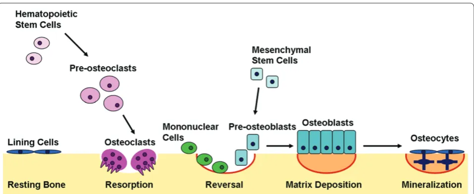
Bone remodeling, or turnover, is mediated by the delicate balance of osteoblast and osteoclast numbers and activities (Figure 3). Osteoclasts resorb bone,
where-as osteoblwhere-asts synthesize new bone. Th e release of
cytokines at the site of remodeling recruits osteoclasts to the bone surface. Th e osteoclasts form a ruffl ed boarder that allows their tight adherence to the bone surface. Th e space between the osteoclast and the underlying bone becomes an isolated microenvironment into which the osteoclast’s proton pump releases ions that generate an acidic environment, dissolving the mineralized compo-nent of the bone matrix. Th e organic matrix becomes exposed and is subsequently degraded by cathepsin K [41]. Th e reversal phase of bone remodeling begins with mononuclear cells preparing the bone surface for new osteoblasts and providing signals to recruit them. Early osteoblasts proliferate and secrete an extracellular matrix abundant in type I collagen. As the osteoblasts continue to diff erentiate, the matrix matures and is mineralized. Once the bone surface is restored, mature osteoblasts can undergo apoptosis or terminally diff erentiate into either bone surface lining cells or osteocytes, which are embedded in the calcifi ed matrix and are responsive to mechanical stresses [42].
Diff erentiation of osteoclasts and osteoblasts from multipotent precursors is a critical component of bone turnover. Osteoclasts are derived from the monocyte/ macrophage lineage. Diff erentiation into osteoclasts is dependent on multiple extracellular signaling molecules, including macrophage colony-stimulating factor (MCSF), receptor activator for nuclear factor κB ligand (RANKL), tumor necrosis factor, interferon gamma, and inter-leukins [41].
Osteoblasts are derived from mesenchymal stem cells (MSCs), which can diff erentiate into osteoblasts, adipo-cytes, chondroadipo-cytes, or myoadipo-cytes, depending on the activation or inhibition of specifi c signaling pathways [43]. Some of the most important signaling molecules regu lat ing osteoblastic diff erentiation include bone
morpho genetic proteins (BMPs), transforming growth
factor (TGF)-β, WNT, Hedgehog, parathyroid hormone, insulin-like growth factor-1, fi broblast growth factors, and Notch.
pathological consequences, including osteoporosis. miRNAs have been shown to regulate osteoblast and osteoclast diff erentiation and function. Th e following section will summarize the known roles of miRNAs in osteoblast and osteoclast biology.
miRNAs in osteoblasts
Cell type-specifi c deletion of Dicer in committed osteo-progenitors results in embryonic lethality at embryonic day (E)14.5 [44]. Th e mutant embryos display a deformed cartilaginous skeleton, and a lack of bone formation. Th is study used the rat 2.3-kb Col1a1 promoter driven-Cre construct to delete the fl oxed Dicer alleles, and this Cre construct is transiently expressed during embryonic develop ment, likely causing the embryonic lethality. Th ese data reinforce the concept that Dicer-mediated miRNA processing is critical for osteogenesis.
Targeted deletion of Dicer in mature osteoblasts, using an osteocalcin-Cre construct, led initially to delayed perinatal bone formation, with a subsequent increase in postnatal bone acquisition [44]. In these mice, signifi cant increases in cortical and trabecular bone volume were evident at 4 months of age, and persisted to 8 months. Long bone from mice lacking Dicer expression in mature osteoblasts had increased levels of mRNA for type I collagens, RANKL, and tartrate-resistant acid phospha-tase (TRAP). Although data on osteoblast and osteoclast number were not reported, these results suggest increased bone remodeling in the mutant mice. In con-trast, another group deleted Dicer in mature osteo blasts using a mouse 2.3-kb Col1 promoter driven-Cre con-struct, and reported no eff ect on bone phenotype [45].
Comparing these two studies, it is likely that diff erences in the degree of Cre-mediated recombination and cell type specifi city had an impact on the resulting mouse phenotypes. Nevertheless, these data suggest that Dicer activity is important for controlling both early and late bone cell diff erentiation programs.
In vitro knockdown of Dicer or Drosha in human MSCs inhibited osteogenic diff erentiation, confi rming similar eff ects in human cells [46]. Indeed, since Dicer is important for the processing of many miRNAs, dramatic eff ects on cell diff erentiation with Dicer ablation are not unexpected. However, Dicer-independent mechanisms for miRNA maturation have recently been described [47]. It will be interesting to consider how these mechanisms may impact skeletal phenotype and to identify which miRNAs are regulated by these alternative pathways.
Th e BMP signaling pathway plays a prominent role in promoting osteoblast diff erentiation and bone formation [48]. Several studies have focused on miRNAs modulated by BMP signaling as a means to understand the role of miRNAs in osteoblasts. From these studies, it is clear that a panel of BMP-regulated miRNAs is important for the
regulation of osteoblast diff erentiation. Since some
[image:5.612.68.547.87.283.2]as alkaline phosphatase (ALP), osteocalcin, and HOXA10. Further studies demonstrated that Runx2 is a target for miR-133, and Smad5 is a target for miR-135. Runx2 is a transcription factor essential for osteoblast diff eren-tiation, whereas Smad5 is an intracellular Runx2 co-receptor [50,51]. Th e concomitant down-regulation of these two miRNAs by BMP-2 likely plays an important role in the up-regulation of Runx2 and Smad5 during osteogenic diff erentiation.
Similarly, miR-206 is decreased in response to BMP-2 in C2C12 cells and during diff erentiation of primary murine osteoblasts [52]. Over-expression of miR-206 inhibits ALP activity, and Connexin 43 (Cx43) was shown to be a target of miR-206. Cx43 is a gap junction protein necessary for osteoblast diff erentiation and function [53]. In vivo, miR-206 is highly expressed in muscle and perichondrial osteoblasts at E14.5, is moderately ex-pressed in bone collar cells at E16.5, and is undetectable in bone at E18.5. Transgenic mice expressing miR-206 in mature osteoblasts display a low bone mass phenotype, particularly evident in the trabecular compartment. Bone formation rate was decreased in the transgenic mice, suggesting a defect in osteoblast function, whereas the osteo clast surface was unchanged. Th ese data confi rm miR-206 as a negative regulator of osteoblast function.
Th e sequences of mature miR-141 and miR-200a are very similar, and these two miRNAs are decreased in BMP-2-treated MC3T3-E1 osteoblastic cells. miR-141 and -200a appear to be negative regulators of osteoblast diff eren tiation, since their over-expression inhibits this process. Dlx5 (Distal-less homeobox 5) is a master osteogenic transcription factor, and the 3’ UTR for Dlx5 has a potential binding site for miR-141 or -200a [54]. In cells over expressing miR-141 or -200a, Dlx5 protein levels were decreased. Since Dlx5 activates the trans crip-tion of osterix, a transcripcrip-tion factor crucial for osteo-blast diff er entiation, it is possible that the BMP-2-induced repres sion of miR-141 and -200a expression may indirectly augment the transcription of osterix, to promote osteo blastogenesis [55].
BMP-2 was also shown to decrease the expression of miR-208 [56]. Over-expression of miR-208 antagonized the BMP-2-mediated osteoblastic diff erentiation of
MC3T3-E1 cells and primary mouse osteoblasts. Th e
osteogenic transcription factor Ets1 was shown to be a target of miR-208. Ets1 stimulates the transcription of osteopontin and Runx2, and this was inhibited by miR-208 over-expression [56,57]. Th us, BMP-2 decreases miR-208 expression, allowing up-regulation of Ets1 and promoting osteoblastic diff erentiation.
In ST2 mouse mesenchymal cells, miR-125b expression is decreased after a 6-day treatment with BMP-4. miR-125b has a negative eff ect on osteoblastic diff erentiation, as over-expression of miR-125b decreased ALP activity
and knockdown of miR-125b increased ALP. Potential targets for miR-125b in osteoblasts have not been validated; however, miR-125b may play a role in regulat-ing cell proliferation [58]. In contrast, miR-210 was found to be increased during BMP-4-induced osteoblastic diff erentiation of ST2 cells [59]. miR-210 plays a positive role in osteoblast diff erentiation. Transcripts for ALP and osterix were increased in ST2 cells over-expressing miR-210. Activin A receptor type 1B (AcvR1b) was identifi ed as a target of miR-210. AcvR1b is essential for activin signaling, which may play a role in repressing osteoblastic diff erentiation [60].
Commitment of mesenchymal cells to a particular lineage depends on intracellular and extracellular cues to guide diff erentiation. miR-204 is induced during the adipo genic diff erentiation of murine C3H10T1/2 cells and human MSCs. Concomitantly, Runx2 is decreased. Runx2 is a target for miR-204. Over-expression of miR-204 negatively regulates Runx2 expression, inhibits osteoblast diff erentiation, and promotes adipocyte diff erentiation [61]. Runx2 can also be negatively regulated by some members of the miR-23a/27a/24-2 cluster in murine osteoblasts [62]. Th e cluster is up-regulated during diff er-en tiation of rat osteoblasts, but only miR-23 appears to directly target the Runx2 3’ UTR.
During terminal osteoblastic diff erentiation of human adipocyte-derived stem cells, miR-26a expression is increased. Further, as miR-26a increases, SMAD1 levels decrease. SMAD1 was shown to be a target for miR-26a, and knockdown of miR-26a increased the expression of osteoblast maker genes, including those encoding COL1A1, osteopontin, and osteocalcin [63]. SMAD1 is a critical downstream mediator of BMP signaling, and increased expression of SMAD1 augments osteoblasto genesis.
miR-199a and -346 are up-regulated during osteoblastic or adipogenic diff erentiation of human MSCs [46]. Th ese miRNAs negatively regulate leukemia inhibitory factor (LIF), which is a marker for human MSC multipotency, and is associated with the uncommitted state of embry-onic and adult stem cells [64]. Indeed, expression of LIF is decreased as stem cell plasticity is decreased. Th e inhibition of LIF by miR-199a and -346 in human MSCs likely contributes to the induction of diff erentiation.
Th e miR-29 family is one of the best characterized miRNA families with regard to osteoblast function, and these miRNAs are important positive regulators of osteoblast diff erentiation. Th e expression of miR-29 family members is low during the early, matrix deposition phases of osteoblastogenesis. A low level of miR-29 expression is important at this time since miR-29 targets
bone matrix RNAs, including COL1A1, COL3A1, and
osteonectin (ON)/SPARC (secreted protein acidic and rich in cysteine) [65,66]. Th e co-regulation of osteonectin
osteo nectin plays a critical role in regulating collagen fi bril formation [67].
Later, the expression of miR-29 is increased as the matrix matures and the osteoblast achieves terminal maturation. Th is increase in miR-29 likely plays a role in the time-dependent suppression of collagen synthesis, which may be necessary to prevent fi brosis, and to allow for subsequent mineral deposition and proper fi bril alignment, both of which are necessary for normal bone turnover [68,69]. Over-expression of miR-29 family members promotes osteoblastic diff erentiation, whereas knockdown of miR-29 decreases diff erentiation markers. It is interesting to note that other validated targets for miR-29, important in osteoblast function, include several proteins that are inhibitors of osteoblast diff erentiation, such as histone deacetylase (HDAC)4, TGFβ3, AcvR2A, CTNNBIP1 (catenin beta interacting protein I), and DUSP2 (dual specifi c phosphatase 2) [70].
It was recently reported that canonical Wnt signaling, a critical positive regulator of osteoblast diff erentiation, rapidly induces the expression of miR-29a and -29c [65]. Since activation of canonical Wnt signaling tends to increase during osteoblast diff erentiation, it is possible that Wnt signaling plays a role in the increased expression of miR-29 observed during osteoblast diff erentiation [71]. Further, miR-29a modulates canonical Wnt signaling in a positive feedback loop, to promote human osteoblast diff erentiation [72]. In human osteoblasts, transcription of miR-29a is induced by canonical Wnt signaling, and two T-cell factor/lymphoid enhancer factor (TCF/LEF) binding sites in the miR-29a promoter region are necessary for this induction. miR-29a was also found to target three inhibitors of Wnt signaling, Dkk1 (dikkopf-1), Kremen2 (kringle domain-containing transmembrane protein), and sFRP2 (secreted frizzled related protein 2). Th us, the induction of miR-29a transcription, in response to canonical Wnt signaling, results in decreased Dkk1, Kremen2, and sFRP2 levels, which potentiates Wnt signal ing. Th is loop provides an additional mechanism by which miR-29 can promote human osteoblast diff eren-tiation, and a mechanism for fi ne tuning the expression of specifi c components in the Wnt signaling pathway [71,72].
A similar study demonstrated that miR-27, which is increased during osteoblast diff erentiation, positively regu lates this process by targeting adenomatous poly-posis coli (APC) [73]. In the absence of canonical Wnt signaling, APC is part of an inhibitor complex that binds β-catenin, preventing β-catenin translocation to the nucleus. Th is study suggests miR-27 promotes osteoblast diff erentiation by down-regulating APC, thus allowing for Wnt signaling. Indeed, over-expression of miR-27 increased ALP and osteocalcin, whereas its inhibition decreased these markers.
In the fi rst study assessing miRNAs and osteoporosis in humans, an inactivating mutation in miR-2861 was found to be associated with decreased serum markers of osteo-blast activity in a kindred of osteoporosis patients [74]. Interestingly, miR-2861 appears to be a bone-enriched miRNA. It is highly expressed in osteoblasts, with a lower level of expression in liver. Knockdown of miR-2861 in mice resulted in decreased bone volume, bone formation rate and osteoblast surface. Conversely, increased expres-sion of this miRNA in vitro augmented osteoblast diff erentiation. miR-2861 was shown to target the protein coding region of HDAC5. HDAC5 mediates deacetylation of Runx2 and deacetylated Runx2 undergoes Smurf1-mediated degradation [75]. Th erefore, HDAC5 inhibition by miR-2861 likely increases the abundance of acetylated Runx2, allowing for augmentation of osteoblast diff eren-tiation. Th is key study demonstrates the importance of miRNA genes and their contribution to human disease phenotypes.
Transcribed in the same pri-miRNA as miR-2861 is another novel miRNA, miR-3960. Th ese miRNAs likely work in concert, promoting osteoblastic diff erentiation by indirectly up-regulating Runx2 [76]. When miR-3960 is over-expressed, ALP, osteocalcin, and Runx2 are increased. Conversely, these diff erentiation markers are decreased when miR-3960 is inhibited. miR-3960 was found to directly target Hoxa2, a negative regulator of Runx2. Th us, the down-regulation of Hoxa2 by miR-3960 results in increased Runx2 expression. Further, Runx2 can bind the miR-3960 promoter, suggesting regulation of this miRNA cluster.
Overall, it is clear that miRNAs play an important role in osteoblast function and diff erentiation (summarized in Table 1). Future work, defi ning the function of particular miRNAs in osteoblast commitment and diff erentiation, could allow for the assembly of a panel of miRNAs that could be used as diff erentiation markers, similar to the panel of markers presently used to annotate the progression of diff erentiation, such as Runx2, ALP, osteocalcin, and osterix.
miRNAs in osteoclasts
resulted in decreased bone formation rate, indicating an overall decrease in bone remodeling. Th ese authors reported that miRNA expression microarray analysis showed little change in the miRNA profi le in bone marrow macrophages (BMMs) treated for 24 hours with MCSF/RANKL to induce osteoclastogenesis. In this study, expression of miR-302c was stimulated to the
greatest extent (approximately 1.8-fold), and this miRNA has been implicated in stem cell pluripotency [77].
Similar results were obtained with Dicer knockdown in osteoclast precursors and mature osteoclasts [78]. Expres sion of a CD11b promoter driven-Cre transgene in mice bearing fl oxed Dicer alleles resulted in increased
[image:8.612.66.549.102.612.2]trabecular bone volume. Th e mice had decreased
Table 1. Summary of microRNAs, their targets, expression, and eff ects on osteoblast diff erentiation
miRNA
Target gene(s)
Endogenous miRNA
expression miRNA over-expression miRNA inhibition Reference
125b NR Decreased in BMP4-induced osteoblastic diff erentiation of ST2 cells
Inhibits ALP activity Increases ALP activity [58]
133 Runx2 Decreased in BMP2-induced
osteoblast diff erentiation of C2C12 cells
Decreases ALP and osteocalcin mRNA
NR [49]
135 Smad5
141, 200a
Dlx5 Repressed by BMP in MC3T3-E1 cells
Inhibits ALP activity Increases ALP activity [54]
199a, 346
LIF Increased during osteoblast diff erentiation of hMSCs
NR NR [46]
204 Runx2 Increased in C3H10T1/2 cells and
hMSCs during adipogenesis
Enhances adipogenesis (increases oil red O staining, and AP2, adipsin, and PPARγ mRNA); inhibits osteoblastogenesis (decreases ALP activity, and ALP, osteopontin, and osteocalcin mRNA)
Enhances osteoblastogenesis (increases ALP activity, and ALP, osteopontin, and osteocalcin mRNA); inhibits adipogenesis (decreases oil red O staining, and AP2, adipsin, and PPARγ mRNA)
[61]
206 Connexin 43 Decreased in BMP2-induced osteoblastic diff erentiation of C2C12 cells; deceased during osteoblastic diff erentiation of primary mouse osteoblasts
Causes osteopenia in vivo; decreases osteocalcin and Runx2 mRNA, and ALP activity; increases myogenic markers (MyoD, Myf5)
Increases ALP activity [52]
208 Ets1 Repressed by BMP treatment in MC3T3-E1 cells
Decreases ALP activity, Alizarin red staining, and osteopontin protein
No eff ect [56]
210 AcvR1b Increased in BMP4-induced
osteoblastic diff erentiation of ST2 cells
Increases ALP, osteocalcin, and osterix mRNA, and ALP activity
Decreases osteocalcin mRNA [59]
23a/27a/ 24-2
Runx2, SATB2 Increased during osteoblastic diff erentiation of rat primary osteoblasts
Decreases ALP activity and mRNA, vonKossa staining, Runx2, osterix, and osteocalcin mRNA
NR [62]
26a SMAD1 Increased during terminal
osteoblastic diff erentiation in hADSCs
NR Increases ALP, osteopontin, and
osteocalcin mRNA
[63]
27 APC Increased during osteoblastic diff erentiation of hFOB 1.19 cells
Increases ALP and osteocalcin mRNA; augments Wnt signaling
Decreases ALP and osteocalcin mRNA
[73]
2861 HDAC5 Increased in BMP2-induced
osteoblastic diff erentiation of ST2 cells; enriched in human bone
Augments BMP-induced increase in osteocalcin mRNA
Decreases bone volume and BFR in vivo; patients homozygous for miR-2861 mutation have very low bone mass
[74]
29a, 29c
COL1A1, COL3A1, Osteonectin/ SPARC, Kremen2,
Dkk1, Sfrp2
Increased during osteoblastic diff erentiation in MC3T3-E1 cells, primary osteoblasts (human and mouse), and hFOB1.19 cells
Increases osteocalcin mRNA; potentiates Wnt signaling
Inhibits ALP and osteocalcin mRNA, and ALP activity; blunts Wnt signaling
[60,68, 72]
29b COL1A1, HDAC4, DUSP2, TGFβ3, AcvR2a, CTNNBIP1
Increased during osteoblastic diff erentiation in MC3T3-E1 cells
Increases ALP mRNA and ALP activity NR [70]
3960 Hoxa2 Increased in response to BMP-2 in
mouse primary osteoblasts
Increases ALP, osteocalcin, and Runx2 mRNA
Decreases ALP, osteocalcin, and Runx2 mRNA
[76]
osteo clast numbers and decreased bone resorption, without alterations in osteoblast parameters. In vitro, small interfering RNA-mediated knockdown (approximately 60% knockdown) of DGCR8, Ago2, or Dicer1 in mouse BMMs antagonized the RANKL-induced stimulation of osteoclast diff erentiation markers, including c-fos, PU.1, and NFATc1 [78].
Th is group also studied the function of miR-223 in osteoclastogenesis, since it is expressed in osteoclast precursors. Knockdown of miR-223 decreased the RANKL-induced formation of osteoclast-like cells in mouse RAW264.7 cells. However, over-expression of miR-223 also had a similar eff ect. Th ese data suggest that appropriate levels of miR-223 expression are important for normal osteoclastogenesis. Interestingly, knockdown of miR-223 resulted in an increase in NFI-A, a suppressor of osteoclastogenesis. Bioinformatics suggests that the 3’ UTR for NFI-A may have a potential miR-223 binding site. Further, two PU.1 binding sites are present in the miR-223 promoter region [79]. Th e authors hypothesize that in osteoclast precursors, PU.1, induced by MCSF, may stimulate transcription of miR-223 and RANK. Mature miR-223 then likely targets NFI-A to increase expression of MCSF receptor, which is critical for osteoclast diff erentiation, function, and survival [80].
Another group demonstrated increased association of c-Fos and PU.1 with the miR-21 promoter during osteo clast diff erentiation of mouse BMMs [81]. In BMMs transduced with a miR-21-antisense lentivirus, expres sion of PDCD4 (programmed cell death 4), a repressor of c-Fos, was increased. Th is eff ect correlated with decreased osteoclast diff erentiation markers, including c-Fos, cathepsin K, NFATc1, and TRAP staining, compared to controls.
In contra st, miR-155 acts as a molecular switch in monocytic cells to repress osteoclast formation and pro-mote the macrophage fate [82]. miR-155 is down-regulated in osteoclasts and up-down-regulated in macro-phages. Over-expression of miR-155 in RAW 264.7
mono cytes blocked the formation of multinucleated
osteo clasts, suppressed TRAP staining, blocked bone-resorb ing activity, and increased macrophage diff eren-tiation. Conversely, knockdown of miR-155 caused an increase in osteoclasts and decrease in macrophages. Th e authors found that microphthalmia-associated trans-cription factor is a direct target of miR-155, and that it activates transcription of GPNMB (glycoprotein NMB, osteoactivin), a transmembrane protein critical for osteoclast diff erentiation. Th erefore, down-regulation of miR-155 is important for osteoclastogenesis.
As with osteoblasts, there is likely a panel of miRNAs important for osteoclast function and diff erentiation. More research will reveal these miRNAs and how they interact with signaling molecules to control osteoclast phenotypes.
Conclusion
Th ese studies represent the fi rst steps in demonstrating the importance of miRNAs in the control of osteoblast and osteoclast diff erentiation and function, and their role in bone turnover. An in-depth understanding of the roles of these regulatory RNAs in the skeleton will be critical for the development of new therapeutics aimed at treating bone loss and perhaps facilitating fracture repair. Although current therapies can be eff ective at preventing bone loss, there are patients who are refractory to treatment. Th e inhibition or over-expression of certain miRNAs in a tissue-specifi c manner has shown some promise in the treatment of tumors in animal models, and miRNA-based therapeutics have recently gone into clinical trials [83]. Th ere are two obvious benefi ts of miRNA-based therapeutics. miRNA oligonucleotide inhibitors or mimics can be targeted to specifi c tissues using relatively non-toxic delivery vehicles. In addition, these molecules can be relatively cost-eff ective compared to current peptide-based therapies. Much more work needs to be done to determine which miRNAs are important in bone, and to defi ne their roles in bone remodeling, fracture repair, and bone-related diseases.
Abbreviations
AcvR, activin A receptor; Ago, Argonaute; ALP, alkaline phosphatase; APC, adenomatous polyposis coli; BMM, bone marrow macrophage; BMP, bone morphogenetic protein; bp, base pair; DGCR8, DiGeorge syndrome critical region gene 8; E, embryonic day; HDAC, histone deacetylase; LIF, leukemia inhibitory factor; MCSF, macrophage colony-stimulating factor; miRNA, microRNA; MSC, mesenchymal stem cell; P-body, processing body; pre-miRNA, precursor miRNA; pri-miRNA, primary miRNA; RANKL, receptor activator for nuclear factor kappa B ligand; RISC, RNA-induced silencing complex; TGF, transforming growth factor; TRAP, tartrate-resistant acid phosphatase; TRBP, Dicer-TAR RNA binding protein; UTR, untranslated region.
Competing interests
The authors declare that they have no competing interests.
Acknowledgements
This work was funded by NIH-NIAMS (AR44877 to AMD).
Author details
1Current address: Cell Biology Department, Room S7-318, University of Massachusetts Medical School, 55 Lake Avenue North, Worcester, MA 01655, USA. 2Center for Molecular Medicine, University of Connecticut Health Center, 263 Farmingtion Ave, Farmington, CT 06030, USA.
Published: 27 May 2011
References
1. Keene JD: RNA regulons: coordination of post-transcriptional events.
Nat Rev Genet 2007, 8:533-543.
2. Ambros V: The functions of animal microRNAs. Nature 2004, 431:350-355. 3. Bartel DP: MicroRNAs: genomics, biogenesis, mechanism, and function.
Cell 2004, 116:281-297.
4. Lee RC, Feinbaum RL, Ambros V: The C. elegans heterochroic gene lin-4 encodes small RNAs with antisense complementarity to lin-14.Cell 1993,
75:843-854.
5. Wightman B, Ha I, Ruvkun G: Posttranscriptional regulation of the heterochroic gene lin-14 by lin-4 mediates temporal pattern formation in
C. elegans. Cell 1993, 75:855-862.
7. Borchert GM, Lanier W, Davidson BL: RNA polymerase III transcribes human microRNAs. Nat Struct Mol Biol 2006, 13:1097-1101.
8. Cai X, Hagedorn CH, Cullen BR: Human microRNAs are processed from capped, polyadenylated transcripts that can also function as mRNAs.RNA
2004, 10:1957-1966.
9. Han J, Lee Y, Yeom KH, Kim YK, Jin H, Kim VN: The Drosha-DGCR8 complex in primary microRNA processing. Genes Dev 2004, 18:3016-3027.
10. Han J, Lee Y, Yeom KH, Nam JW, Heo I, Rhee JK, Sohn SY, Cho Y, Zhang BT, Kim VN: Molecular basis for the recognition of primary microRNAs by the Drosha-DGCR8 complex.Cell 2006, 125:887-901.
11. Lund E, Guttinger S, Calado A, Dahlberg JE, Kutay U: Nuclear export of microRNA precursors. Science 2004, 303:95-98.
12. Berezikov E, Chung WJ, Willis J, Cuppen E, Lai EC: Mammalian mirtron genes.
Mol Cell 2007, 28:328-336.
13. Chendrimada TP, Gregory RI, Kumaraswamy E, Norman J, Cooch N, Nishikura K, Shiekhattar R: TRBP recruits the Dicer complex to Ago2 for microRNA processing and gene silencing.Nature 2005, 436:740-744.
14. Kawamata T, Seitz H, Tomari Y: Structural determinants of miRNAs for RISC loading and slicer-independent unwinding.Nat Struct Mol Biol 2009,
16:953-960.
15. Zhang X, Zeng Y: The terminal loop region controls microRNA processing by Drosha and Dicer. Nucleic Acids Res 2010, 38:7689-7697.
16. Landthaler M, Gaidatzis D, Rothballer A, Chen PY, Soll SJ, Dinic L, Ojo T, Hafner M, Zavolan M, Tuschl T: Molecular characterization of human Argonaute-containing ribonucleoprotein complexes and their bound target mRNAs.
RNA 2008, 14:2580-2596.
17. Takeda A, Iwasaki S, Watanabe T, Utsumi M, Watanabe Y: The mechanism selecting the guide strand from small RNA duplexes is diff erent among argonaute proteins. Plant Cell Physiol 2008, 49:493-500.
18. Ro S, Park C, Young D, Sanders KM, Yan W: Tissue-dependent paired expression of miRNAs. Nucleic Acids Res 2007, 35:5944-5953. 19. Kai ZS, Pasquinelli AE: MicroRNA assassins: factors that regulate the
disappearance of miRNAs. Nat Struct Mol Biol 2010, 17:5-10. 20. Bail S, Swerdel M, Liu H, Jiao X, Goff LA, Hart RP, Kiledjian M: Diff erential
regulation of microRNA stability. RNA 2010, 16:1032-1039.
21. Lytle JR, Yario TA, Steitz JA: Target mRNAs are repressed as effi ciently by microRNA-binding sites in the 5’ UTR as in the 3’ UTR.Proc Natl Acad Sci U S A 2007, 104:9667-9672.
22. Duursma AM, Kedde M, Schrier M, le Sage C, Agami R: miR-148 targets human DNMT3b protein coding region. RNA 2008, 14:872-877. 23. Gu S, Jin L, Zhang F, Sarnow P, Kay MA: Biological basis for restriction of
microRNA targets to the 3’ untranslated region in mammalian mRNAs.
Nat Struct Mol Biol 2009, 16:144-150.
24. Lewis BP, Burge CB, Bartel DP: Conserved seed pairing, often fl anked by adenosines, indicates that thousands of human genes are microRNA targets.Cell 2005, 120:15-20.
25. Brenneke J, Stark A, Russell RB, Cohen SM: Principles of microRNA-target recognition. PLoS Biol 2005, 3:e85.
26. Long D, Lee R, Williams P, Chang CY, Ambros V, Ding Y: Potent eff ect of target structure on microRNA function. Nat Struct Mol Biol 2007, 14:287-294. 27. Saetrom P, Heale BS, Snove O, Aagaard L, Alluin J, Rossi JJ: Distance
constraints between microRNA target sites dictate effi cacy and cooperativity.Nucleic Acids Res 2007, 35:2333-2342.
28. Liu J, Valencia-Sanchez MA, Hannon GJ, Parker R: MicroRNA-dependent localization of targeted mRNAs to mammalian P-bodies.Nat Cell Biol 2005,
7:719-723.
29. Eulalio A, Behm-Ansmant I, Izaurralde E: P bodies: at the crossroads of the post-transcriptional pathways.Nat Rev Mol Cell Biol 2007, 8:9-22. 30. Valencia-Sanchez MA, Liu J, Hannon GJ, Parker R: Control of translation and
mRNA degradation by miRNAs and siRNAs. Genes Dev 2006, 20:515-524. 31. Merrick WC: Cap-dependent and cap-independent translation in
eukaryotic systems. Gene 2004, 332:1-11.
32. Mathonnet G, Fabian MR, Svitkin YV, Parsyan A, Huck L, Murata T, Biff o S, Merrick WC, Darzynkiewicz E, Pillai RS, Filipowicz W, Duchaine TF, Sonenberg N: MicroRNA inhibition of translation initiation in vitro by targeting the cap-binding complex eIF4F.Science2007, 317:1764-1767.
33. Pillai RS, Bhattacharyya SN, Artus CG, Zoller T, Cougot N, Basyuk E, Bertrand E, Filipowicz W: Inhibition of translational initiation by Let-7 MicroRNA in human cells. Science 2005, 309:1573-1576.
34. Kiriakidou M, Tan GS, Lamprinaki S, De Plannell-Saquer M, Nelson PT, Mourelatos Z: An mRNA m7G cap binding-like motif within human Ago2
represses translation.Cell 2007, 129:1141-1151.
35. Olsen PH, Ambros V: The lin-4 regulatory RNA controls developmental timing in Caenorhabditis elegans by blocking LIN-14 protein synthesis after the initiation of translation. Dev Biol 1999, 216:671-680. 36. Petersen CP, Bordeleau ME, Pelletier J, Sharp PA: Short RNAs repress
translation after initiation in mammalian cells. Mol Cell 2006, 21:533-542. 37. Giraldez AJ, Mishima Y, Rihel J, Grocock RJ, Van Dongen S, Inoue K, Enright AJ,
Schier AF: Zebrafi sh MiR-430 promotes deadenylation and clearance of maternal mRNAs. Science 2006, 312:75-79.
38. Fabian MR, Mathonnet G, Sundermeier T, Mathys H, Zipprich JT, Svitkin YV, Rivas F, Jinek M, Wohlschlegel J, Doudna JA, Chen CY, Shyu AB, Yates Jr 3rd, Hannon GJ, Filipowicz W, Duchaine TF, Sonenberg N: Mammalian miRNA RISC recruits CAF1 and PABP to aff ect PABP-dependent deadenylation.
Mol Cell 2009, 35:868-880.
39. Bhattacharyya SN, Habermacher R, Matrine U, Closs EL, Filipowicz W: Stress-induced reversal of microRNA repression and mRNA P-body localization in human cells.Cold Spring Harb Symp Quant Biol 2006, 71:513-521.
40. Raisz LG: Physiology and pathophysiology of bone remodeling. Clin Chem
1999, 45:1353-1358.
41. Ross FP: Osteoclast biology and bone resorption. In Primer on the Metabolic Bone Diseases and Disorders of Mineral Metabolism. 7th edition. Edited by Rosen CJ, Compston J, Lian JB. Washington, DC: American Society for Bone and Mineral Research; 2008:16-21.
42. Bonewald L: Osteocytes. In Primer on the Metabolic Bone Diseases and Disorders of Mineral Metabolism. 7th edition. Edited by Rosen CJ, Compston JE, Lian JB. Washington, DC: American Society of Bone and Mineral Research; 2008:22-26.
43. Krause C, de Gorter DJJ, Karperien M, ten Dijke P: Signal transduction cascades controlling osteoblast diff erentiation. In Primer on the Metabolic Bone Diseases and Disorders of Mineral Metabolism. 7th edition. Edited by Rosen CJ, Compston J, Lian JB. Washington, DC: American Society for Bone and Mineral Research; 2008:10-16.
44. Gaur T, Hussain S, Mudhasani R, Parulkar I, Colby JL, Frederick D, Kream BE, van Wijnen AJ, Stein JL, Stein GS, Jones SN, Lian JB: Dicer inactivation in osteoprogenitor cells compromises fetal survival and bone formation, while excision in diff erentiated osteoblasts increases bone mass in the adult mouse. Dev Biol 2010, 340:10-21.
45. Mizoguchi F, Izu Y, Hayata T, Hemmi H, Nakashima K, Nakamura T, Kato S, Miyasaka N, Ezura Y, Noda, M: Osteoclast-specifi c Dicer gene defi ciency suppresses osteoclastic bone resorption. J Cell Biochem 2010, 109:866-875. 46. Oskowitz AZ, Lu J, Penfornis P, Ylostalo J, McBride J, Flemington EK, Prockop DJ, Pochampally R: Human multipotent stromal cells from bone marrow and microRNA: regulation of diff erentiation and leukemia inhibitory factor expression.Proc Natl Acad Sci U S A 2008, 105:18372-18377.
47. Cheloufi S, Dos Santos CO, Chong MM, Hannon GJ: A dicer-independent miRNA biogenesis pathway that requires Ago catalysis. Nature 2010,
465:584-589.
48. LeBoy P: Regulating bone growth and development with bone morphogenetic proteins. Ann N Y Acad Sci 2006, 1068:14-18.
49. Li Z, Hassan MQ, Volinia S, van Wijnen AJ, Stein JL, Croce CM, Lian JB, Stein GS:
A microRNA signature for a BMP2-induced osteoblast lineage commitment program.Proc Natl Acad Sci U S A 2008, 105:13906-13911. 50. Lee KS, Kim HJ, Li QL, Chi XZ, Ueta C, Komori T, Wozney JM, Kim EG, Choi JY,
Ryoo HM, Bae SC: Runx2 is a common target of transformation growth factor beta1 and bone morphogenetic protein 2, and cooperation between Runx2 and Smad5 induces osteoblast-specifi c gene expression in the pluripotent mesenchymal precursor cell line C2C12. Mol Cell Biol
2000, 20:8783-8792.
51. Javed A, Bae JS, Afzal F, Gutierrez S, Pratap J, Zaidi SK, Lou Y, van Wijnen AJ, Stein JL, Stein GS, Lian, JB: Structural coupling of Smad and Runx2 for execution of the BMP2 osteogenic signal. J Biol Chem 2008, 283:8412-8422. 52. Inose H, Ochi H, Kimura A, Fujita K, Xu R, Sato S, Iwasaki M, Sunamura S,
Takeuchi Y, Fukumoto S, Saito K, Nakamura T, Siomi H, Ito H, Arai Y, Shinomiya KI, Takeda S: A microRNA regulatory mechanism of osteoblast
diff erentiation. Proc Natl Acad Sci U S A 2009, 106:20794-20799. 53. Lecanda F, Warlow PM, Sheikh S, Furlan F, Steinberg TH, Civitelli R:
55. Ulsamer A, Ortuno MJ, Ruiz S, Susperregui AR, Osses N, Rosa JL, Ventura F:
BMP-2 induces Osterix expression through up-regulation of Dlx5 and its phosphorylation by p38.J Biol Chem 2008, 283:3816-3826.
56. Itoh T, Takeda S, Akao Y: MicroRNA-208 modulates BMP-2 stimulated mouse preosteoblast diff erentiation by directly targeting V-ets erythroblastosis virus E26 oncogene homolog 1. J Biol Chem 2010, 285:27745-27752. 57. Raouf A, Seth A: Ets transcription factors and targets in osteogenesis.
Oncogene 2000, 19:6455-6463.
58. Mizuno Y, Yagi K, Tokuzawa Y, Kanesaki-Yatsuka Y, Suda T, Katagiri T, Fukuda T, Maruyama M, Okuda A, Amemiua T, Kondoh Y, Tashiro H, Okazaki Y: miR-125b inhibits osteoblastic diff erentiation by down-regulation of cell
proliferation.Biochem Biophys Res Commun 2008, 368:267-272.
59. Mizuno Y, Tokuzawa Y, Ninomiya Y, Yagi K, Yatsuka-Kanesaki Y, Suda T, Fukuda T, Katagiri T, Kondoh Y, Amemiya T, Tashiro H, Okazaki Y: miR-210 promotes osteoblastic diff erentiation through inhibition of AcvR1b. FEBS Lett2009,
583:2263-2268.
60. Miyazono K, Maeda S, Imamura T: BMP receptor signaling: transcriptional targets, regulation of signals, and signaling cross-talk.Cytokine Growth Factor Res 2005, 16:251-263.
61. Huang J, Zhao L, Xing L, Chen D: MicroRNA-204 regulates Runx2 protein expression and mesenchymal progenitor cell diff erentiation.Stem Cells
2010, 28:357-364.
62. Hassan MQ, Gordon JA, Beloti MM, Croce CM, van Wijnen AJ, Stein JL, Stein GS, Lian JB: A network connecting Runx2, SATB2, and the miR-23a~27a~24-2 cluster regulates the osteoblast diff erentiation program.
Proc Natl Acad Sci U S A 2010, 107:19879-19884.
63. Luzi E, Marini F, Sala SC, Tognarini I, Galli G, Brandi ML: Osteogenic diff erentiation of human adipose tissue-derived stem cells is modulated by the miR-26a targeting of the SMAD1 transcription factor. J Bone Miner Res 2008, 23:287-295.
64. Williams RL, Hilton DJ, Pease S, Willson TA, Stewart CL, Gearing DP, Wagner EF, Metcalf D, Nicola NA, Gough NM: Myeloid leukaemia inhibitory factor maintains the developmental potential of embryonic stem cells.Nature
1988, 336:684-687.
65. Kapinas K, Kessler CB, Delany AM: miR-29 suppression of osteonectin in osteoblasts: regulation during diff erentiation and by canonical Wnt signaling.J Cell Biochem 2009, 108:216-224.
66. Sengupta S, den Boon JA, Chen IH, Newton MA, Stanhope SA, Cheng YJ, Chen CJ, Hildesheim A, Sugden B, Ahlquist P: MicroRNA 29c is down-regulated in nasopharyngeal carcinomas, up-down-regulated mRNAs encoding extracellular matrix proteins.Proc Natl Acad Sci U S A 2008, 105:5874-5878. 67. Bradshaw AD: The role of SPARC in extracellular matrix assembly.J Cell
Commun Signal 2009, 3:239-246.
68. Van Rooij E, Sutherland LB, Thatcher JE, DiMaio JM, Naseem RH, Marshall WS, Hill JA, Olson EN: Dysregulation of microRNAs after myocardial infarction reveals a role of miR-29 in cardiac fi brosis.Proc Natl Acad Sci U S A 2008,
105:13027-13032.
69. Golub EE: Role of matrix vesicles in biomineralization.Biochim Biophys Acta
2009, 1790:1592-1598.
70. Li Z, Hassan MQ, Jaff erji M, Aqeilan RI, Garzon R, Croce CM, van Wijnen AJ, Stein JL, Stein GS, Lian JB: Biological functions of miR-29b contribute to positive regulation of osteoblast diff erentiation. J Biol Chem 2009,
284:15676-15684.
71. Hartmann C: A Wnt canon orchestrating osteoblastogenesis.Trends Cell Biol
2006, 16:151-158.
72. Kapinas K, Kessler CB, Ricks T, Gronowicz G, Delany AM: miR-29 modulates Wnt signaling in human osteoblasts through a positive feedback loop.
J Biol Chem 2010, 285:25221-25231.
73. Wang T, Xu Z: miR-27 promotes osteoblast diff erentiation by modulating Wnt signaling. Biochem Biophys Res Commun 2010, 402:186-189.
74. Li H, Xie H, Liu W, Hu R, Huang B, Tan YF, Xu K, Sheng ZF, Zhou HD, Wu XP, Luo XH: A novel microRNA targeting HDAC5 regulates osteoblast
diff erentiation in mice and contributes to primary osteoporosis in humans.J Clin Invest 2009, 119:3666-3677.
75. Jeon EJ, Lee KY, Choi NS, Lee MH, Kim HN, Jin YH, Ryoo HM, Choi JY, Yoshida M, Nishino N, Oh BC, Lee KS, Lee YH, Bae SC: Bone morphogenetic protein-2 stimulates Runx2 acetylation. J Biol Chem 2006, 281:16502-16511. 76. Hu R, Liu W, Li H, Yang L, Chen C, Xia ZY, Guo LJ, Xie H, Zhou HD, Wu XP, Luo
XH: A RUNX2/MIR-3960/MIR-2861 regulatory feedback loop during mouse osteoblast diff erentiation. J Biol Chem 2011, 286:12328-12339.
77. Barroso-del Jesus A, Lucena-Aguilar G, Menendez, P: The miR-302-367 cluster as a potential stemness regulator in ESCs. Cell Cycle 2009, 8:394-398. 78. Sugatani T, Hruska KA: Impaired micro-RNA pathways diminish osteoclast
diff erentiation and function. J Biol Chem 2009, 284:4667-4678.
79. Fukao T, Fukuda Y, Kiga K, Sharif J, Hino K, Enomoto Y, Kawamura A, Nakamura K, Takeuchi T, Tanabe M: An evolutionarily conserved mechanism for microRNA-223 expression revealed by microRNA gene profi ling. Cell 2007,
129:617-631.
80. Karsenty G, Wagner EF: Reaching a genetic and molecular understanding of skeletal development. Dev Cell 2002, 2:389-406.
81. Sugatani T, Vacher J, Hruska KA: A microRNA expression signature of osteoclastogenesis. Blood 2011, 117:3648-3657.
82. Mann M, Barad O, Agami R, Geiger B, Hornstein E: miRNA-based mechanism for the commitment of multipotent progenitors to a single cellular fate.
Proc Natl Acad Sci U S A 2010, 107:15804-15809.
83. Tiemann K, Rossi JJ: RNAi-based therapeutics-current status, challenges and prospects. EMBO Mol Med 2009, 1:142-151.
doi:10.1186/ar3325