N A N O E X P R E S S
Open Access
Amperometric Biosensor of Matrix
Metalloproteinase-7 Enhanced by
Pd-Functionalized Carbon Nanocomposites
Zheng Wei, Huiqiang Wang, Zhanfang Ma
*and Hongliang Han
*Abstract
Matrix metalloproteinase-7 plays a pivotal role in tumour progression and metastasis as an enzyme that can degrade the cell-matrix composition and cleave peptides between alanine and leucine in various biomolecular activation processes. In this work, a Pd-functionalised carbon nanocomposite was designed as a new impedance enhancer for an amperometric sensor of MMP-7. Pd nanoparticles in the enhancer can catalyse the oxidation of 4-chloro-1-naphthol with H2O2to generate insoluble precipitation in situ, forming high-resistance precipitation on electrodes. In
addition, poorly conductive carbon nanospheres of the nanocomposite increased the precipitation resistance, further causing a dramatic increase in resistivity of the enhancer and, subsequently, a significant decrease in current. This can significantly promote the current signal difference between the biosensor treated with and without the target analyte, which is directly related to the sensitivity of the amperometric biosensor. Overall, electrochemical biosensor can sensitively detect MMP-7 in the range of 100 fg mL−1to 100 ng mL−1with a limit of detection for MMP-7 of 17.38 fg mL−1.
Keywords:Matrix metalloproteinase-7, Tumour marker, Peptide cleavage, Impedance enhancer, Amperometric biosensor
Background
Matrix metalloproteinase-7 (MMP-7), an enzyme that can degrade the composition of extracellular matrix [1], is highlighted for its pivotal role in tumour progression and
metastasis [2, 3]. The content of MMP-7 in serum
sam-ples is associated with lymph node metastasis in patients with some cancers, such as salivary gland cancer [4], colon adenocarcinoma [5] and high-grade renal cell carcinoma [6]. Due to its various roles in physiological processes, high sensitive and accurate detection of MMP-7 has attracted intensive research attention [7], leading to the development of several approaches, including colorimetry [8], electrochemiluminescence (ECL) [9] and fluorescence resonance energy transfer (FRET) analysis [10]. Neverthe-less, the limit of detection (LOD) of these approaches is typically in the picogram range and, thus, not low enough. Compared to these methods, electrochemical biosensors offer much more advanced MMP-7 detection capacities with lower LOD in femtogram level [11]. Moreover, some analytic methods have been constructed to meet the
ur-gent requirement for ultrasensitive electrochemical
detection of MMP-7 using electrochemical assays due to their low cost and miniaturisation [12].
In many electrochemical protocols, enzymatic biocata-lytic reaction is applicable in signal amplification to pro-mote the performance of the amperometric biosensor [13, 14]. However, it was well known that enzyme, the most widely used catalyst in catalytic reactions, had obvious shortcomings in both environmental-sensitive activity and low stabilities [15–19]. Therefore, developing a high effi-cient and stable catalyst is a top priority in constructing an ultrasensitive electrochemical assay for MMP-7 detec-tion. Pd is a noble metal material with superior catalytic properties and possesses high chemical stability in cata-lytic reactions [20, 21]. In addition, carbon material can act as a chemical inert support in catalysts to adsorb noble metal materials and retain catalytic properties [22,23].
Considering above situations, we designed a
Pd-functionalised carbon nanocomposite as an imped-ance enhimped-ancer to dramatically increase the sensitivity of an amperometric assay of MMP-7, which has the follow-ing two functions. (1) Carbon nanospheres are a poor-conductive material [24]; (2) Pd nanoparticles can catalyse the oxidation of 4-chloro-1-naphthol with H2O2
* Correspondence:[email protected];[email protected] Department of Chemistry, Capital Normal University, Beijing 100048, China
to generate insoluble precipitation in situ, forming high-resistance precipitation on electrodes [25]. These two factors increase resistivity and significantly reduce current, which can remarkably improve the sensitivity of the biosensor to have a low LOD of 17.38 fg mL−1. The constructed Pd-functionalized nanocomposite for cata-lytic precipitation reaction is practical in amperometric assays of MMP-7 with high selectivity and sensitivity.
Methods
Material
HAuCl4·3H2O, H2PtCl4, 4-CN, glucose, H2O2 (30%),
thrombin of bovine serum (TBS) were purchased from Alfa Aesar (Tianjin, China). Graphene oxide (GO) was purchased from JCNANO (Nanjing, China). Bovine serum albumin (BSA, standard grade) was commercially achieved from Beijing Xinjingke Biotechnologies Co., Ltd. (Beijing,
China). The peptide (NH2-KKKRPLALWRSCCC-SH) was
obtained from Science Peptide Biological Technology Co., Ltd. (Shanghai, China). Neuron-specific enolase (NSE) and prostate-specific antigen (PSA) were purchased from Shanghai Linc-Bio Science Co. Ltd. (Shanghai). Matrix metalloproteinase-2 (MMP-2) was purchased from Yeasen Biotechnologies Co., Ltd. (Shanghai, China). MMP-7 was provided from Sino Biological Inc. (Beijing, China). Clinical human serum samples were purchased by ZhongKe Che-nYu Biotech (Beijing, China). All aqueous solutions were prepared with ultrapure water (resistivity > 18 MΩ cm). The phosphate buffer solution (PBS) contains 0.1 M KCl and 10 mM phosphate buffer (pH = 7.4).
Apparatus
The microwave synthesis was carried out through CEM Discover® SP Microwave reactor (CEM, USA). Scanning electron microscope (SEM) images were obtained by using HITACHI S-4800 SEM (HITACHI, Japan). Transmission electron microscope (TEM) images were obtained on HITACHI H7650 TEM (HITACHI, Japan). Energy disper-sive X-ray spectroscopy (EDS) was determined on HITA-CHI SU8010 SEM (HITAHITA-CHI, Japan). All electrochemical measurements were carried out on CHI600 electrochem-ical workstation (Chenhua Instruments Co., Shanghai, China). A glassy carbon electrode (GCE) (4 mm in diam-eter) was used as working electrode, a platinum wire and a Ag/AgCl electrode were as counter electrode and refer-ence electrode, thus a three electrochemical system in ex-periment was constructed. High-resolution transmission electron microscope (HR-TEM) images were performed
by Tecnai G2F30 TEM under 300 kV accelerating.
Synthesis of the Pd-Functionalized Carbon Nanocomposites
Pd-functionalized carbon nanocomposites (Pd-CNCs) were synthesised according to a previously reported litera-ture [26]. Briefly, 4 g of glucose was dissolved into 40 mL
of ultrapure water to form a clear solution. The above so-lution was then transferred into a 50 mL Teflon-lined stainless-steel autoclave and kept at 170 °C for 5 h. After reaction, the transparent solution turned to dark brown. The obtained carbon nanospheres were collected via cen-trifugation and washed by ultrapure water/ethanol for sev-eral times. The resulted products were redisposed in 8 mL of ultrapure water.
To functionalize Pd nanoparticles on carbon nano-spheres [24], 1 mL carbon nanonano-spheres suspension was
mixed with 25 μL HPdCl4 solution. The mixture was
reacted in microwave reaction instrument (250 W) at 100 °C for 15 min and then cooled down to the room temperature naturally. Next, the obtained Pd-carbon nanospheres were centrifuged, washed by ultrapure water and redisposed in 1 mL of ultrapure water.
To avoid the unspecific adsorption of MMP-7, BSA was further modified on the Pd-carbon nanospheres. The
nanospheres suspension was stirred with 100μL BSA
so-lution (1 wt%) for 1 h under room temperature. After sev-eral centrifugation and washing steps, the BSA-modified Pd-carbon nanospheres were redisposed in ultrapure water and stored in 4 °C for further experiments.
Electrodeposition Au-rGO on GCE
Before modification, the GCE was polished with
0.05μm alumina slurry and sonication washed in
ultra-pure water and ethanol, respectively. The electrodepos-iting solution was configured by the following steps [27]. Firstly, 8 mg GO powder was dispersed in 20 mL
ultrapure water under sonication for 2 h. Then, 200μL
HAuCl4 solution (4 wt%) was added into the GO
sus-pension Subsequently, the electrodeposition of Au-rGO on GCE by cyclic voltammetry (CV) technique in the
range between 1.5 and −1.5 V with the scan rate of
50 mV s−1in the above electrodepositing solution. Fi-nally, the Au-rGO deposited GCE (Au-rGO/GCE) was washed by ultrapure water to remove the residual elec-trodepositing solution and then blow dried by nitrogen at room temperature.
Fabrication of the Biosensor
The Au-rGO/GCE was incubated with 40 μL peptide
solution (50 μM) for 40 min at 37 °C. Subsequently, the
modified peptide on the GCE was activated with 50 μL
glutaraldehyde solution (0.10 wt%) for another 30 min
(peptides/Au-rGO/GCE). Then, 20μL Pd-CNCs suspension
was dropped on the peptide-modified electrode for 1 h (Pd-CNCs/peptides/Au-rGO/GCE). After each modification step, the modified electrode was washed by ultrapure water.
Electrochemical Measurement
Eighty microliters of MMP-7 solution (1 ng mL−1) was
1 h at 37 °C and sufficiently washed by ultrapure water.
Then, 50 μL of 1.0 mM 4-CN solution containing
10 mM H2O2was dropped to proceed precipitation
re-action for 50 min. Finally, the square wave voltammetry
(SWV) measurement was conducted from−0.2 to 0.6 V
in 5 mM [Fe(CN)6]3−/4− phosphate-buffered solution
(0.1 M, pH = 7.4) with pulse amplitude of 25 mV and an increase potential of 4 mV s−1.
Results and Discussion
Principle of the Peptide Cleavage Biosensor
The construction process of the biosensor based on
pep-tide cleavage is illustrated in Scheme 1. First, Au-rGO
was deposited on the glassy carbon electrode (GCE) through an electrochemical method, then peptides were
immobilised on Au-rGO through Au-S bonding.
Pd-CNCs were composed as a catalytic enhancer, and glutaraldehyde was selected as the chemical linker
be-tween the peptide and enhancers. 4-CN and H2O2were
employed as catalysed precipitation substrates to en-hance impedance. Particularly, MMP-7 was chosen as the peptide-cleavage enzyme between alanine (A) and leucine (L) in peptides [8], which caused an indistinctive current signal change measured by SWV. Electrodeposi-tion of Au-rGO on the GCE has two advantages: (1) Au-rGO can effectively improve the conductivity of the
sensing interface; and (2) it allows the sites to immobil-ise peptides. Taking advantage of the high impedance
and catalytic performance of the Pd-CNCs,ΔI was then
amplified by replacing the Pd-CNCs with the specific peptide (NH2-KKKRPLALWRSCCC-SH) cleavage. This phenomenon is contributed to the poor electrical con-ductivity of Pd-CNCs and high impedance of
precipita-tion generated from the oxidaprecipita-tion of 4-CN with H2O2
catalysed by Pd-CNCs.
The catalytic precipitation reaction on the GCE was fur-ther characterised by scanning electron microscopy (SEM) by incubating Pd-CNCs on the surface of the peptides/
Au-rGO/GCE (Fig.1a). Pd-CNCs were coated with an
in-soluble layer after the precipitation reaction, indicating that the insoluble poor-conductivity layer was formed on the modified electrode (Fig.1b). Hence, a biosensor for ul-trasensitive detection of MMP-7 was successfully estab-lished from the sensitivity amplification attracted by both peptide cleavage and catalytic precipitation.
Characterisation of the Pd-CNCs
By a typical hydrothermal method, homogeneous
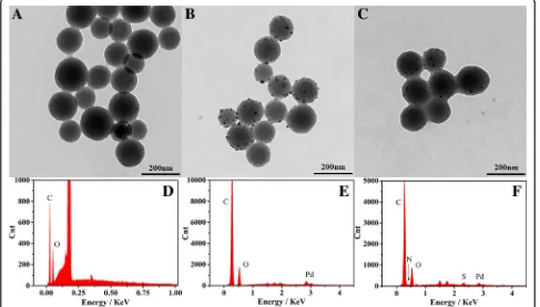
Pd-CNCs were successfully prepared. The morphologies of carbon nanospheres, Pd-carbon nanospheres and Pd-CNCs were characterised by transmission electron
microscopy (TEM) and energy dispersive X-ray
spectrometry (EDS). Pd nanoparticles were formed and distributed on the carbon nanospheres following the microwave reaction (Fig.2b), demonstrating the success-ful preparation of Pd-carbon nanospheres with
diame-ters of 150 nm (Fig. 2a). The morphologies of Pd-CNCs
are shown in Fig.2c. The EDS results confirm the
chem-ical compositions of carbon nanoparticles, Pd-carbon nanospheres and Pd-CNCs, which shows the carbon nanoparticles contain C and O (Fig.2d). After the func-tionalization of Pd nanoparticles, Pd also existed in the
spectra of Pd-carbon nanospheres (Fig. 2e), while S and
N found in the spectra of Pd-CNCs originate from BSA, which was used to block Pd-carbon nanospheres
(Fig.2f ). These results intuitively reveal the successful syn-thesis of the Pd-CNC catalytic enhancers. HR-TEM im-ages of Pd-carbon nanospheres (Fig.3a) illustrate that the functionalised Pd nanoparticles (Fig.3b) are in face-center cubic (FCC) phase with an observable (1,1,1) lattice plane (Fig.3c). Corresponding fast Fourier transformation (FFT) images (Fig.3d) also support the FCC crystalline nature of Pd nanoparticles.
Characterisation of Construction Procedures of the Biosensor
The construction procedures of the biosensor were
monitored by SWV measurements (Fig. 4a) and
Fig. 2TEM micrographs of carbon nanosphere (a), Pd-carbon nanospheres (b) and Pd-CNCs (c) EDS of carbon nanosphere (d), Pd-carbon nanospheres (e) and Pd-CNCs (f)
[image:4.595.58.539.87.221.2] [image:4.595.56.543.426.703.2]electrochemical impedance spectroscopy (EIS) (Fig. 4b). Compared to the current signal of bare GCE (curve bare GCE), the peak current in the potential range increased
to about 420 μA (curve Au-rGO/GCE) after
electrode-position of Au-rGO, which may be attributed to the ex-cellent conductivity of Au-rGO. Then, the signal current peak decreased after the immobilisation of peptides on the electrode (curve peptides) and was further reduced after the linkage between peptides and Pd-CNCs due to the high impedance of the peptides and enhancers (curve Pd-CNCs). After incubating with MMP-7, the peak current increased (curve MMP-7 cleavage), which may be caused by the specific cleavage of peptides by MMP-7 and partial removal of enhancers from the elec-trode surface. Under the same conditions, the current
change (ΔI1) between curves “Pd-CNCs” and “MMP-7
cleavage” was calculated to be 48.7 μA. Then, the peak current was reduced after the catalytic precipitation reaction of 4-CN, with a current peak of 136.1μA (curve MMP-7 cleavage precipitation). In contrast, the biosen-sor without MMP-7 induced a lower current peak (54.9 μA, curve precipitation), which was stated as the blank signal, after the catalysed precipitation reaction. In the circumstances, ΔI2was the current signal difference
between curves“precipitation”and curve“MMP-7 cleav-age precipitation”, increasing from 48.7 to 81.2 μA. EIS was also used to monitor the fabrication of the biosen-sor. The electron transfer resistance corresponded to the
semicircle diameter of the Nyquist plots (Fig. 4b). For
comparison, the plot of the peptides/Au-rGO/GCE
[image:5.595.57.539.88.347.2] [image:5.595.58.539.584.714.2](curve peptides) displays a larger semicircle, and thus larger resistance, than those of Au-rGO/GCE (curve Au-rGO/GCE) and bare GCE (curve bare GC) which has the smallest circle, indicating that peptides were suc-cessfully modified on the GCE. The resistance increased when peptides were linked with the enhancer via glutar-aldehyde (curve Pd-CNCs). The diameter of semicircle slightly decreased (curve MMP-7 cleavage) after incuba-tion with MMP-7, which may be ascribed to the MMP-7 specifically cleaved the peptides. In contrast, the resist-ance significantly increased when the biosensor was dipped in the solution of 4-CN and H2O2(curve
precipi-tation). Undergoing both peptide-cleavage and catalytic
precipitation reaction, the resistance decreased obviously (curve MMP-7 cleavage precipitation).
Optimization of Detection Conditions
The quantity and incubation time of peptides, as import-ant factors for the detection performance of the biosen-sor, were further optimised. It was found that the current signal decreased accordingly when the peptide
concentration increased from 20 to 40 μM and kept
constant between 50 and 80μM (Fig.5a). To avoid
non-specific adsorption, we chose 50 μM for subsequent
measurements and then optimised the incubation time
of the 50 μM peptide (Fig. 5b). The SWV current
Fig. 5Effects of peptide concentration (a), incubation time of 50μM peptide (b), peptide cleavage time (c) and precipitation reaction time (d) on the current responses of the biosensor
Fig. 6aSWV responses of electrochemical detection for MMP-7 in 5 mM [Fe(CN)6]3−/4−phosphate buffered (0.1 M, pH = 7.4) at the
concentrations from 100 fg mL−1to 100 ng mL−1.bLinear calibration curve of current and the logarithm of the concentration of MMP-7. The
[image:6.595.58.536.87.343.2] [image:6.595.58.539.557.694.2]decreased gradually from 20 to 40 min, then remained constant after 40 min. Thus, 40 min was chosen as the in-cubation time for peptides. The cleavage reaction time is also a significant factor for the analytical performance of the biosensor (Fig. 5c). The signal of the biosensor in-creased when the cleavage time ranged between 30 and 50 min, then remained constant after 50 min, suggesting that the peptide was completely cleaved. Thus, 50 min was chosen as the cleavage time for the following experi-ments. The precipitation reaction, which also influences the sensitivity of the biosensor (Fig.5d), was found to in-crease within 50 min, and the electrochemical signal de-creased continuously. Since the current signal maintained constant with a further increase in reaction time, 50 min was selected as the precipitation reaction time for the fol-lowing assay.
Performance of Proposed Biosensor
After incubation with different concentrations of MMP-7 under the optimal conditions, the performance of the pro-posed biosensor was determined. As shown in Fig.6, with decreasing concentration of MMP-7, the SWV signal de-creased. The calibration plot reveals a good linear
relationship between the current peaks and the logarithm of the analyte concentrations in the range from 0.1 pg mL−1 to 100 ng mL−1. The linear equation was determined to be
(I=−16.53lgCMMP-7-137.26) with a correlation
coefficient of 0.9967. The detection limit of the biosensor was 17.38 fg mL−1for MMP-7 at a signal-to-noise ratio of 3 (S/N= 3; is the standard deviation of the signal in a blank solution). Compared with recent reports on peptide-cleavage detection of MMP-7, the biosensor ex-hibited a better analytical performance (Table1).
Evaluation of Performance of Immunosensor
To assess the reproducibility of the biosensor, three modified electrodes were incubated with 0.01, 0.1, 1, 10
and 100 ng mL−1MMP-7. The corresponding standard
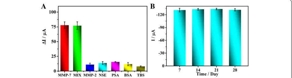
deviations were calculated to be 1.3%, 1.4%, 4.0%, 1.0% and 3.0%, respectively, indicating good reproducibility of the biosensor. MMP-2, NSE, PSA, TBS and BSA were used as distractions to analyse the specificity of this bio-sensor. No obvious responses were observed when the biosensor was incubated with individual mixtures of
1 ng mL−1 MMP-7 with (100 ng mL−1 each) MMP-2,
NSE, PSA, TBS and BSA. Compared to the biosensor
Fig. 7SWV current responses (a) about anti-interference ability of the biosensor (the error bars are standard deviations forn= 3). The concentrations of distractions: MMP-2 (100 ng mL−1), NSE (100 ng mL−1), PSA (100 ng mL−1), THR (100 ng mL−1) and BSA (100 ng mL−1), respectively. The mixture
contains all those distractions (100 ng mL−1) and MMP-7 (1 ng mL−1). SWV responses (b) of the proposed biosensors stored at 4 °C for different time
[image:7.595.59.539.98.251.2]for the detection of 1 ng mL−1MMP-7
Table 1Comparison of some recently reported biosensor for detection of MMP-7
Material and method Detection limit Detection range Reference
DNA enzyme-decorated DNA nanoladders Differential pulse voltammetry
0.05 pg mL−1 2 × 10⁻⁴–20 ng mL−1 Kou et al. 2016 [14]
Au/Ag@SiO2
Fluorescence resonance energy transfer
0.22 pg mL−1 1 × 10⁻²–100 ng mL−1 Lei et al. 2017 [10]
Gold nanoparticles Colorimetry
0.1μg mL−1 0–2μg mL−1 Chen et al. 2013 [8]
Magnetic peptide−DNA probe Square wave voltammetry
0.02 pg mL−1 1 × 10⁻⁴–50 ng mL−1 Wang et al. 2017 [28]
BSA-Pd-SAM-PDA probe Square wave voltammetry
3.1 fg mL−1 1 × 10⁻⁵–10 ng mL−1 Zheng et al. 2018 [11]
Pd-CNCs enhancer Square wave voltammetry
[image:7.595.60.539.557.685.2]incubated with only 1 ng mL−1MMP-7, the current
sig-nals of each mixture were almost the same (Fig. 7a),
which demonstrates that the biosensor displays excellent specificity for MMP-7. To investigate stability, the con-structed biosensor was stored at 4 °C for 28 days, and its performance for MMP-7 detection was evaluated every 7 days (Fig. 7b). The relative standard deviations (RSD) between parallel experiments were all less than 10%, im-plying remarkable stability of the biosensor.
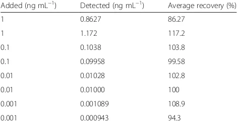
To investigate the practicability of the proposed method, recovery experiments were carried out. The recoveries ranged from 86.3 to 117.2% (Table2), further indicating the promising practicability of the biosensor in clinical analyses.
Conclusions
In summary, a Pd-functionalized carbon nanocomposite was fabricated as a novel impedance enhancer, which re-veals promising H2O2catalytic performance for 4-CN
oxi-dation. Utilising the high impedance of insoluble oxidised 4-CN precipitation on the electrode, an amperometric biosensor for detection of MMP-7 was constructed. The biosensor possesses comparable sensitivity, a broad detec-tion range, good practicability and outstanding selectivity to the detection of MMP-7, which suggests it potential ap-plication in various bio-apap-plications. Our work further highlights the importance of the impedance enhancer in improving the performance of amperometric assays, en-couraging the fabrication of new enhancers with advanced catalytic activity and high resistance.
Abbreviations
4-CN:4-Chloro-1-naphthol; BSA: Bovine serum albumin; EDS: Energy dispersive X-ray spectroscopy; FCC: Face-center cubic; FFT: Fast Fourier transformation; GCE: Glassy carbon electrode; GO: Graphene oxide; HR-TEM: High-resolution transmission electron microscope; LOD: Limit of detection; MMP-2: Matrix metalloproteinase-2; MMP-7: Matrix metalloproteinase-7; NSE: Neuron specific enolase; PBS: Phosphate buffer solution; Pd-CNCs: Pd-functionalized carbon nanocomposites; PSA: Prostate-specific antigen; RSD: Relative standard deviations; SEM: Scanning electron microscope; SWV: Square wave voltammetry; TBS: Thrombin of bovine serum; TEM: Transmission electron microscope
Acknowledgments
This research was financed by grants from the National Natural Science Foundation of China (21673143, 21273153), Natural Science Foundation of Beijing Municipality (2172016, 2132008), High-level Teachers in Beijing
Municipal Universities in the Period of 13th Five-year Plan (IDHT20180517) and Capacity Building for Sci-Tech Innovation—Fundamental Scientific Research Funds (025185305000/195).
Availability of Data and Materials
All data are fully available without restriction.
Authors’Contributions
ZW carried out the experiments and drafted the manuscript. HQW designed the measurement study. ZFM and HLH supervised the overall study and polished the manuscript. All authors read and approved the final manuscript.
Competing Interests
The authors declare that they have no competing interests.
Publisher’s Note
Springer Nature remains neutral with regard to jurisdictional claims in published maps and institutional affiliations.
Received: 26 September 2018 Accepted: 9 November 2018
References
1. Szarvas T, Becker M, vom Dorp F, Gethmann C, Totsch M, Bankfalvi A et al (2010) Matrix metalloproteinase-7 as a marker of metastasis and predictor of poor survival in bladder cancer. Cancer Sci 101(5):1300–1308.https://doi. org/10.1111/j.1349-7006.2010.01506.x
2. Miwa S, Miyagawa S, Soeda J, Kawasaki S (2002) Matrix metalloproteinase-7 expression and biologic aggressiveness of cholangiocellular carcinoma. Cancer 94(2):428–434.https://doi.org/10.1002/cncr.10235
3. Tanioka Y, Yoshida T, Yagawa T, Saiki Y, Takeo S, Harada T et al (2003) Matrix metalloproteinase-7 and matrix metalloproteinase-9 are associated with unfavourable prognosis in superficial oesophageal cancer. Br J Cancer 89(11):2116–2121.https://doi.org/10.1038/sj.bjc.6601372
4. Luukkaa H, Klemi P, Hirsimaki P, Vahlberg T, Kivisaari A, Kahari VM et al (2010) Matrix metalloproteinase (MMP)-7 in salivary gland cancer. Acta Oncol 49(1):85–90.https://doi.org/10.3109/02841860903287197 5. Zucker S, Vacirca J (2004) Role of matrix metalloproteinases (MMPs) in
colorectal cancer. CANCER METAST REV 23(1–2):101–117.https://doi.org/10. 1023/A:1025867130437
6. Lu H, Yang Z, Zhang H, Gan M, Zhou T, Wang S (2013) The expression and clinical significance of matrix metalloproteinase 7 and tissue inhibitor of matrix metalloproteinases 2 in clear cell renal cell carcinoma. Exp Ther Med 5(3):890–896.https://doi.org/10.3892/etm.2012.859
7. Cheng W, Chen Y, Yan F, Ding L, Ding S, Ju H et al (2011) Ultrasensitive scanometric strategy for detection of matrix metalloproteinases using a histidine tagged peptide-Au nanoparticle probe. Chem Commun (Camb) 47(10):2877–2879.https://doi.org/10.1039/c0cc04441e
8. Chen P, Selegard R, Aili D, Liedberg B (2013) Peptide functionalized gold nanoparticles for colorimetric detection of matrilysin (MMP-7) activity. Nanoscale 5(19):8973–8976.https://doi.org/10.1039/c3nr03006g 9. Gao H, Dang Q, Xia S, Zhao Y, Qi H, Gao Q et al (2017) Highly selective
electrogenerated chemiluminescence biosensor for simultaneous detection of matrix metalloproteinase-2 and matrix metalloproteinase-7 in cell secretions. Sensors Actuators B Chem 253:69–76.https://doi.org/10.1016/j.snb.2017.05.142 10. Lei Z, Zhang H, Wang Y, Meng X, Wang Z (2017) Peptide microarray-based
metal enhanced fluorescence assay for multiple profiling of matrix metalloproteinases activities. Anal Chem 89(12):6749–6757.https://doi.org/ 10.1021/acs.analchem.7b01037
11. Zheng Y, Ma Z (2018) Dual-reaction triggered sensitivity amplification for ultrasensitive peptide-cleavage based electrochemical detection of matrix metalloproteinase-7. Biosens Bioelectron 108:46–52.https://doi.org/10.1016/ j.bios.2018.02.045
12. Tang Z, Ma Z (2017) Multiple functional strategies for amplifying sensitivity of amperometric immunoassay for tumor markers: a review. Biosens Bioelectron 98:100–112.https://doi.org/10.1016/j.bios.2017.06.041 13. Tang Z, Wang L, Ma Z (2017) Triple sensitivity amplification for ultrasensitive
electrochemical detection of prostate specific antigen. Biosens Bioelectron 92:577–582.https://doi.org/10.1016/j.bios.2016.10.057
14. Kou BB, Zhang L, Xie H, Wang D, Yuan YL, Chai YQ et al (2016) DNA enzyme-decorated DNA Nanoladders as enhancer for peptide
cleavage-Table 2Recovery tests for MMP-7 in human serum samples
Added (ng mL−1) Detected (ng mL−1) Average recovery (%)
1 0.8627 86.27
1 1.172 117.2
0.1 0.1038 103.8
0.1 0.09958 99.58
0.01 0.01028 102.8
0.01 0.01000 100
0.001 0.001089 108.9
[image:8.595.56.292.100.224.2]based electrochemical biosensor. ACS Appl Mater Interfaces 8(35):22869– 22874.https://doi.org/10.1021/acsami.6b07017.
15. Sun Z, Glebe U, Charan H, Boker A, Wu C (2018) Enzyme-polymer conjugates as robust Pickering interfacial biocatalysts for efficient biotransformations and one-pot Cascade reactions. Angew Chem Int Ed Engl 57(42):13810–13814.https://doi.org/10.1002/anie.201806049 16. Wang X, Liu X, Yan X, Zhao P, Ding Y, Xu P (2011) Enzyme-nanoporous gold
biocomposite: excellent biocatalyst with improved biocatalytic performance and stability. PLoS One 6(9):e24207.https://doi.org/10.1371/journal.pone.0024207 17. Cheng MB, Wang JC, Li YH, Liu XY, Zhang X, Chen DW et al (2008)
Characterization of water-in-oil microemulsion for oral delivery of earthworm fibrinolytic enzyme. J Control Release 129(1):41–48.https://doi. org/10.1016/j.jconrel.2008.03.018
18. Chiu NF, Chen CC, Yang CD, Kao YS, Wu WR (2018) Enhanced Plasmonic Biosensors of hybrid gold nanoparticle-graphene oxide-based label-free immunoassay. Nanoscale Res Lett 13(1):152. https://doi.org/10.1186/s11671-018-2565-7.
19. He G, Tian L, Cai Y, Wu S, Su Y, Yan H et al (2018) Sensitive nonenzymatic electrochemical glucose detection based on hollow porous NiO. Nanoscale Res Lett 13(1):3.https://doi.org/10.1186/s11671-017-2406-0.
20. Stahl SS (2004) Palladium oxidase catalysis: selective oxidation of organic chemicals by direct dioxygen-coupled turnover. Angew Chem Int Ed Engl 43(26):3400–3420.https://doi.org/10.1002/anie.200300630
21. Goodman ED, Dai S, Yang A-C, Wrasman CJ, Gallo A, Bare SR et al (2017) Uniform Pt/Pd bimetallic nanocrystals demonstrate platinum effect on palladium methane combustion activity and stability. ACS Catal 7(7):4372– 4380.https://doi.org/10.1021/acscatal.7b00393.
22. Xu F, Tang Z, Huang S, Chen L, Liang Y, Mai W et al (2015) Erratum: facile synthesis of ultrahigh-surface-area hollow carbon nanospheres for enhanced adsorption and energy storage. Nat Commun 6:7863.https://doi. org/10.1038/ncomms8863
23. Liao X, Ju H (2015) In situ quantitation of intracellular microRNA in the whole cell cycle with a functionalized carbon nanosphere probe. Chem Commun (Camb) 51(11):2141–2144.https://doi.org/10.1039/c4cc09097g 24. Xu T, Liu N, Yuan J, Ma Z (2015) Triple tumor markers assay based on
carbon-gold nanocomposite. Biosens Bioelectron 70:161–166.https://doi. org/10.1016/j.bios.2015.03.036
25. Tang Z, Han G-H, Jung E, M-g S, Lee K-Y, Kim W-S et al (2018) Synthesis of Cu-Pd nanoplates and their catalytic performance for H2O2generation
reaction. Mol Catal 452:117–122.https://doi.org/10.1016/j.mcat.2018.04.010 26. Cui R, Liu C, Shen J, Gao D, Zhu J-J, Chen H-Y (2008) Gold
nanoparticle-colloidal carbon nanosphere hybrid material: preparation, characterization, and application for an amplified electrochemical immunoassay. Adv Funct Mater 18(15):2197–2204.https://doi.org/10.1002/adfm.200701340 27. Chen Y, Liu X, Zhang S, Yang L, Liu M, Zhang Y et al (2017) Ultrasensitive
and simultaneous detection of hydroquinone, catechol and resorcinol based on the electrochemical co-reduction prepared Au-Pd nanoflower/ reduced graphene oxide nanocomposite. Electrochim Acta 231:677–685. https://doi.org/10.1016/j.electacta.2017.02.060
![Fig. 4 SWV (a) and EIS (b) responses of modification process of electrode in 5 mM [Fe(CN)6]3−/4- phosphate buffered (0.1 M, pH = 7.4)](https://thumb-us.123doks.com/thumbv2/123dok_us/8844532.932326/5.595.57.539.88.347/fig-swv-responses-modification-process-electrode-phosphate-buffered.webp)
![Fig. 6 a SWV responses of electrochemical detection for MMP-7 in 5 mM [Fe(CN)6]3−/4− phosphate buffered (0.1 M, pH = 7.4) at theconcentrations from 100 fg mL−1 to 100 ng mL−1](https://thumb-us.123doks.com/thumbv2/123dok_us/8844532.932326/6.595.58.536.87.343/fig-swv-responses-electrochemical-detection-phosphate-buffered-theconcentrations.webp)