IJPSR (2020), Volume 11, Issue 1 (Research Article)
Received on 22 November 2019; received in revised form, 08 December 2019; accepted, 14 December 2019; published 01 January 2020
ANTIOXIDATIVE AND HYPOLIPIDEMIC EFFECT OF PUERARIA TUBEROSA WATER EXTRACT (PTWE) IN RATS WITH HIGH FAT DIET INDUCED NON-ALCOHOLIC FATTY LIVER DISEASE (NAFLD)
Prerana Aditi and Yamini Bhushan Tripathi *
Department of Medicinal Chemistry, Institute of Medical Sciences, Banaras Hindu University, Varanasi-221005, Uttar Pradesh, India.
ABSTRACT: Non-alcoholic fatty liver disease (NAFLD) is chronic liver diseases ensuing from excessive fat accumulation in liver attributed to high dietary fat and carbohydrate and lower hepatic activity. It is associated with obesity and high BMI (body mass index). The excess carbohydrate-rich diet activates its conversion to TG (triglycerides), which gets accumulated in adipose and non-adipose tissues in the body. Insulin resistance also plays an important role in its pathogenesis. This work is focused on antioxidant and hypolipidemic potentials of water extract of Pueraria tuberosa (PTWE) in rats with high fat diet-induced non-alcoholic fatty liver disease. NAFLD was induced in rats with a high-fat diet (HFD) for 100 days and grouped into HFD control, HFD+PT 50 mg/100 g bw, HFD+PT 100 mg/100 g bw. Blood triglycerides, cholesterol, glucose, serum glutamate oxaloacetate trans-aminases (SGOT), serum glutamate pyruvate transtrans-aminases (SGPT), Superoxide dismutase (SOD), Catalase activity and lipid peroxides (LPO) levels were assessed. The histological studies in the liver were done by H&E staining. PTWE shows the antioxidant properties by decreasing lipid peroxidation and increasing the activity of SOD and catalase enzymes. It also shows the hypolipidemic effect on NAFLD rats by decreasing the TG and cholesterol levels in serum. It also prevented the progression of liver damage, as evident by lowering the activity of SGOT and SGPT and decreased hepatocyte ballooning, inflammatory infiltration and microvascular steatosis. All the results demonstrate that PT can exhibit a therapeutic effect on NAFLD by way of through its antioxidant, anti-inflammatory, and hypolipidemic potentials.
INTRODUCTION: Non-alcoholic fatty liver diseases (NAFLD) was firstly exposed by Ludwig et al. (1980) in people with no history of alcohol intake 1.
QUICK RESPONSE CODE
DOI:
10.13040/IJPSR.0975-8232.11(1).378-86
This article can be accessed online on
www.ijpsr.com
DOI link: http://dx.doi.org/10.13040/IJPSR.0975-8232.11(1).378-86
Nowadays, NAFLD is considered as the most common form of chronic liver disease. It affects high populations (more than 30%) in western countries and identified as one of the most common causes of liver dysfunction worldwide 2. This is a type of metabolic syndrome, which encompasses a spectrum of liver diseases like hepatic steatosis (fatty liver), non-alcoholic steatohepatitis (NASH), fibrosis, cirrhosis and hepatocellular carcinoma which occur without any intake of alcohol and absence of genetic, viral and any other autoimmune component 3.
Keywords:
NAFLD, Pueraria tuberosa, Medicinal plants, Insulin resistant,
Antioxidant and hypolipidemic potential
Correspondence to Author: Prof. Y. B. Tripathi
Department of Medicinal Chemistry, Institute of Medical Sciences, Banaras Hindu University, Varanasi-221005, Uttar Pradesh, India.
The prevalence of NAFLD increases to 58% in overweight individuals and can be as high as 98% in non-diabetic obese individuals 4. NAFLD is considered as the hepatic manifestation of metabolic syndrome, which is defined by the occurrence of central obesity, insulin resistance, hyperlipidemia, hyperglycemia and hypertension 5,
6, 7
. In NAFLD there is an accumulation of excess fatty acids (> 5% weight or volume) in the liver. Deposition of the ―bad fat‖ (such as fatty acids, triacylglycerides, cholesterol) in hepatocytes in NASH causes insulin resistance, which in turn contributes to type 2 diabetes 8.
There is a theory called ―two-hit theory‖ proposed by Day et al., in 1998, which is the most widely
accepted theory for NAFLD development.
According to this theory, NAFLD is a progressive disease in which insulin resistance (―the first hit‖) leads to increased FFA flux to the liver. If FFAs are not oxidized or secreted, hepato-steatosis develops
5. Hepatic steatosis predisposes the liver to ―second
hits‖ such as mitochondrial dysfunction, cytokines, adipokines, ER stress and bacterial endotoxins. This original has been modified to suggest that NAFLD may be the consequence of parallel ―multiple hits‖ 9
.
The biological mechanism related to NAFLD happening and its progression to NASH is not entirely understood and is probably due to a number of factors that are expressed in the context of genetic tendency. The primary source of cellular
reactive oxygen species in mitochondria
(mitochondrial electron transport chain).
Mitochondrial dysfunction impairs fat homeostasis in the liver and also leads to an overproduction of reactive oxygen species (ROS). It has already been reported that approximately 0.2%-2 of the oxygen consumed by the cell is converted by mitochondria to ROS, mainly through the production of superoxide anion (O2.-) 10. Mitochondria consume
around 90 % of a cell’s oxygen to produce ATP through the process of oxidative phosphorylation. The primary oxygen radical species generated by mitochondria is superoxide anion which is converted to hydrogen peroxide (H2O2) by
spontaneous dismutation or by superoxide
dismutase (SOD), present both within the mitochondria and in the cytosol. Hydrogen peroxide, in turn, is converted into water by
glutathione peroxidase or catalase, otherwise, in the presence of divalent cations such as iron, H2O2 can
undergo Fenton’s reaction to produce hydroxyl radical OH.
It has been confirmed that high generation of reactive oxygen species (ROS) may cause damage at the cellular level, such as membrane lipid peroxidation, cell degeneration and necrosis, cell death by apoptosis 11, 12, 13 proinflammatory cytokine expression, liver stellate cell activation and fibrogenesis 14, 15, 16, 17. Subsequently, increased accumulation of free fatty acids and triglycerides formation leads to the progression of the diseases in the liver 18. So, fatty acid accumulation, Oxidative stress, inflammation and insulin resistance are major contributors to the pathogenesis of NAFLD.
Ayurveda is a traditional system of medicine in India, Pueraria tuberosa (PT) DC, Fabaceaefamily, is a medicinal plant, also known as ―Indian kudzu‖ or Vidarikand. It is uses as health promotive and also for disorders of kidney diseases, as diuretics, antiaging, galactagogue, cardiotonic.
Recent studies in our lab have shown that the PT behaves like DPP-4 inhibitor, nephroprotective, antioxidant and antiapoptotic etc. 19, 20, 21, 22. It is distributed in the tropical parts of India 23. The
voluminous literature showed its
anti-hyperglycemic, hepatoprotective, anti-inflammatory, antioxidant properties and anti-fertility in male rats
24, 25, 26, 27, 28, 29
. About 113 phytochemicals have been identified by LCMS of PT extract but the major compounds of PT are daidzin, tuberosin, puerarin, genistein, pterocarpintuberosin, puerarone,
coumarin, hydroxytuberosone, anthocyanin,
lupinoside, puetuberosanol 30, 31.
In the present study, we have checked the antioxidative and hypolipidemic role of PTWE in the case of high-fat diet-induced NAFLD in rats. We have seen the accumulation of fat in liver tissue by H & E (Hematoxyline and Eosin).
MATERIALS AND METHODS:
Materials: Biochemical kits were purchased from Accurex Biomedical Pvt. Ltd., Biosar, Thane.
Nitroblue tetrazolium chloride, Riboflavin,
Methods:
Preparation of Water Extract of PT Tuber (PTWE): PT tuber was purchased from Ayurvedic Pharmacy, IMS, BHU. First, the coarse powder of PT tuber was formed and 120 g of coarse powder was extracted with 8 volumes of distilled water by boiling. When the volume reduced to 1/4th, it was filtered and boiled again with distilled water. The filtrates were pooled and further concentrated at reduced temperature (60-75 °C) by a rotary evaporator and lyophilized. The dried powder was weighed and the % extraction was calculated. Its fine powder was made with the help of a juicer grinder mixer.
Animal: The experimental protocol was approved by an animal welfare ethics committee of the Institute of Medical Sciences, Banaras Hindu University, Varanasi (ethical committee letter # No Dean/2017/CAEC/720). We have taken healthy male Charles Foster strain rats (32) from the central animal house of our institute (Institute of Medical Sciences) of average weight ranging between 100– 120 gm. The intention of taking male rats is only to evade the variation like different metabolic rates of gender including sex hormones, pregnancy, lactation which is inherent in a female. Rats were kept separately in propylene cage, fed standard diet
and kept in hygiene conditions. The entire animals were kept at 25 °C with standard condition 12 h light/dark cycle. Before treatments start rats were kept under the standard condition for 1 week for acclimatization with free access to standard chow and tap water.
[image:3.612.314.567.311.424.2]Induction of NAFLD: The 24 rats were randomly selected from the inbred animal colony and put to a high fat diet (HFD) for 100 days. The composition of a high fat diet was decided according to literature published earlier 32 Table 1. The 8 rats were fed with a normal diet and they served as normal control. The weight of each animal was taken every 20 days and blood was also collected to check changes in biochemical parameters. Blood biochemistry during the induction of NAFLD was shown in Table 2 and Table 3.
TABLE 1:COMPOSITION OF HIGH FAT DIET
S. no. Ingredients Diet gm/kg
1 Powdered normal diet 365
2 Lard 310
3 Casein 250
4 Cholesterol 10
5 DL-methionine 03
6 Vitamins and minerals 06
7 Yeast powder 01
8 NaCl 01
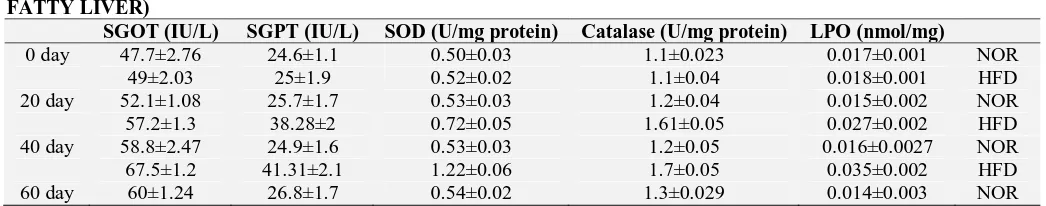
TABLE 2: INDUCTION OF NAFLD MODEL (BLOOD BIOCHEMISTRY OF RATS DURING INDUCTION OF FATTY LIVER)
Weight (gm) Hb (g/dl) Protein (mg/ml) Glucose (mg%) Cholesterol (mg%) TG (mg %) 0 day 118.1±6.7 11.8±0.8 5.8±0.4 98.5±3.0 52.2±3.7 45.9±2.1 NOR
118.6±5 12.5±0.7 6.1±0.4 100.5±2 55.2±4.7 48.9±3.1 HFD 20 day 132.5±5.4 12.0±0.7 5.9±0.7 104.0±3.2 55.5±3.0 53±3.0 NOR 156.6±4.9 13.0±0.8 6.4±0.3 128.3±5.0 59.6±5.1 65.9±2.9 HFD 40 day 150±6.3 12.3±0.6 6.1±0.9 106.9±2.6 57.2±4.4 55.5±3.3 NOR 186.6±7.1 12.4±0.6 6.4±0.4 149.0±4.0 68.3±5.8 66.3±3.25 HFD 60 day 177±6.5 12.6±0.8 6.2±0.8 108.4±1.6 59.0±4.1 60±3.2 NOR 198±6.0 12.1±0.9 6.5±0.4 161.4±5.8 76.1±5.3 82.2±2.09 HFD 80 day 182±7.2 12.1±0.7 5.9±0.8 109.7±2.5 61.0±4.5 64±2.7 NOR 240±5.7 12.1±1.0 6.6±0.2 167.3±5.8 81.1±2.3 89.9±1.97 HFD 100 day 207±8.1 12.2±0.8 6.5±1.0 111.9±1.8 63.9±4.4 67.1±2.6 NOR 252±9.4 12.3±0.8 7.0±0.2 170.3±5.6 89.5±4.0 93.5±2.4 HFD
TABLE 3: INDUCTION OF NAFLD MODEL (BLOOD BIOCHEMISTRY OF RATS DURING INDUCTION OF FATTY LIVER)
SGOT (IU/L) SGPT (IU/L) SOD (U/mg protein) Catalase (U/mg protein) LPO (nmol/mg)
[image:3.612.53.567.470.622.2] [image:3.612.45.568.645.748.2]68.4±1.5 47.49±2.3 0.72±0.06 0.84±0.04 0.046±0.003 HFD 80 day 63±1.93 27.6±1.5 0.5±0.04 1.3±0.026 0.016±0.002 NOR 72.2±2.5 49.67±2.3 0.59±0.04 0.64±0.03 0.055±0.003 HFD 100 day 64.3±1.49 29.3±1.6 0.51±0.04 1.2±0.03 0.015±0.0026 NOR 77.7±2.6 53.79±2 0.46±0.06 0.57±0.05 0.062±0.004 HFD NOR = Normal, HFD = High fat diet
Treatment: When we found that (after 100-day high fat diet feeding) the weight of rat was increased and the blood biochemical parameters like TG, cholesterol, glucose, SGOT, SGPT and LPO Were also increased more than 20%, as well as level of antioxidant enzymes, were decreased then we started the treatment of PTWE in two different dose. These rats were randomly divided into three group (n=8), HFD control, HFD + PT 50 mg/100 gm body weight (HFD+PT50), HFD + PT 100 mg/100 gm body weight (HFD+PT100) and accordingly, PT water extract was given for 20 days orally and fatty diet was continued.
On the 10th and 20th days, blood was collected in EDTA tubes. Hemolysate was prepared and plasma was isolated after centrifugation of blood in cooling centrifuge at 5000 rpm for 15 min. All biochemical parameters mentioned above were checked in blood plasma and hemolysate. On the 20th day, the rats of all groups were sacrificed. Liver tissue was collected and washed in 1x phosphate buffer saline (pH 7.4). Half of the liver tissue was stored at -20 °C for further experiments. Half of the liver tissue was collected in 10% formalin for histological study.
Biochemical Parameters: Hemoglobin was estimated in blood by using cyanmethaemoglobin method 33. Plasma protein was estimated by using Bradford’s reagent. The triglycerides, cholesterol, glucose, SGOT, SGPT were estimated by standard methods and kits (Accurex biomedical, Mumbai, India).
Superoxide Dismutase: Superoxide dismutase (SOD) activity was measured in terms of inhibition of reduction of nitro blue tetrazolium (NBT) in the presence of riboflavin as described 34 with slight modification 35. Blood hemolysate was prepared by adding 25 µl of blood in 975 µl of cold water. Three tubes were prepared namely sample tube, control tube and blank. Reaction mixture in sample tube contains SOD buffer (pH 7.8), 130 mM L-methionine (300 µl), 10 times diluted hemolysate
(250 µl), 750 µM NBT (150 µl), 0.5 mM EDTA (75 µl) and 60 µM riboflavin (100 µl). The control tube was devoid of hemolysate and riboflavin was not added in the blank tube. The tubes were kept in front of fluorescent light for 10 min. NBT in the presence of riboflavin under fluorescent light gets reduced by photochemical reaction and formed a blue color product formazan. In the presence of the enzyme, the reaction was inhibited to a different extent depending on the activity of the enzyme and the color produced was read at 560 nm.
Catalase: Catalase enzyme activity was measured by Aebi’s method by monitoring H2O2 breakdown
at 240 nm 36. In this method, hemolysate was diluted 10 times in catalase buffer. The reaction was initiated by adding 10 µl of diluted hemolysate in 900 µl of 30 mM H2O2 and the absorbance was
measured at 240 nm for 3 min. The activity of the catalase enzyme was expressed in U/mg.
Lipid Peroxidation: Lipid peroxidation was measured by thiobarbituric acid method 37. Briefly, 0.5 ml of hemolysate and 0.5 ml of 10% trichloroacetic acid was added and centrifuged at 1000 g for 10 min. To 0.5 ml of supernatant 1.5 ml of 0.67% of thiobarbituric acid prepared in 50% glacial acetic acid was added, and the test tubes were kept in a boiling water bath for 20 min. After cooling, absorbance was measured at 532 nm.
Histological Study: Histological study (H & E) in liver tissue was done in the Department of Pathology, IMS, BHU according to standard method 38.
RESULTS:
a similar manner, we found the significance changes in level of glucose Fig. 1, triglycerides
Fig. 2, cholesterol Fig. 2, SGOT Fig. 2, SGPT Fig. 2 and LPO Fig. 3 in blood. We found that the level of antioxidant enzyme (SOD and catalase) level
was significantly decreased in the high fat diet-fed rats, and after PTWE treatment the level was significantly increased compared to high-fat diet-fed rats Fig. 3.
FIG. 1: BIOCHEMICAL PARAMETER: A. WEIGHT (gm), B. GLUCOSE (mg%), C. HEMOGLOBIN (g/dl), D. PROTEIN (mg/ml). *P<0.05 comparison between normal and other groups, #p<0.05 comparison between HFD control and PT treated group
FIG. 2: EFFECT OF PTWE ON LIPID PROFILE AND LFT ENZYMES. A. TRIGLYCERIDES (mg %), B. CHOLESTEROL (mg %), C. SGOT (IU/L), D. SGPT (IU/L). *P<0.05 comparison between normal and other groups, #p<0.05 comparison between HFD control and PT treated group
A B
C D
D
A B
[image:5.612.56.558.132.406.2] [image:5.612.60.559.453.715.2]FIG. 3: EFFECT OF PTWE ON ANTIOXIDANT ENZYMES AND LIPID PEROXIDATION. A. SOD (U/mg protein), B. CTALASE (U/mg protein), C. LPO (nmol/mg protein). *P<0.05 comparison between normal and other groups. #p<0.05 comparison between HFD control and PT treated group
Histology Result: Fat droplet accumulation was found in high-fat diet rats liver tissue and it was decreased in PTWE treated rats liver tissue. Hepatocyte ballooning, inflammatory infiltration and microvesicular steatosis was found in the high
fat diet-fed rat liver tissue and this was decreased in PTWE treated rats liver tissue. Sinusoidal dilution was found in PTWE treated rat which was decreased in HFD treated rat because increased fat accumulation Fig. 4.
FIG. 4: HISTOLOGY RESULTS. A = NORMAL SHOWING NORMAL ARCHITECTURE OF LIVER, B = HFD CONTROL SHOWING HEPATOCYTE BALLOONING, LESS SINUSOIDAL DILATION, INFLAMMATORY INFILTRATION, MICRO VESICULAR STEATOSIS, C = HFD+PT50 SHOWING MORE SINUSOIDAL DILATION AND STRUCTURE LIKE NORMAL LIVER TISSUE, D = HFD+PT100 SHOWING MORE SINUSOIDAL DILATION AND DECREASED INFLAMMATION AND STEATOSIS H&E (20x)
B C
[image:6.612.54.560.54.329.2] [image:6.612.78.536.453.688.2]Statistical Analysis: All experimental data were expressed as mean and standard deviation and all the results were evaluated by one way ANOVA by using SPSS and the differences between the mean were performed by using Tukey HSD multiple test range at p<0.05.
DISCUSSION: In this study, we have investigated the effect of PTWE in the liver of high-fat diet-induced NAFLD. In fact, the actual pathogenesis of NAFLD in rats is difficult because these animals have high BMR (basal metabolic rate) and it is always difficult to induce hyperlipidemia in rats, which is one of the basic causes of the pathogenesis of NAFLD. However, a high-fat diet did produce high blood TG and cholesterol and fat deposition in the liver which induce simple NAFLD. We did not find the liver fibrosis in our model. We found the gradual increase in body weight and lipids in blood in a span of 100 days treatment of a high-fat diet. The histological picture also showed micro-vesicular steatosis, hepatocyte ballooning in experimental control rats.
In this study, we found that the body weight and liver weight of PTWE treated rats were significantly decreased compared to the high fat diet-fed experimental control rats in time and but not the dose-dependent manner in 20 days treatment (after 100 days of HFD feeding). The blood triglyceride and cholesterol levels were also decreased indicating the hypolipidemic potential of PTWE. However, the mechanism behind this hypolipidemia could not be worked out in this paper. Still, literature available with some of the secondary metabolites of PT extract, suggests its action may be through puerarin, daidzin, tuberosin, puerarone,coumarin, genistein, hydroxytuberosone, anthocyanin, pterocarpintuberosin, lupinoside, puetuberosanol etc. 39, 40, 41.
This inflammation in the liver is also associated with enhanced oxidative stress and reduced antioxidant enzymes in liver tissue. This abnormal state in the liver induces cellular damages, which can be measured by raised SGOT, SGPT. Our results are similar to earlier findings. There was a rise in blood SGPT and SGOT in experimental control rats, which was significantly reduced in PTWE treated rats. Decreased level of antioxidant enzymes superoxide dismutase and catalase is the
symbol of high generation of reactive oxygen
species in mitochondria which is further
responsible for the lipid peroxidation of liver tissue. These are the common indication of oxidative stress found in liver tissue.
In this present study, we found that the level of SOD and catalase were significantly increased in the blood of PTWE treated rats and considerably decreased the level of lipid peroxidation in blood of PTWE treated rats which enlighten the role of PTWE as antioxidative and decrease oxidative stress in high fat diet-induced non-alcoholic fatty liver diseases. In our study, blood SGOT and SGPT levels were significantly decreased in PTWE treated rats, suggesting that reducing inflammation in the liver tissue. In the case of high-fat diet-induced fatty liver diseases, the accumulation of fat is increased in liver tissue which we reported as
inflammatory infiltration and microvascular
steatosis which increases hepatocyte ballooning followed by decreasing sinusoidal dilation. Here we found that the fat accumulation was significantly decreased after PTWE treatment
means decreased inflammatory infiltration,
microvascular steatosis and hepatocyte ballooning.
CONCLUSION: All the results demonstrate that PT can exhibit a therapeutic effect on NAFLD by improving antioxidant enzyme activity, decreasing TG, cholesterol level, improving the liver function test enzymes SGOT and SGPT indicating lesser damage to hepatocytes, as evidenced by a histological picture showing lesser hepatocyte
ballooning, inflammatory infiltration and
microvascular steatosis. Thus, it could be concluded that PT water extract shows multi-targeted action to prevent the pathogenesis of NAFLD through its antioxidant, anti-inflammatory, and hypolipidemic potentials, which have earlier been reported with this plant extract and its several secondary metabolites in some other diseases.
Chaudhary mam, Department of Pathology, IMS, BHU for taking the image of H&E stained liver tissue slides.
CONFLICTS OF INTEREST: All authors declare they have no conflicts of interest.
REFERENCES:
1. Ludwig J, Viggiano TR, McGill DB and Oh BJ: Nonalcoholic steatohepatitis: Mayo Clinic experiences with a hitherto unnamed disease. Mayo Clin Proc 1980; 55(7): 434-38.
2. Chalasani N, Younossi Z and Lavine JE: The diagnosis and management of non-alcoholic fatty liver disease: Practice Guideline by the American Association for the Study of Liver Diseases, American College of Gastroenterology, and the American Gastroenterological Association. Hepatology 2012; 55(6): 2005-23.
3. Cortez-Pinto H, de Moura MC and Day CP: Non-alcoholic steatohepatitis: From cell biology to clinical practice. J Hepatol 2006; 44(1): 197-08.
4. Machado M, Marques-Vidal P and Cortez-Pinto H: Hepatic histology in obese patients undergoing bariatric surgery. J Hepatol 2006; 45(4): 600-06.
5. NNT: A concise review of non-alcoholic fatty liver disease. Atherosclerosis 2015; 239(1): 192-02.
6. Parikh R and Mohan V: Changing definitions of metabolic syndrome. Indian J Endocrinol Metab 2012; 16(1): 7. 7. Rahman R, Hammoud GM, Almashhrawi AA, Ahmed KT
and Ibdah JA: Primary hepatocellular carcinoma and metabolic syndrome: An update. World J Gastrointest Oncol 2013; 5(9): 186.
8. Takamura T, Misu H, Ota T and Kaneko S: Fatty liver as a consequence and cause of insulin resistance: lessons from type 2 diabetic liver. Endocr J 2012; 59(9): 745-63. 9. Tilg H and Moschen AR: Evolution of inflammation in
nonalcoholic fatty liver disease: The multiple parallel hits hypothesis. Hepatology 2010; 52(5): 1836-46.
10. Boveris A and Chance B: The mitochondrial generation of hydrogen peroxide general properties and effect of hyperbaric oxygen. 1973; 134. https://pdfs. semanticscholar.org/442c/615a4cae1f5e2316e0dde229360 e2eceb30c.pdf. Accessed February 20, 2019.
11. Benoiˆ B, Malassagne B and Ferret PJ: The superoxide dismutase mimetic MnTBAP prevents Fas-induced acute liver failure in the mouse. 2001. doi: 10.1053/gast. 2001.29590
12. Alkhouri N, Carter-Kent C and Feldstein AE: Apoptosis in nonalcoholic fatty liver disease: diagnostic and therapeutic implications 2011. doi:10.1586/egh.11.6
13. Syn WK, Mrcp M, Choi SS and Diehl AM: Apoptosis and Cytokines in Nonalcoholic Steatohepatitis 2009. doi:10.1016/j.cld.2009.07.003
14. Begriche K, Massart J, Robin MA, Bonnet F and Fromenty B: Mitochondrial adaptations and dysfunctions in nonalco-holic fatty liver disease. Hepatology 2013; 58(4): 1497-07. 15. Chitturi S and Farrell GC: Etiopathogenesis of nonalcoholic steatohepatitis. Semin Liver Dis 2001; 21(1): 27-41.
16. Begriche K, Igoudjil A, Pessayre D and Fromenty B: Mitochondrial dysfunction in NASH: Causes, conse-quences and possible means to prevent it. Mitochondrion. 2006; 6(1): 1-28.
17. Pessayre D and Fromenty B: NASH: a mitochondrial disease. J Hepatol 2005; 42(6): 928-40.
18. Malhi H and Gores GJ: Molecular mechanisms of lipotoxicity in nonalcoholic fatty liver disease 2008. doi:10.1055/s-0028-1091980
19. Srivastava S, Yadav D and Tripathi YB: DPP-IV inhibitory potential of methanolic extract of Pueraria tuberosa in liver of alloxan-induced diabetic model. Biosci Biotechnol Res Asia 2018; 15(1): 01-04.
20. Srivastava S, Koley TK, Singh SK and Tripathi YB: The tuber extract of Pueraria tuberosa Linn. competitively inhibits DPP-IV activity in normoglycemic rats. Int J Pharm Pharm Sci 2015; 7(9): 227-31.
21. Srivastava S, Shree P and Tripathi YB: Active phytochemicals of Pueraria tuberosa for DPP-IV inhibition: in-silico and experimental approach. J Diabetes Metab Disord 2017; 16: 46.
22. Shukla R, Banerjee S and Tripathi YB: Antioxidant and antiapoptotic effect of aqueous extract of Pueraria tuberosa (Roxb. Ex Willd.) DC. on streptozotocin-induced diabetic nephropathy in rats. BMC Complement Altern Med 2018; 18(1).
23. Khare CP: Indian Medicinal Plants—An Illustrated Dictionary, 1st Ed. New York springer; (2007)
24. Srivastava S, Pandey H, Singh SK and Tripathi YB: Anti-oxidant, anti-apoptotic, anti-hypoxic and anti-inflammatory conditions induced by PTY-2 against STZ-induced stress in islets. BioSc Trends 2019; 13(5): 382-93. 25. Srivastava S, Shree P, Pandey H and Tripathi YB: Incretin hormones receptor signaling plays the key role in antidiabetic potential of PTY-2 against STZ-induced pancreatitis. Biomed Pharmacother 2018; 97: 330-38. 26. Chauhan NS, Sharma V, Thakur M, Helena C, Sawaya AF
and Dixit VK: Pueraria tuberosa DC Extract Improves Androgenesis and Sexual Behavior via FSH LH Cascade. Sci World J 2013. doi:10.1155/2013/780659
27. Hsu FL, Liu IM, Kuo DH, Chen WC, Su HC and Cheng JT: Antihyperglycemic effect of puerarin in streptozotocin-induced diabetic rats. 2003. doi:10.1021/np0203887 28. Ma JQ, Ding J, Xiao ZH and Liu CM: Puerarin
ameliorates carbon tetrachloride-induced oxidative DNA damage and inflammation in mouse kidney through ERK/ Nrf2/ARE pathway. Food Chem Toxic 2014; 71: 264-71. 29. Tripathi YB, Shukla R, Pandey N, Pandey V and Kumar
M: An extract of Pueraria tuberosa tubers attenuates diabetic nephropathy by upregulating matrix metallo-proteinase-9 expression in the kidney of diabetic rats. J Diabetes 2017; 9(2): 123-32.
30. Maji AK, Pandit S, Banerji P and Banerjee D: Pueraria tuberosa: a review on its phytochemical and therapeutic potential. Nat Prod Res 2014; 28(23): 2111-27.
31. Rastogi S, Katara A, Pandey MM, Arora S, Singh RRB and Rawat AKS: Physical stability and HPLC analysis of Indian Kudzu (Pueraria tuberosa Linn.) fortified milk. Evidence-based Complement Altern Med 2013. doi:10.1155/2013/368248
32. Srinivasan K, Viswanad B, Asrat L, Kaul CL and Ramarao P: Combination of high-fat diet-fed and low-dose strepto-zotocin-treated rat: A model for type 2 diabetes and phar-macological screening. Pharmac Res 2005; 52(4): 313-20. 33. van Kampen EJ and Zijlstra WG: Standardization of
hemoglobinometry II. The hemiglobincyanide method. Clin Chim Acta 1961; 6(4): 538-44.
34. Beauchamp C and Fridovich I: Superoxide dismutase: Improved assays and an assay applicable to acrylamide gels. Anal Biochem 1971; 44(1): 276-87.
36. Aebi H. Catalase in-vitro. Methods Enzymol 1984; 105: 121-26.
37. Matsunami T, Sato Y, Sato T, Yukawa M and Sato Y: Antioxidant status and lipid peroxidation in diabetic rats under hyperbaric oxygen exposure. 2010. www.biomed. cas.cz/physiolresPhysiol.Res.59:97-104,2010.
38. Bancroft JD, Stevens A. Theory and Practice of Histological Technique. Ed. 2nd London: Churchill Livingstone 1996; 320-1.
39. Zheng P, Ji G and Ma Z: Therapeutic effect of puerarin on non-alcoholic rat fatty liver by improving leptin signal
transduction through JAK2/STAT3 pathways. Am J Chin Med 2009; 37(1): 69-83.
40. Atmaca M, Bilgin HM, Obay BD, Diken H, Kelle M and Kale E: The hepatoprotective effect of coumarin and coumarin derivates on carbon tetrachloride-induced hepatic injury by antioxidative activities in rats. J Physiol Biochem 2011; 67(4): 569-76.
41. Park CE, Yun H and Lee EB: The antioxidant effects of genistein are associated with AMP-activated protein kinase activation and PTEN induction in prostate cancer cells. J Med Food 2010; 13(4): 815-20.
All © 2013 are reserved by the International Journal of Pharmaceutical Sciences and Research. This Journal licensed under a Creative Commons Attribution-NonCommercial-ShareAlike 3.0 Unported License.
This article can be downloaded to Android OS based mobile. Scan QR Code using Code/Bar Scanner from your mobile. (Scanners are available on Google Playstore)
How to cite this article: