0095-1137/04/$08.00⫹0 DOI: 10.1128/JCM.42.11.4988–4995.2004
Copyright © 2004, American Society for Microbiology. All Rights Reserved.
Development and Evaluation of a Quality-Controlled Ribosomal
Sequence Database for 16S Ribosomal DNA-Based
Identification of
Staphylococcus
Species
Karsten Becker,
1* Dag Harmsen,
2Alexander Mellmann,
2Christian Meier,
1Peter Schumann,
3Georg Peters,
1and Christof von Eiff
1Institute of Medical Microbiology1and Institute for Hygiene,2University Hospital of Mu¨nster, Mu¨nster, and
DSMZ—German Collection of Microorganisms and Cell Cultures, Braunschweig,3Germany
Received 17 May 2004/Returned for modification 8 July 2004/Accepted 21 July 2004
To establish an improved ribosomal gene sequence database as part of the Ribosomal Differentiation of Microorganisms (RIDOM) project and to overcome the drawbacks of phenotypic identification systems and publicly accessible sequence databases, both strands of the 5ⴕend of the 16S ribosomal DNA (rDNA) of 81 type and reference strains comprising all validly described staphylococcal (sub)species were sequenced. Assuming a normal distribution for pairwise distances of all unique staphylococcal sequences and choosing a reporting criterion of>98.7% similarity for a “distinct species,” a statistical error probability of 1.0% was calculated. To evaluate this database, a 16S rDNA fragment (corresponding to Escherichia colipositions 54 to 510) of 55 clinicalStaphylococcus isolates (including those of the small-colony variant phenotype) were sequenced and analyzed by the RIDOM approach. Of these isolates, 54 (98.2%) had a similarity score above the proposed threshold using RIDOM; 48 (87.3%) of the sequences gave a perfect match, whereas 83.6% were found by searching National Center for Biotechnology Information (NCBI) database entries. In contrast to RIDOM, which showed four ambiguities at the species level (mainly concerning Staphylococcus intermedius versus
Staphylococcus delphini), the NCBI database search yielded 18 taxon-related ambiguities and showed numerous matches exhibiting redundant or unspecified entries. Comparing molecular results with those of biochemical procedures, ID 32 Staph (bioMe´rieux, Marcy I’Etoile, France) and VITEK 2 (bioMe´rieux) failed to identify 13 (23.6%) and 19 (34.5%) isolates, respectively, due to incorrect identification and/or categorization below acceptable values. In contrast to phenotypic methods and the NCBI database, the novel high-quality RIDOM sequence database provides excellent identification of staphylococci, including rarely isolated species and phenotypic variants.
The emerging role of coagulase-negative staphylococci (CoNS), along withStaphylococcus aureus, in connection with the expanding number of staphylococcal (sub)species de-scribed necessitates their comprehensive and accurate identi-fication. A variety of methods have been proposed for the identification of staphylococcal species, including conventional identification schemes based on publications of Kloos and Schleifer (18) and commercial identification tests. In general, methods based on phenotypic characteristics are hampered by the fact that they are dependent on the expression of metabolic activities and/or morphological features. Furthermore, com-mercial systems often offer two or more suggestions for iden-tification with a comparable safety level.
Nowadays, with the advent of molecular biology-based tech-niques, investigations based on comparative DNA sequence analysis of the genes of conserved macromolecules have be-come commonplace in microbiology as a tool for classification of microbial organisms. The most useful and extensively inves-tigated taxonomic marker molecules are the larger rRNAs and their corresponding genes, respectively, especially 16S rRNAs
and to a lesser extent 23S rRNAs (14). While molecular iden-tification based on sequence analysis of universal targets offers several advantages, such as improved accuracy and short turn-around time, some drawbacks in currently available databases should be noted, such as the presence of faulty and/or redun-dant sequence entries, ragged sequence ends, outdated no-menclature, and unavailable quality assurance.
Since the 5⬘end of the gene encoding 16S rRNA (16S rDNA) contains enough information for the identification of almost all staphylococcal species, we established a new quality-controlled part of the Ribosomal Differentiation of Microorganisms (RI-DOM) sequence database based on 81 type and reference strains encompassing all species and subspecies of the genus
Staphylococcusrecognized as valid. In addition, the recently proposed candidate species “S. pettenkoferi” was included (36). Using a broad spectrum of clinical human and veteri-nary staphylococcal isolates, the 16S rDNA-derived results were analyzed by using the RIDOM database in comparison to the entries of the sequence database of the National Center for Biotechnology Information (NCBI). Further-more, the molecular biology-based results were compared to those obtained biochemically using the ID 32 Staph gallery and the VITEK 2 system. Since phenotypic variants are known to cause diagnostic difficulties, isolates exhibiting the small-colony variant (SCV) phenotype were also included.
* Corresponding author. Mailing address: Institute of Medical Mi-crobiology, University of Mu¨nster, D-48149 Mu¨nster, Germany. Phone: (49) 251 83-55375. Fax: (49) 251 83-55350. E-mail: kbecker @uni-muenster.de.
4988
on May 15, 2020 by guest
http://jcm.asm.org/
(The results of this study were presented in part at the 104th General Meeting of the American Society for Microbiology, New Orleans, La., in May 2004.)
MATERIALS AND METHODS
Bacterial strains. Altogether, 81 type and other culture collection reference strains encompassing all hitherto validly described species and subspecies of the genusStaphylococcusand the recently proposed candidate species“S. pettenkoferi” (36) (Table 1) were used to establish a quality-controlled ribosomal sequence data-base for 16S rDNA-data-based differentiation of staphylococci. Using this datadata-base, 55 clinical staphylococcal isolates collected from human (n⫽52) and veterinary (S. intermedius, n⫽2;S. felis, n⫽1) specimens during the course of several German multicenter studies (5, 6, 38, 41) were analyzed and the molecular biology-based results were compared to those obtained by conventional biochemical methods. Six of the human isolates exhibit the SCV phenotype (39, 42).
Biochemical identification. Gallery API ID 32 Staph (bioMe´rieux, Marcy I’Etoile, France) and the ID-GBP card of the VITEK 2 system (bioMe´rieux) were used for biochemical identification as recommended by the manufacturer. Analysis of the results was based on the report provided by the APILAB Plus software (version 3.3.3) and the computerized software of VITEK 2 (version 3.01) according to the percent identification accuracy (%ID), an estimate of how closely the profile corresponds to the taxon relative to all other taxa in the database, and theTindex, an estimate of how closely the profile corresponds to the most typical set of reactions for the stated taxon. Identifications were cate-gorized into one of several confidence levels: excellent (%IDⱖ99.9,Tⱖ0.75), very good (%IDⱖ99.0,Tⱖ0.5), good (%IDⱖ90.0,Tⱖ0.25), and acceptable (%IDⱖ80.0,Tⱖ0). Results below these levels were categorized as unaccept-able.
To differentiate betweenS. aureusand other staphylococcal species, a PCR amplification targeting thenucgene (8) was additionally applied. The SCV phenotype was identified as described elsewhere (37).
Chemotaxonomic characterization.Cell wall analysis was performed as previ-ously described (12). For gas chromatographic analysis of cellular fatty acids, 40-mg samples of cells were saponified, methylated, extracted, and analyzed using the microbial identification system described by Miller (25). The pepti-doglycan types were classified by the system proposed by Schleifer and Kandler (33) using abbreviations as listed in theDSMZ Catalogue of Strains, 7th ed. (http://www.dsmz.de/species/murein.htm).
Riboprinting.Riboprinting analysis was done as described elsewhere, using the restriction enzyme EcoRI (1).
DNA preparation and amplification.DNA isolation and amplification proce-dures were performed as previously described (4). Briefly, the thermal cycling conditions were 30 cycles of denaturation at 94°C for 45 s (300 s for the first cycle), annealing at 53°C for 60 s, and polymerization at 72°C for 90 s (600 s for the last cycle). For amplification, the broad-range primers SSU-bact-27f (5⬘ -AGA GTT TGA TCM TGG CTC AG-3⬘) and SSU-bact-907r (5⬘-CCG TCA ATT CMT TTR AGT TT-3⬘) reported by Lane (21) were applied.
DNA sequencing.As described elsewhere (16), the broad-range primers SSU-bact-27f and SSU-bact-519r (5⬘-GWA TTA CCG CGG CKG CTG-3⬘) were used for sequencing of both strands of the 5⬘end of the 16S rDNA genes. Briefly, sequencing was performed with a total volume of 10l containing 0.5l of premix from the ABI Prism BigDye Terminator v3.0 ready-reaction cycle-se-quencing kit (Applied Biosystems), 1.8l of Tris-HCl/MgCl2buffer (400 mM
Tris-HCl, 10 mM MgCl2), 10 pmol of sequencing primer, and 2l of the cleaned
PCR product. The sequencing products were purified using Centri-Sep spin columns (Princeton Separations, Adelphia, N.J.) and analyzed on an ABI Prism 310 or Avant Genetic Analyzer 3100 as specified by the manufacturer (Applied Biosystems).
Analysis of the rDNA sequences.The region from base position 54 to 510 (corresponding toE. coli16S rDNA positions) of the 16S rDNA was used for further analysis. The pairwise distance matrix was calculated using the CLUSTAL W program. A Kolmogorov-Smirnov normality test was used to test the goodness of fit to a normal distribution of all pairwise distances. QAlign program version 1.10 was used for multiple simultaneous alignments (32). To evaluate the quality and accuracy of reference sequence services, we queried the RIDOM (version 1.2; Ridom GmbH, Wu¨rzburg, Germany) (15) and GenBank (search dated 12 January 2004) databases (7) with the sequences of the 55 clinical staphylococcal isolates.
Nucleotide sequence accession numbers. All staphylococcal partial rDNA reference sequences determined in this study were deposited in GenBank under accession numbers AY688029 to AY688109.
RESULTS
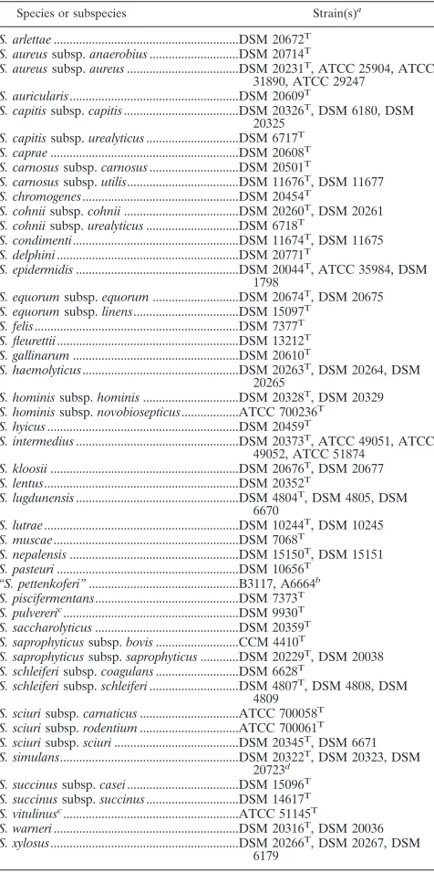
[image:2.585.302.540.87.565.2]Establishing the staphylococcal part of the RIDOM data-base service.A total of 81 staphylococcal type and reference strains including all validly described and recently proposed
TABLE 1. List of 5⬘16S rDNA-sequenced staphylococcal culture collection strains (n⫽81)
Species or subspecies Strain(s)a
S. arlettae...DSM 20672T
S. aureussubsp.anaerobius...DSM 20714T
S. aureussubsp.aureus...DSM 20231T
, ATCC 25904, ATCC 31890, ATCC 29247
S. auricularis...DSM 20609T
S. capitissubsp.capitis...DSM 20326T, DSM 6180, DSM
20325 S. capitissubsp.urealyticus...DSM 6717T
S. caprae...DSM 20608T
S. carnosussubsp.carnosus...DSM 20501T
S. carnosussubsp.utilis...DSM 11676T
, DSM 11677 S. chromogenes...DSM 20454T
S. cohniisubsp.cohnii...DSM 20260T
, DSM 20261 S. cohniisubsp.urealyticus...DSM 6718T
S. condimenti...DSM 11674T
, DSM 11675 S. delphini...DSM 20771T
S. epidermidis...DSM 20044T
, ATCC 35984, DSM 1798
S. equorumsubsp.equorum...DSM 20674T, DSM 20675
S. equorumsubsp.linens...DSM 15097T
S. felis...DSM 7377T
S. fleurettii...DSM 13212T
S. gallinarum...DSM 20610T
S. haemolyticus...DSM 20263T, DSM 20264, DSM
20265 S. hominissubsp.hominis...DSM 20328T
, DSM 20329 S. hominissubsp.novobiosepticus...ATCC 700236T
S. hyicus...DSM 20459T
S. intermedius...DSM 20373T
, ATCC 49051, ATCC 49052, ATCC 51874
S. kloosii...DSM 20676T, DSM 20677
S. lentus...DSM 20352T
S. lugdunensis...DSM 4804T, DSM 4805, DSM
6670 S. lutrae...DSM 10244T
, DSM 10245 S. muscae...DSM 7068T
S. nepalensis...DSM 15150T
, DSM 15151 S. pasteuri...DSM 10656T
“S. pettenkoferi”...B3117, A6664b
S. piscifermentans...DSM 7373T
S. pulvereric
...DSM 9930T
S. saccharolyticus...DSM 20359T
S. saprophyticussubsp.bovis...CCM 4410T
S. saprophyticussubsp.saprophyticus...DSM 20229T
, DSM 20038 S. schleiferisubsp.coagulans...DSM 6628T
S. schleiferisubsp.schleiferi...DSM 4807T
, DSM 4808, DSM 4809
S. sciurisubsp.carnaticus...ATCC 700058T
S. sciurisubsp.rodentium...ATCC 700061T
S. sciurisubsp.sciuri...DSM 20345T, DSM 6671
S. simulans...DSM 20322T, DSM 20323, DSM
20723d
S. succinussubsp.casei...DSM 15096T
S. succinussubsp.succinus...DSM 14617T
S. vitulinusc
...ATCC 51145T
S. warneri...DSM 20316T
, DSM 20036 S. xylosus...DSM 20266T
, DSM 20267, DSM 6179
a
T, type strain; ATCC, American Type Culture Collection, Manassas, Va.; CCM, C˘ eska´ sbirka mikroorganismu˚ (Czech Collection of Microorganisms); DSM, Deutsche Sammlung von Mikroorganismen und Zellkulturen (German Collection of Microorganisms and Cell Cultures), Braunschweig, Germany.
b
Courtesy of the authors, both strains were used to acknowledge the recently proposed but not yet validly described staphylococcal species “S. pettenkoferi” (36).
c
It was recently proposed that strains (including the type strain) ofS. pulvereri actually belong to the speciesS. vitulinus(26).
d
Strain NRRL B-2628 (DSM 20723), originally described as“S. staphylolyticus,” was classified as a biovar ofS. simulans(34).
on May 15, 2020 by guest
http://jcm.asm.org/
members of theStaphylococcusgenus were analyzed by 5⬘- 16S rDNA sequencing and used to establish a quality-controlled ribosomal sequence database. Most staphylococcal sequences exhibited a length of 464 bp, analyzing the nucleotides at po-sitions 54 to 510 (corresponding to the Escherichia coli 16S rDNA positions), except those of theS. sciuricluster (S. sciuri
subspecies,S. pulvereri,S. vitulinus,S. fleurettii, andS. lentus), which contained one additional guanine (E. coliposition 214). The mean pairwise distance of 52 sequences (all 37 type strains and otherStaphylococcusculture collection strains with unique sequences) was 4.7%, and the standard deviation was 1.5%. Assuming a normal distribution for the distances and choosing a reporting criterion ofⱖ98.70% similarity for a “distinct spe-cies” correlates with a statistical error probability of 1.0%.
Analyses of these sequences revealed that all of the type and reference strains were distinguishable at the species level, with the exception ofS. intermediusandS. delphini. In addition,S. pulvereri and S. vitulinus, recently proposed to be identical species (26), showed the same nucleotide sequences. At the subspecies level, the established subspecies ofS. carnosus,S. cohnii,S. hominis,S. schleiferi, andS. succinuscould be distin-guished. The type strains ofS. aureussubsp.aureusversusS. aureussubsp.anaerobius,S. capitissubsp.capitisversusS. ca-pitissubsp. urealyticus, S. equorum subsp. equorumversus S. equorum subsp. linens, S. saprophyticus subsp. saprophyticus
versusS. saprophyticus subsp.bovis, and S. sciuri subsp. car-naticusversusS. sciurisubsp.rodentiumwere indistinguishable in the range of the 5⬘16S rDNA fragment used. In contrast,S. sciurisubsp.sciuriwas distinguishable from both other subspe-cies of this spesubspe-cies.
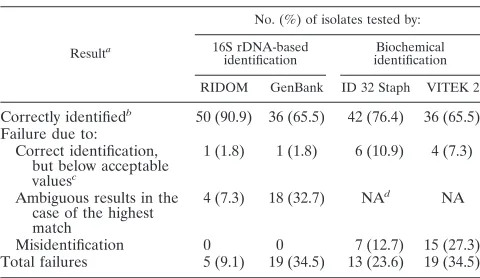
Comparison of RIDOM and NCBI database queries.A total of 55 clinical staphylococcal isolates were analyzed by 16S rDNA sequencing. Using the newly established RIDOM data-base, the isolates were assigned to 20 different species and subspecies (Tables 2 and 3). Except for isolate M08, with 98.49% similarity, all other isolates (98.2%) had a similarity score above the proposed threshold in the RIDOM database. Of the 55 isolates, 48 (87.3%) had sequences which gave a perfect match. Performing a similarity search against the NCBI database, a perfect match was yielded in 83.6% of cases.
At the species level, the RIDOM database contained a total of four ambiguities (Tables 3 and 4). In three of these cases, ambiguous results were caused by the nucleotide sequence identity ofS. intermediusandS. delphiniin the fragment ana-lyzed. In addition, isolate M55 yielded an ambiguous result in the RIDOM analysis, showing an identical percent similarity to two species (S. xylosusand S. saprophyticus; 99.35%). Minor ambiguities were found at the subspecies level forS. aureus,S. capitis, S. equorum, and S. saprophyticus. Analyzing the se-quences of the clinical isolates tested by the GenBank nucle-otide database at NCBI, a total of 18 ambiguities were ob-served: (i) S. aureus versus S. haemolyticus (n ⫽ 7), (ii) S. saprophyticusversusS. cohnii(n⫽2), (iii)S. cohniiversusS. succinus(n⫽2), (iv)S. intermediusversusS. delphini(n⫽3), (v)S. capitisversusS. capraeversusS. epidermidis(n⫽2), and (vi)S. capitis versusS. capraeversusS. arlettae(n⫽2). The NCBI results were not analyzed at the subspecies level because of a frequent lack of appropriate taxonomic indications in the database entries.
Isolates yielding RIDOM results ranging between the
threshold (98.7%) and a 100% match (n⫽6) were classified by the RIDOM database search asS. capitissubsp.capitis(isolate M13),S. caprae(isolate M14),S. warneri(isolate M52), andS. xylosus(isolates M54 and M56) (each with 99.78% similarity). In contrast, the NCBI database search resulted in ambiguous results for isolates M13 and M14 (Table 4). Regarding isolates M52, M54, and M56, the NCBI database search reached 99% similarity, exhibiting the same species affiliation as found by RIDOM.
Analyzing ambiguous or below-the-threshold RIDOM re-sults by chemotaxonomy and riboprinting.Concerning isolate M55, two results with the same similarity (99.35%) were of-fered by RIDOM, i.e.,S. xylosus and S. saprophyticussubsp.
saprophyticus(Table 4). The NCBI database showed a perfect match with S. xylosus. Major fatty acids were ai-C15:0 (12-methyltetradecanoic acid) followed by i-C15:0(13-methyl tet-radecanoic acid), i-C17:0 (15-methylhexadecanoic acid), and ai-C17:0 (14-methylhexadecanoic acid), indicating S. xylosus (similarity index, 0.779) or, with lower likelihood,S. gallinarum
(similarity index, 0.538). The peptidoglycan type of this isolate was A3␣L-Lys-Gly5–6(A11.2), which is in accordance withS.
xylosus. Also, riboprinting analysis confirmed the species to be
S. xylosus(data not shown).
Isolate M08, found to be marginally below the threshold, showed the highest similarity to S. haemolyticus when the RIDOM database was used (Table 4). Also, the NCBI data-base search yielded this species with 98.5% similarity. Further-more, both phenotypic test systems used resulted inS. haemo-lyticus. However, when the fatty acid profile was analyzed (main fatty acid components, ai-C15:0, C20:0, C18:0, and i-C15:0), an ambiguous result indicating eitherS. epidermidis(similarity index, 0.696) orS. haemolyticus (similarity index, 0.536) was found. In addition, the riboprinting pattern revealed grouping of this isolate into theS. aureus-S. epidermidis-S. warnericluster (data not shown). The peptidoglycan of this isolate was based on the diamino acid lysine and contained glycine and serine residues in its interpeptide bridge, which corresponds to the type A3␣L-Lys–Gly5–6–L-Ser1–2–Gly (A11.3), which is known
to be associated withS. haemolyticusbut also withS. epider-midisandS. warneri(not withS. aureus).
Comparison of RIDOM results with biochemical identifica-tion results. In a second assessment, the results of the 16S rDNA sequencing were compared to two phenotypic ap-proaches comprising the commercially available differentiation systems ID 32 Staph and VITEK 2 (Table 3).
Analyzing results obtained by the ID 32 Staph system, 39 (70.9%) of the isolates were identified at confidence levels from excellent (n⫽12; 21.8%) to very good (n⫽13; 23.6%) to good (n⫽14; 25.4%). Another six (10.9%) were identified but at a low level of discrimination categorized as acceptable. The ID 32 Staph system could not identify 10 (18.2%) of the 55 isolates tested because the %ID and/orTwas below acceptable levels. In comparison with the RIDOM results, three (5.4%) of the isolates categorized as good to acceptable by ID 32 Staph were incorrectly identified (Table 3). Overall, ID 32 Staph failed in the case of 13 (23.6%) of the isolates due to incorrect identification and/or categorization below acceptable values, although 11 results were at least in the correct genus (Table 3). However, three of them categorized as questionable by the software representedS. felisisolates, which are not part of the
on May 15, 2020 by guest
http://jcm.asm.org/
ID 32 Staph identification table. OneS. cohniiisolate and one
S. capitis (SCV phenotype) isolate were misidentified at the genus level as Micrococcus lylae and Kocuria rosea, respec-tively.
VITEK 2 identified 40 (72.7%) of the isolates at confidence levels of excellent (n⫽30; 54.5%), very good (n⫽5; 9.1%), and good (n⫽5; 9.1%). Two isolates (3.6%) were categorized as acceptable. Because of the %ID and/orT being below ac-ceptable levels, the VITEK 2 system could not identify 13 (23.6%) of the isolates. Although categorized as excellent to acceptable by VITEK 2, seven (12.7%) of the isolates were incorrectly identified compared with the RIDOM results (Ta-ble 3). Due to the lack of appropriate species data in the VITEK 2 database, allS. arlettae,S. caprae,S. equorum, andS. felis isolates (n ⫽ 8) were misidentified. Overall, VITEK 2 failed for 19 (34.5%) of the isolates due to incorrect identifi-cation and/or categorization below acceptable value, but 17 of the misidentifications were placed at least into the correct genus. Two S. epidermidis isolates both exhibiting the SCV phenotype were misidentified at the genus level as Kocuria varians.
Six SCV isolates included were found to belong toS. aureus
(n ⫽ 2), S. epidermidis (n ⫽ 3), and S. capitis (n ⫽ 1) by RIDOM analyses. Compared with the RIDOM results, both ID 32 Staph and VITEK 2 failed to identify two (S. aureusand
S. capitis) and four (S. aureus,S. capitis, andS. epidermidis[n
⫽2]) SCVs, respectively. The slow growth of SCVs and their reduced biochemical activity were responsible for three of the four misidentifications at the genus level when the commercial kits were used. The remaining case of genus misidentification using ID 32 Staph was caused by anS. cohniiisolate exhibiting a normal morphotype.
DISCUSSION
Since the first taxonomic description of the genus Staphylo-coccusby Rosenbach in 1884 (31), the division of this genus— based on colony pigment—into the two speciesS.(pyogenes)
aureusandS. albus(now referred to asS. epidermidis) primar-ily has been the basis to differentiate between the opportunistic pathogenS. aureus, which causes high morbidity and mortality, and other staphylococci generally considered to be harmless commensals or saprophytic bacteria. Using a variety of mor-phological criteria and physiological and biochemical tests, Baird-Parker created several schemes to differentiate between
S. epidermidisandS. saprophyticus, subdivided into subgroups or biotypes (2, 3). Despite the subsequent introduction of some more species considered to be part of the family Micrococ-caceae(19) and their differentiation in particular by Kloos and Schleifer (18), it soon became obvious that more staphylococ-cal species are associated with humans and animal species than had been recognized so far. Therefore, the genus Staphylococ-cuswas enlarged to 37 validly described species, including 21 subspecies, according to the currentList of Bacterial Names with Standing in Nomenclature, updated 24 January 2004 (9). To take into account the recent results of molecular phyloge-netic classification, it was proposed to reclassify the genus
Staphylococcusinto a family“Staphylococcaceae”(order Bacil-lales, class“Bacilli,”phylumFirmicutes) (10).
While conventional differentiation schemes based on physi-ological and biochemical tests are relatively cumbersome and time-consuming and require various approaches, the
commer-TABLE 2. Species and subspecies of the clinical staphylococcal isolates (n⫽55) as determined by the RIDOM database
Species or subspecies tested
(no. of isolates) Species or subspecies indistinguishable(no. of isolates)
S. arlettae(1)
S. aureussubsp.aureus(7) ...S. aureussubsp.anaerobius(7)a
S. capitissubsp.capitis(2) ...S. capitissubsp.urealyticus(2)b
S. caprae(2) S. chromogenes(1) S. cohniisubsp.cohnii(2) S. epidermidis(10)
S. equorumsubsp.equorum(2)...S. equorumsubsp.linens(2)c
S. felis(3) S. haemolyticus(3)
S. hominissubsp.novobiosepticus (1)
S. hyicus(1)
S. intermedius(3) ...S. delphini(3)d
S. lugdunensis(2)
S. saprophyticussubsp.bovis(2) S. schleiferisubsp.schleiferi(2) S. sciurisubsp.sciuri(3) S. simulans(1) S. warneri(3)
S. xylosus(4)...S. saprophyticussubsp.saprophyticus(1)e
aSince the isolates tested were collected from human specimens, showing a
positive catalase reaction and growing aerobically without exception, the pres-ence of isolates of the sheep-adapted, catalase-negative, anaerobic subspeciesS. aureussubsp.anaerobiuscould be excluded.
bSince isolates tested showed negative urease activity and were unable to
produce acid aerobically from maltose, the presence ofS. capitissubsp. urealyti-cusisolates is improbable.
cSince isolates tested produced acid aerobically from maltose,D-lactose,
D-trehalose, andD-ribose, the presence ofS. equorumsubsp.linens isolates is improbable.
dSince isolates tested were collected from human and canine specimens, the
presence of isolates of the strictly dolphin-adapted subspeciesS. delphiniis most improbable.
eOne isolate (M55), which gave the same similarity (99.35%) forS. xylosusand
[image:4.585.44.281.86.279.2]S. saprophyticussubsp.saprophyticus, was chemotaxonomically and by riboprint-ing confirmed asS. xylosus.
TABLE 3. Results of identification of clinical staphylococcal isolates (n⫽55) by 16S rDNA differentiation-based systems
compared to biochemical assays
Resulta
No. (%) of isolates tested by: 16S rDNA-based
identification
Biochemical identification RIDOM GenBank ID 32 Staph VITEK 2
Correctly identifiedb 50 (90.9) 36 (65.5) 42 (76.4) 36 (65.5) Failure due to:
Correct identification, but below acceptable valuesc
1 (1.8) 1 (1.8) 6 (10.9) 4 (7.3)
Ambiguous results in the case of the highest match
4 (7.3) 18 (32.7) NAd NA
Misidentification 0 0 7 (12.7) 15 (27.3) Total failures 5 (9.1) 19 (34.5) 13 (23.6) 19 (34.5)
aBased on species level.
bFor biochemical assays, classification is based on a %ID that is acceptable or
better (%IDⱖ80.0) and on aTindex value that is acceptable or better (Tⱖ0); for 16S rDNA-based identification, classification is based on percent similarity (%Sⱖ98.5) to the 16S rDNA sequence of the type and reference strains.
cFor biochemical assays, classification is based on a %ID that is below
ac-ceptable (%ID⬍80.0) and on aTvalue that is below acceptable (T⫽0); for 16S rDNA-based identification, classification is based on a %S of⬍98.5%.
dNA, not applicable.
on May 15, 2020 by guest
http://jcm.asm.org/
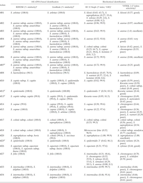
[image:4.585.301.541.96.235.2]TABLE 4. Clinical staphylococcal isolates with dissenting or ambiguous results in identification
Isolate
16S rDNA-based identification Biochemical identification
RIDOM (% similarity)a GenBank (% similarity)b ID 32 Staph (Tindex; %ID)c VITEK 2 (Tindex;
selectivity)c
M01 S. arlettae(100.0) S. arlettae(100.0) S. klosii(0.65; 63.7),S. saprophyticus(0.27; 33.4),
S. xylosus(0.29; 2.6),S. warneri(0.00; 0.2)
S. kloosii(0.37; good)
M02 S. aureussubsp.aureus(100.0),
S. aureussubsp.anaerobius
(100.0)
S. aureussubsp.aureus(100.0),
S. aureus(100.0),S. haemolyticus(100.0)
S. aureus(0.96; 99.9) S. aureus(0.97; excellent)
M03 S. aureussubsp.aureus(100.0),
S. aureussubsp.anaerobius
(100.0)
S. aureussubsp.aureus(100.0),
S. aureus(100.0),S. haemolyticus(100.0)
S. aureus(0.63; 99.9) S. aureus(1.0; excellent)
M04 S. aureussubsp.aureus(100.0),
S. aureussubsp.anaerobius
(100.0)
S. aureussubsp.aureus(100.0),
S. aureus(100.0),S. haemolyticus(100.0)
S. aureus(0.52; 93.0) S. aureus(0.65; very good)
M05d S. aureussubsp.aureus(100.0),
S. aureussubsp.anaerobius
(100.0)
S. aureussubsp.aureus(100.0),
S. aureus(100.0),S. haemolyticus(100.0)
S. cohniisubsp.cohnii
(0.23; 64.7),S. caprae
(0.21; 30.6),S. hominis2e (0.0; 3.2)
S. hyicus(0.42; poor),S. chromogenes(0.33; poor)
M06 S. aureussubsp.aureus(100.0),
S. aureussubsp.anaerobius
(100.0)
S. aureussubsp.aureus(100.0),
S. aureus(100.0),S. haemolyticus(100.0)
S. aureus(0.72; 99.9) S. aureus(0.86; excellent)
M07d S. aureussubsp.aureus(100.0),
S. aureussubsp.anaerobius
(100.0)
S. aureussubsp.aureus(100.0),
S. aureus(100.0),S. haemolyticus(100.0)
S. aureus(0.72; 99.9) S. aureus(0.25; good)
M08 S. haemolyticus(98.5) S. haemolyticus(98.5) S. haemolyticus(0.45; 76.9),
S. warneri(0.37; 22.4),S. hominis(0.04; 0.6)
S. haemolyticus(0.89; excellent)
M10 S. capitissubsp.S. capitis
(100.0)
S. capitis(100.0),S. epidermidis
(100.0),S. caprae(100.0)
S. capitis(0.82; 99.9) S. capitis(0.59; poor),
Kocuria rosea(0.50; poor),S. cohniisubsp.
cohnii(0.49; poor) M12d S. epidermidis(100.0) S. epidermidis(100.00) S. epidermidis1e(0.54; 83.3) Kocuria varians(0.44;
good) M13d S. capitissubsp.capitis(99.8) S. capitis(99.8),S. epidermidis
(99.8),S. caprae(99.8)
Kocuria rosea(0.95; 81.5) S. chromogenes(0.49; poor),S. auricularis
(0.43; poor) M14 S. caprae(99.8) S. caprae(99.8),S. capitis
(99.8),S. arlettae(99.8)
S. caprae(0.58; 99.6) S. chromogenes(0.44; good)
M15 S. caprae(100.0) S. caprae(100.0),S. capitis
(100.0),S. arlettae(100.0)
S. caprae(0.22; 97.4) S. chromogenes(0.36; poor),S. aureus(0.35; poor),S. warneri(0.29; poor)
M17 S. cohniisubsp.cohnii(100.0) S. cohnii(100.0),S. saprophyticus(100.0)
S. cohniisubsp.cohnii
(0.33; 99.3)
S. cohniisubsp.cohnii
(0.56; poor),S. saprophyticus(0.54; poor)
M18 S. cohniisubsp.cohnii(100.0) S. cohnii(100.0),S. saprophyticus(100.0)
Micrococcus lylae(0.52; 96.9)
S. cohniisubsp.urealyticus
(0.77; excellent) M20 S. saprophyticussubsp.bovis
(100.0)
S. cohnii(99.8),S. succinus
(99.8)
S. saprophyticus(1.00; 99.9) S. saprophyticus(1.00; excellent)
M25d S. epidermidis(100.0) S. epidermidis(100.0) S. epidermidis2e(0.62; 86.1) Kocuria varians(0.24; poor)
M26 S. equorumsubsp.equorum
(100.0),S. equorumsubsp.
linens(100.0)
S. equorum(100.0),S. equorum
subsp.linens(100.0)
S. equorum(0.31; 97.6) S. xylosus(0.44; good)
M27 S. felis(100.0) S. felis(100.0) S. intermedius(0.31; 48.6),
S. chromogenes(0.32; 25.9),S. xylosus(0.43; 15.4),S. simulans(0.29; 10.1),S. aureus(0.00; 0.1)
S. chromogenes(0.78; poor),S. schleiferi
(0.70; poor)
M33 S. intermedius(100.0),S. delphini(100.0)
S. intermedius(100.0),S. delphini(100.0)
S. intermedius(0.99; 99.7) S. intermedius(0.66; poor),S. chromogenes
(0.61; poor) M34 S. intermedius(100.0),S.
delphini(100.0)
S. intermedius(100.0),S. delphini(100.0)
S. intermedius(0.46; 95.4) S. intermedius(0.66; poor),S. chromogenes
(0.61; poor)
Continued on following page
on May 15, 2020 by guest
http://jcm.asm.org/
cial “rapid” identification systems share the problems of failure to identify commonly encountered bacteria, uselessness in identifying uncommon isolates, and lack of adequate (refer-ence) strains in the accompanying databases (30, 35). Further-more, commercial systems may provide ambiguous results, pre-senting two or more suggestions for identification with a comparable safety level.
With the advent of molecular biology techniques, investiga-tions based on comparative DNA sequence analysis of the genes of conserved macromolecules have become common-place in microbiology as a tool for classification of microbial organisms. Presently, the most useful and extensively investi-gated taxonomic marker molecules are the larger rRNAs and their corresponding genes, respectively, especially 16S rRNAs and to a lesser extent 23S rRNAs. However, analysis of rDNA sequences does not necessarily show agreement with charac-terizations based on classical taxonomic methods. While mo-lecular identification based on sequence analysis of universal targets offers several advantages, such as improved accuracy and short turnaround time, some drawbacks in currently avail-able databases should be noted. The deficiencies of publicly accessible databases without controlled input in the contents include the presence of faulty and/or redundant sequence en-tries, ragged sequence ends, outdated nomenclature, and un-available quality assurance. Since “gold standard” sequences are lacking, we established a quality-controlled sequence
da-tabase based on 81 type and reference strains encompassing all species and subspecies of the genusStaphylococcusrecognized as valid and the recently proposed candidate species“S. pet-tenkoferi”(36).
Analyses of the newly determined sequences of the 5⬘end of the 16S rDNA revealed that all type and reference strains were distinguishable at the species level, with two exceptions: (i)S. intermedius versus S. delphini and (ii) S. pulvereri versus S. vitulinus. Whereas it has been recently proposed that the strains assigned to the last two species belong to the same species (26), the coagulase-positive speciesS. intermediusand
S. delphinirepresent well-accepted different species. Compar-ing their 16S rDNA regions deposited in the EMBL database (accession numbers AB009938 and D83369, respectively), downstream of the fragment analyzed here, five base ex-changes between both sequences were noted, which may be useful for differentiation between these species if needed. Whereas the chaperonin 60 gene (hsp60) as sequencing target does not allow discrimination betweenS. intermedius and S. delphini, the sequencing of a manganese-dependent superox-ide dismutase gene (sodA) fragment was able to differentiate between them (11, 29).
[image:6.585.48.541.78.362.2]At the subspecies level, sequencing of the 5⬘ 16S rDNA fragment was discriminative enough to differ specifically be-tween the type strains of the established subspecies ofS. car-nosus, S. cohnii,S. hominis,S. schleiferi, andS. succinus. Our
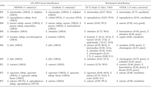
TABLE 4—Continued
Isolate
16S rDNA-based identification Biochemical identification
RIDOM (% similarity)a GenBank (% similarity)b ID 32 Staph (Tindex; %ID)c VITEK 2 (Tindex; selectivity)c
M35 S. intermedius(100.0),S. delphini
(100.0)
S. intermedius(100.0),S. delphini
(100.0)
S. intermedius(0.67; 98.0) S. intermedius(0.75; excellent)
M39 S. saprophyticussubsp.bovis
(100.0)
S. cohnii(99.8),S. succinus(99.8) S. saprophyticus(0.83; 99.9) S. saprophyticus(0.91; excellent)
M40 S. aureussubsp.aureus(100.0),S. aureussubsp.anaerobius
(100.0)
S. aureussubsp.aureus(100.0),S. aureus(100.0),S. haemolyticus
(100.0)
S. aureus(0.69; 99.3) S. aureus(0.50; very good)
M46 S. simulans(100.0) S. simulans(100.0) S. simulans(0.73; 98.6) S. haemolyticus(0.48; poor),S. simulans(0.46; poor) M47 S. hominissubsp.novobiosepticus
(100.0)
S. hominis(100.0) S. hominis1e(0.21; 65.9),S.
warneri(0.18; 17.0),S. epidermidis2 (0.15; 10.9)
S. capitis(0.86; excellent)
M48 S. felis(100.0) S. felis(100.0) S. xylosus(0.58; 60.8),S. intermedius(0.30; 22.6),
S. simulans(0.36; 14.8),
S. chromogenes(0.16; 1.7),S. aureus(0.00; 0.1)
S. simulans(0.60; poor),S. chromogenes(0.53; poor)
M49 S. felis(100.0) S. felis(100.0) S. simulans(0.65; 83.3) S. chromogenes(0.53; poor),S. schleiferi(0.45; poor) M51 S. warneri(100.0) S. warneri(100.0) S. warneri(0.76; 99.9) S. warneri(0.46; poor),S.
haemolyticus(0.46; poor),S. kloosii(0.39; poor) M53 S. equorumsubsp.equorum
(100.0),S. equorumsubsp.
linens(100.0)
S. equorum(100.0),S. equorum
subsp.linens(100.0)
S. equorum(0.68; 66.0),S. xylosus(0.74; 34.0),S. sciuri(0.03; 0.1)
S. xylosus(0.88; excellent)
M55 S. xylosus(99.35),S. saprophyticus
subsp.saprophyticus(99.35)
S. xylosus(100.0) S. xylosus(0.89; 99.2) S. xylosus(0.90; excellent)
aPercent similarity compared to the 16S rDNA sequence of the type strain of the staphylococcal taxon. bResults of redundant database entries as well as of entries without taxonomic classification were not included.
cIn the case of identification with low discrimination below acceptable values as given by the biochemical identification kits, i.e., a percent identification accuracy
(%ID) of⬍80.0 (as specified by ID 32 Staph) and poor selectivity (as specified by VITEK 2), all results are listed; otherwise, only results with the highest %ID and best selectivity, respectively, are shown.
dIsolate exhibiting the SCV phenotype.
eThe numeral identifies the biotype.
on May 15, 2020 by guest
http://jcm.asm.org/
results confirm that 16S rDNA might be a more discriminative target sequence than other ribosomal regions or target genes to differentiate among staphylococcal subspecies. Neither the 16S-23S rDNA intergenic spacer region nor genes such as
hsp60orsodAallow discrimination at the subspecies level (20, 24, 29).
Using 55 clinical staphylococcal isolates, the 16S rDNA-derived results obtained by using the RIDOM database were queried with NCBI database entries and compared to results obtained by using commercial “rapid” identification systems. The range of clinical isolates was chosen in such a manner that a broad diversity of different human and animal species was covered and examples of diagnostically challenging isolates such as phenotypic variants were acknowledged. However, this selection does not reflect the typical distribution of staphylo-coccal (sub)species in a human or veterinary routine microbi-ological laboratory.
Overall, the RIDOM database was proven to be particularly suitable for differentiation of staphylococcal isolates belonging to common as well as rare species and different phenotypes. When analyzing the sequence of isolate M08, both RIDOM and NCBI database queries resulted in the same percent sim-ilarity below the threshold with a best match indicating S. haemolyticus. Whereas the VITEK 2 system displayedS. hae-molyticus, the ID 32 Staph result was ambiguous, as were the results of chemotaxonomic and riboprinting analyses. Thus, the diversity of the 16S rDNA sequence of this species might be broader than is currently appreciated or the proposal of new subspecies to accommodate this isolate should be considered. Searching the NCBI database gave numerous ambiguous results not only for CoNS but also forS. aureus, which dis-played a perfect match with anS. haemolyticusentry (accession no. Z26896). This obviously incorrect database entry highlights the common presence of faulty sequence entries.
Apart from staphylococcal isolates with the SCV phenotype, phenotypically normal clinical isolates were also misidentified or showed ambiguous results if the commercial kits were used. This was shown for species seldom associated with infections and was also shown for commonly encountered staphylococcal species. This is of particular importance, as shown here, if the kits suggested good or better identification accuracy but indeed failed in species (and in one case also in genus) identification. Comparing the results of the two commercial kits used, the VITEK 2 kit showed more misidentifications, mainly due to the limited database of gram-positive cocci (19 staphylococcal species) included in the ID-GBP card, whereas ID 32 Staph covers 24 Staphylococcus species. Beside the utmost impor-tance ofS. aureusidentification, assured identification results, at least at the species level, are vital for differentiation of the expanding number of CoNS (sub)species increasingly charac-terized as emerging pathogens (22, 40) and also recently re-covered as food-fermenting microorganisms, which may have to be considered relevant to food safety and quality (27, 28). Differentiation up to the species level may also have substantial consequences for the management of patients in the case of CoNS. Since infectious endocarditis caused byS. lugdunensisis usually associated with left-side valvular disruption and life-threatening embolic complications, correct identification of this aggressive species is critical to distinguish it from other CoNS (17).
In general, the RIDOM approach (http://www.ridom -rdna.de) based on quality-controlled sequence entries offers a comprehensive and validated sequence database (15) which has already been shown to be useful for species belonging to theNeisseriaceaeandMoraxellaceaeas well as for Mycobacte-riumand Nocardiaspecies (13, 16, 23). The RIDOM system has aNeisseriaceaeandMycobacteriumdata set for demonstra-tion purposes, and an increase in the number of entries for other microorganisms is in progress.
In summary, the 5⬘16S rDNA fragment used to establish the staphylococcal part of the RIDOM database contains enough information suitable for the identification of almost all staph-ylococci at the species level and, with some exceptions, also at the subspecies level. Furthermore, recognition of hitherto un-described species may be possible. The increasing feasibility of high-throughput sequencing, accompanied by falling costs, suggests that 16S rDNA sequencing holds promise as a rapid alternative to biochemical and other phenotypic procedures for differentiation of pathogens. In contrast to public data-bases, which are characterized by a policy of unchecked se-quence input (especially disadvantageous for diagnostic pur-poses), quality-controlled sequence databases such as RIDOM, based only on type and well-characterized reference strains, may prove to be of maximum benefit for identification purposes. A substantial increase in the number of RIDOM entries and their continuing updates will further improve the value of this database and will broaden its possible fields of application.
ACKNOWLEDGMENTS
We are grateful to Martina Schulte, Susanne Weber, Ursula Keck-evoet, and Anika Vester for excellent technical assistance.
This work was supported by a grant from BMBF (Pathogenomic Network).
REFERENCES
1.Allerberger, F., and S. J. Fritschel.1999. Use of automated ribotyping of AustrianListeria monocytogenesisolates to support epidemiological typing. J. Microbiol. Methods35:237–244.
2.Baird-Parker, A. C.1963. A classification of micrococci and staphylococci based on physiological and chemical tests. J. Gen. Microbiol.30:409–427. 3.Baird-Parker, A. C.1974. The basis for the present classification of
staphy-lococci and micrococci. Ann. N. Y. Acad. Sci.236:7–14.
4.Becker, K., A. W. Friedrich, G. Lubritz, M. Weilert, G. Peters, and C. von Eiff.2003. Prevalence of genes encoding pyrogenic toxin superantigens and exfoliative toxins among strains ofStaphylococcus aureusisolated from blood and nasal specimens. J. Clin. Microbiol.41:1434–1439.
5.Becker, K., G. Haverka¨mper, C. von Eiff, R. Roth, and G. Peters.2001. Survey of staphylococcal enterotoxin genes, exfoliative toxin genes, and toxic shock syndrome toxin 1 gene in non-Staphylococcus aureusspecies. Eur. J. Clin. Microbiol. Infect. Dis.20:407–409.
6.Becker, K., B. Keller, C. von Eiff, M. Bru¨ck, G. Lubritz, J. Etienne, and G. Peters.2001. Enterotoxigenic potential ofStaphylococcus intermedius. Appl. Environ. Microbiol.67:5551–5557.
7.Benson, D. A., I. Karsch-Mizrachi, D. J. Lipman, J. Ostell, and D. L. Wheeler.2004. GenBank: update. Nucleic Acids Res.32(Database issue):
D23–D26.
8.Brakstad, O. G., K. Aasbakk, and J. A. Maeland.1992. Detection of Staph-ylococcus aureusby polymerase chain reaction amplification of thenucgene. J. Clin. Microbiol.30:1654–1660.
9.Euze´by, J. P.1997. List of bacterial names with standing in nomenclature: a folder available on the Internet. Int. J. Syst. Bacteriol.47:590–592. 10.Garrity, G. M., and J. G. Holt.2001. The road map to the manual, p.
119–166. InD. R. Boone, R. W. Castenholz, and G. M. Garrity (ed.), Bergey’s manual of systematic bacteriology, 2nd ed. Springer-Verlag, New York, N.Y.
11.Goh, S. H., Z. Santucci, W. E. Kloos, M. Faltyn, C. G. George, D. Driedger, and S. M. Hemmingsen.1997. Identification ofStaphylococcusspecies and subspecies by the chaperonin 60 gene identification method and reverse checkerboard hybridization. J. Clin. Microbiol.35:3116–3121.
on May 15, 2020 by guest
http://jcm.asm.org/
12.Groth, I., P. Schumann, K. Martin, B. Schuetze, K. Augsten, I. Kramer, and E. Stackebrandt.1999.Ornithinicoccus hortensisgen. nov., sp. nov., a soil actinomycete which containsL-ornithine. Int. J. Syst. Bacteriol.49:1717– 1724.
13.Harmsen, D., S. Dostal, A. Roth, S. Niemann, J. Rothga¨nger, M. Sammeth, J. Albert, M. Frosch, and E. Richter.2003. RIDOM: comprehensive and public sequence database for identification ofMycobacteriumspecies. BMC Infect. Dis.3:26.
14.Harmsen, D., and H. Karch.2004. 16S rDNA for diagnosing pathogens: a living tree. ASM News70:19–24.
15.Harmsen, D., J. Rothga¨nger, M. Frosch, and J. Albert.2002. RIDOM: ribosomal differentiation of medical micro-organisms database. Nucleic Ac-ids Res.30:416–417.
16.Harmsen, D., C. Singer, J. Rothga¨nger, T. Tønjum, G. S. de Hoog, H. Shah, J. Albert, and M. Frosch.2001. Diagnostics ofNeisseriaceaeand Moraxel-laceaeby ribosomal DNA sequencing: ribosomal differentiation of medical microorganisms. J. Clin. Microbiol.39:936–942.
17.Jones, R. M., M. A. Jackson, C. Ong, and G. K. Lofland.2002. Endocarditis caused byStaphylococcus lugdunensis. Pediatr. Infect. Dis. J.21:265–268. 18.Kloos, W. E., and K. H. Schleifer. 1975. Simplified scheme for routine
identification of humanStaphylococcusspecies. J. Clin. Microbiol.1:82–88. 19.Kloos, W. E., and K. H. Schleifer.1986. Genus IV.Staphylococcus Rosen-bach 1884, 18AL(Nom. Cons. Opin. 17 Jud. Comm. 1958, 153), p. 1013–1035.
InP. H. A. Sneath, N. S. Mair, M. E. Sharpe, and J. G. Holt (ed.), Bergey’s manual of systematic bacteriology, vol. 2. The Williams & Wilkins Co., Baltimore, Md.
20.Kwok, A. Y., S. C. Su, R. P. Reynolds, S. J. Bay, Y. Av-Gay, N. J. Dovichi, and A. W. Chow. 1999. Species identification and phylogenetic relationships based on partial HSP60 gene sequences within the genusStaphylococcus. Int. J. Syst. Bacteriol.49:1181–1192.
21.Lane, D. J.1991. 16S/23S rRNA sequencing, p. 115–175.InE. Stackebrandt and M. Goodfellow (ed.), Nucleic acid techniques in bacterial systematics. John Wiley & Sons, Inc., Chichester, United Kingdom.
22.McGahee, W., and F. D. Lowy.2000. Staphylococcal infections in the inten-sive care unit. Semin. Respir. Infect.15:308–313.
23.Mellmann, A., J. L. Cloud, S. Andrees, K. Blackwood, K. C. Carroll, A. Kabani, A. Roth, and D. Harmsen.2003. Evaluation of RIDOM, MicroSeq, and Genbank services in the molecular identification ofNocardiaspecies. Int. J. Med. Microbiol.293:359–370.
24.Mendoza, M., H. Meugnier, M. Bes, J. Etienne, and J. Freney.1998. Iden-tification ofStaphylococcusspecies by 16S-23S rDNA intergenic spacer PCR analysis. Int. J. Syst. Bacteriol.48:1049–1055.
25.Miller, L. T.1982. Single derivatization method for routine analysis of bacterial whole-cell fatty acid methyl esters, including hydroxy acids. J. Clin. Microbiol.16:584–586.
26.Petra´s˘, P.1998.Staphylococcus pulvereri⫽Staphylococcus vitulus? Int. J. Syst. Bacteriol.48:617–618.
27.Place, R. B., D. Hiestand, S. Burri, and M. Teuber.2002.Staphylococcus succinussubsp.caseisubsp. nov., a dominant isolate from a surface ripened cheese. Syst. Appl. Microbiol.25:353–359.
28.Place, R. B., D. Hiestand, H. R. Gallmann, and M. Teuber.2003. Staphylo-coccus equorumsubsp.linens, subsp. nov., a starter culture component for surface ripened semi-hard cheeses. Syst. Appl. Microbiol.26:30–37. 29.Poyart, C., G. Quesne, C. Boumaila, and P. Trieu-Cuot.2001. Rapid and
accurate species-level identification of coagulase-negative staphylococci by using thesodAgene as a target. J. Clin. Microbiol.39:4296–4301. 30.Renneberg, J., K. Rieneck, and E. Gutschik.1995. Evaluation of Staph ID 32
system and Staph-Zym system for identification of coagulase-negative staph-ylococci. J. Clin. Microbiol.33:1150–1153.
31.Rosenbach, F. J.1884. Micro-Organismen bei Wund-Infections-Krankheiten des Menschen, p. 1–122. J. F. Bergmann, Wiesbaden, Germany. 32.Sammeth, M., J. Rothga¨nger, W. Esser, J. Albert, J. Stoye, and D. Harmsen.
2003. QAlign: quality-based multiple alignments with dynamic phylogenetic analysis. Bioinformatics19:1592–1593.
33.Schleifer, K. H., and O. Kandler.1972. Peptidoglycan types of bacterial cell walls and their taxonomic implications. Bacteriol. Rev.36:407–477. 34.Sloan, G. L., J. M. Robinson, and W. E. Kloos. 1982. Identification of
“Staphylococcus staphylolyticus”NRRL B-2628 as a biovar ofStaphylococcus simulans. Int. J. Syst. Bacteriol.32:170–174.
35.Spanu, T., M. Sanguinetti, D. Ciccaglione, T. D’Inzeo, L. Romano, F. Leone, and G. Fadda.2003. Use of the VITEK 2 system for rapid identification of clinical isolates of staphylococci from bloodstream infections. J. Clin. Micro-biol.41:4259–4263.
36.Tru¨lzsch, K., H. Rinder, J. Trcek, L. Bader, U. Wilhelm, and J. Heesemann.
2002.“Staphylococcus pettenkoferi,”a novel staphylococcal species isolated from clinical specimens. Diagn. Microbiol. Infect. Dis.43:175–182. 37.von Eiff, C., and K. Becker.2003. Small colony variants: another mechanism
by whichStaphylococcus aureuscan evade the immune response and anti-microbial therapy, p. 253–273.InA. C. Fluit and F.-J. Schmitz (ed.), MRSA: current perspectives. Caister Academic Press, Wymondham, United King-dom.
38.von Eiff, C., K. Becker, K. Machka, H. Stammer, and G. Peters for the Study Group.2001. Nasal carriage as a source ofStaphylococcus aureusbacteremia. N. Engl. J. Med.344:11–16.
39.von Eiff, C., D. Bettin, R. A. Proctor, B. Rolauffs, N. Lindner, W. Winkel-mann, and G. Peters.1997. Recovery of small colony variants of Staphylo-coccus aureusfollowing gentamicin bead placement for osteomyelitis. Clin. Infect. Dis.25:1250–1251.
40.von Eiff, C., G. Peters, and C. Heilmann.2002. Pathogenesis of infections due to coagulase-negative staphylococci. Lancet Infect. Dis.2:677–685. 41.von Eiff, C., R. R. Reinert, M. Kresken, J. Brauers, D. Hafner, and G. Peters
for the Multicenter Study on Antibiotic Resistance in Staphylococci and Other Gram-Positive Cocci Study (MARS) Group.2000. Nationwide Ger-man multicenter study on prevalence of antibiotic resistance in staphylococ-cal bloodstream isolates and comparative in vitro activities of quinupristin-dalfopristin. J. Clin. Microbiol.38:2819–2823.
42.von Eiff, C., P. Vaudaux, B. C. Kahl, D. Lew, S. Emler, A. Schmidt, G. Peters, and R. A. Proctor.1999. Bloodstream infections caused by small-colony variants of coagulase-negative staphylococci following pacemaker implanta-tion. Clin. Infect. Dis.29:932–934.