0095-1137/05/$08.00⫹0 doi:10.1128/JCM.43.12.6189–6193.2005
Copyright © 2005, American Society for Microbiology. All Rights Reserved.
Use of Microelectronic Array Technology for Rapid Identification
of Clinically Relevant Mycobacteria
Maurizio Sanguinetti,
1*§ Linda Novarese,
1§ Brunella Posteraro,
1§ Stefania Ranno,
1Elena De Carolis,
1Giovanni Pecorini,
1Barbara Lucignano,
2Fausta Ardito,
1and Giovanni Fadda
1Istituto di Microbiologia, Universita` Cattolica del Sacro Cuore,1and Laboratorio di Microbiologia, Ospedale Pediatrico “Bambino Gesu`,”2Rome, Italy
Received 30 May 2005/Returned for modification 1 August 2005/Accepted 6 September 2005
We developed a new method based on the Nanochip microelectronic array technology for identification of various clinically relevant mycobacterial species. PCR-amplified rRNA genes obtained from 270 positive Mycobacteria Growth Indicator Tube cultures were successfully tested by hybridizing them with species-selective probes, and the results agreed with those of conventional identification methods. The system is rapid and accurate and opens new perspectives in clinical diagnostics.
Mycobacteria are slow-growing microorganisms causing a va-riety of infections that involve lung, skin, soft tissue, skeletal sys-tem, and lymph nodes in immunocompromised and immunocom-petent patients (5). Among them,Mycobacterium tuberculosisis the most common pathogen, causing 1.7 million deaths a year worldwide according to 2003 World Health Organization esti-mates (http://www.who.int/mediacenter/factsheets/fs104/en/print .html). Rapid and accurate identification of these microorganisms at the species level is important to establish a prompt and appro-priate therapy, considering that antibiotic treatment may vary according to the species encountered (2). Molecular assays that offer an alternative approach to time-consuming culture-based methods have recently been developed to improve identification of mycobacteria (14).
In this study, we developed a new method, based on micro-electronic chip array technology, to rapidly identify a variety of clinically relevant mycobacterial species by use of the Nano-chip Molecular Biology Workstation (Nanogen, Inc., San Di-ego, CA). The instrument uses a proprietary semiconductor microchip, which incorporates a 10-by-10 array of microelec-trodes coated with a permeation layer containing streptavidin, for the rapid transport and concentration of negatively charged nucleic acid molecules through the selective application of a positive electronic bias to selected test sites. The nucleic acid may then be immobilized by direct attachment to the permeation layer or by hybridization to a previously addressed nucleic acid. This technique has been used for single-nucleotide polymorphism assays using fluorescently labeled reporter probes and biotinyl-ated amplified DNA samples (4).
A 205-bp highly conserved region of theMycobacterium16S rRNA gene was amplified from genomic DNA samples using the pMyc14 (5⬘-biotinylated) and pMyc7 primers and PCR conditions as previously described (7). A small amount of biotinylated PCR product was electronically deposited and
anchored by streptavidin linkage on the microchip surface, denatured in situ, and hybridized with 13 dye-labeled oligonu-cleotide probes, seven of which were selective forM. tubercu-losiscomplex,M. avium,M. xenopi,M. gordonae,M. terrae,M. triplex, andM. malmoense, respectively. The other six probes were each selective for two or multiple mycobacterial species, due to the high similarity between their PCR-amplified 16S rRNA gene fragments (Table 1).
Almost of these species are very closely related:M. intracel-lulareversusM. chimaera(17),M. chelonaeversusM. abscessus
andM. massiliense(1); andM. kansasiiversusM. gastri(8), or belong to the same taxonomic group:M. fortuitumversusM. porcinum,M. neworleansense, andM. boenickei(12) (Table 1). Probes were designed by the use of Vector NTI Advance 9.0 software (Invitrogen, Inc., Milan, Italy), taking into account their secondary structure, C⫹G content, length, and melting temperature. In addition, a general probe, specific for the genusMycobacterium, was included as a positive control. The specificity of these probes was assessed by hybridization with DNA from bacterial species belonging to the genera Coryne-bacterium, Nocardia, and Rhodococcus, which are closely re-lated to the genusMycobacterium. Probes were coupled on the basis of their melting temperature and synthesized so that the oligonucleotides of each pair contained either a 5⬘-Cy3 or 5⬘-Cy5 fluorophore (Table 1). The probe pairs were used in consecutive hybridization runs.
Amplicon addressing (the process by which each amplicon was electronically placed in the microchip) was performed following the Nanogen User’s Guide instructions. Briefly, a 96-well microtiter plate was filled as follows: one well contained 60l of 0.3 M NaOH for sample passive (not electronic) pre-treatment; another well contained 60l of 50 mM histidine (30l of 100 mM histidine plus 30l of high-performance liquid chro-matography-grade water) as the background control; the other wells each contained one sample to be processed (15l of PCR product, 15l of high-performance liquid chromatography-grade water, and 30l of 100 mM histidine); the last well contained 60
l of 0.1 M NaOH for sample passive denaturation. Then, the microtiter plate and Nanochip cartridge were put into the
* Corresponding author. Mailing address: Istituto di Microbiologia, Largo F. Vito, 1, 00168 Rome, Italy. Phone: 39 630154964. Fax: 39 63051152. E-mail: [email protected].
§ These authors contributed equally to this work.
6189
on May 15, 2020 by guest
http://jcm.asm.org/
loader, and each purified amplicon was addressed to a specific test site on the cartridge and analyzed.
Prior to starting, the conductivity of the histidine buffer and each purified amplicon was checked; samples with a conductivity not exceeding 100S/cm were passed for load-ing. When addressing was completed, the cartridge was re-moved from the loader and processed for hybridization. Hybridization solutions were prepared by mixing in ice 2l each of Cy3- and Cy5-labeled probes (0.5M, final concen-tration) and 196l of high-salt buffer (50 mM sodium phos-phate, pH 7.4, and 500 mM NaCl). Before hybridization, the Nanochip cartridge was washed four times with 200 l of high-salt buffer.
The hybridization solution (200l) was accurately pipet-ted on the microchip surface. The cartridge was then put into the reader, heated at 56°C for 1 min, cooled until the discrimination temperature of each probe pair (Table 1), and left at this temperature for 3 min. Heating at 56°C was introduced to avoid the formation of secondary structures within the amplicons. After hybridization, three washing steps were performed with low-salt buffer (50 mM sodium phosphate, pH 7.0), and the cartridge was then cooled to 24°C and scanned using a two-laser system. Data were an-alyzed, and samples with a signal-to-noise ratio of⬎5 were registered as positive. To perform a new hybridization step with a different probe pair, bound probes were removed with NaOH (0.3 M). A single cartridge could be stripped and rehybridized at least 10 times.
To set up the assay, to evaluate probes efficacy and to dis-criminate hybridization thermal stringency, we performed pre-liminary experiments using mycobacterial reference strainsM. tuberculosisATCC 27294 and ATCC 35801;M. aviumATCC 25291;M. chelonaeATCC 35752;M. fortuitumATCC 6841;M.
intracellulareATCC 13950;M. kansasiiATCC 12478;M. ma-rinum ATCC 927; M. xenopi ATCC 19250; M. smegmatis
ATCC 607; andM. terraeATCC 15755 and clinical isolates of
M. gordonae, M. triplex, andM. malmoense. Specific fluores-cence signals were obtained for most of the probes tested in a series of independent experiments. Probes that failed to give reproducible hybridization results were redesigned by choosing another target sequence within the 16S rRNA gene region (Table 1).
We tested PCR products obtained from 270 primary liquid cultures of clinical specimens from patients with suspected tuberculosis or other mycobacterial infections that were de-tected as positive by the BACTEC Mycobacteria Growth In-dicator Tube 960 system (Becton Dickinson, Sparks, MD). Briefly, a 1-ml aliquot from each Mycobacteria Growth Indi-cator Tube culture was incubated at 100°C to inactivate myco-bacteria. Genomic DNA was then extracted as previously de-scribed (11). After amplification, the PCR products were purified and desalted by QIAquick PCR purification kit (QIAGEN, Hilden, Germany), according to the manufacturer’s instruc-tions with the following modificainstruc-tions: in step 5, two washes with buffer PE plus 1 wash with 0.75 ml of 70% ethanol were performed; in step 8, elution was performed using 30l of high-performance liquid chromatography-grade water instead of buffer EB.
Of the 270 mycobacterial PCR products tested, the Nano-chip assay was able to identify 141M. tuberculosiscomplex, 36
[image:2.585.45.541.81.291.2]M. avium, 24M. xenopi, 18M. gordonae, 5M. terrae, 1M. triplex, and 1 M. malmoense. The results were compared to those obtained with the matched isolates by conventional biochem-ical tests (8) and molecular methods, such as PCR-restriction enzyme analysis and PCR-reverse hybridization (11, 16). A complete agreement was observed among these methods (data
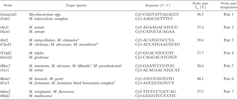
TABLE 1. Oligonucleotides used in the study
Probe Target species Sequence (5⬘–3⬘) Probe pair
Tm(°C)
Probe pair designation
pGeneral3 Mycobacteriumspp. Cy5-CGGTATTAGACCC 38.5 Pair 1
pTub2 M. tuberculosiscomplex Cy3-AAGCGCTTTCC
pAvi1 M. avium Cy5-AGAAGACATGCG 37.4 Pair 2
pXen1 M. xenopi Cy3-CATGCGCAGAA
pInt2 M. intracellulare,M. chimaeraa Cy5-ACATGCGCCTA 39.4 Pair 3
pCheI3 M. chelonae,M. abscessus,M. massilienseb Cy3-ACCATGAAGTGTG
pTripl2 M. triplex Cy5-GGACATGCGTC 37.7 Pair 4
pGord2 M. gordonae Cy3-CAGGACATGTGT
pMar2 M. marinum,M. ulcerans,M. liflandii,cM. pseudoshottsiic Cy5-GAATCCCGTGG 38.4 Pair 5
pTer1 M. terrae Cy3-ACAGAACATGCAT
pKan3 M. kansasii,M. gastri Cy5-ATCCCGGTGTG 40.1 Pair 6
pFor3 M. fortuitum,M. fortuitumthird biovariant complexd Cy3-AGCGCGGTCCT
pSme2 M. smegmatis,M. flavescens Cy5-TTCCCCTACCAG 37.5 Pair 7
pMal2 M. malmoense Cy3-GGGGTCCTATTC
aIncluded in theM. aviumcomplex,M. chimaerahas recently been proposed as a new species (17).
bHaving identical 16S rRNA gene hypervariable region sequences, likeM. abscessus,M. massiliensecan be discriminated fromM. chelonaeby biochemical tests
(1, 8) or by PCR restriction analysis of thehsp65Telenti fragment (16).
cThese species are pathogenic for animals (9, 10).
dAmong the species included in this group, only 16S rRNA gene hypervariable region sequences ofM. porcinum,M. neworleansense, andM. boenickeimatched probe
pFor3 (12).
6190 NOTES J. CLIN. MICROBIOL.
on May 15, 2020 by guest
http://jcm.asm.org/
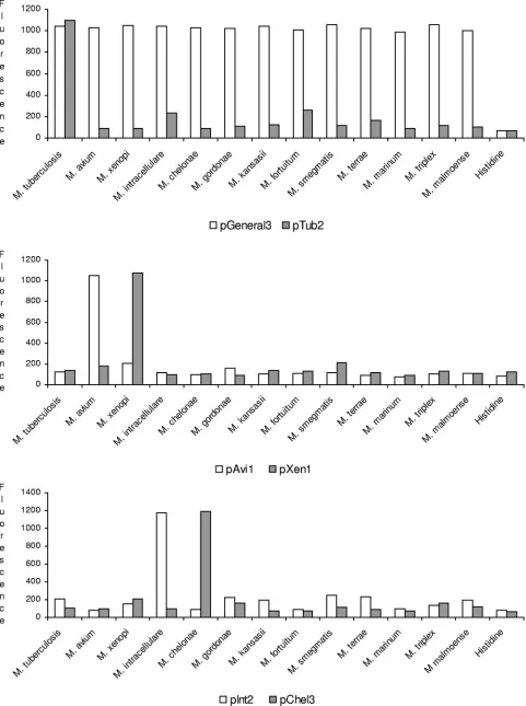
FIG. 1. Examples of Nanochip hybridization results for three sequence-selective fluorescently labeled probe pairs against the 13 mycobacterial species tested. Graphs show fluorescence signal values obtained in each of the three experiments. Histidine was used as a negative control.
6191
on May 15, 2020 by guest
not shown). Among the remaining 44 PCR products, 12 hy-bridized with probe pInt2, 11 with probe pChel3, 9 with probe pFor3, 6 with probe pKan3, 5 with probe pSme2, and 1 with probe pMar2; correspondingly, the culture isolates were iden-tified as M. intracellulare (12 isolates), M. chelonae (11 iso-lates), M. fortuitum (9 isolates), M. kansasii (6 isolates), M. smegmatis(5 isolates), andM. marinum(1 isolate), using bio-chemical and molecular tests (8, 16). Figure 1 shows the results of hybridization obtained with PCR products from 13 different mycobacterial species using three probe pairs. As expected, strong signals were observed only between the PCR products and their selective oligonucleotide probes.
Microelectronic chips or arrays are innovative methods for DNA hybridization analysis of point mutations, single-nucle-otide polymorphism, short tandem repeats, and gene expres-sion. Electronic molecule addressing and hybridization are car-ried out on these devices, where the electric field and the use of low-conductance buffers (histidine, etc.) greatly accelerate the hybridization reactions on the selected test sites (4, 15). The feasibility of these devices, initially developed for geno-typing purposes (6, 13, 20), makes them adaptable for other clinically important applications (3, 19).
In this work, we applied the Nanogen Nanochip Molecular Biology Workstation to the field of bacterial diagnostics for the first time, focusing on the species identification of mycobacteria. We adapted this technology to our purpose by creating a hybrid-ization assay in which multiple species-selective probes, matching a highly polymorphic region of the Mycobacterium16S rRNA gene, were used in consecutive reactions on a single platform. This gene has been used in several molecular methods (11, 14), and, recently, in a microarray system developed by Troesch et al. (18), for the species identification of mycobacteria. Like the last method, the microelectronic chip has the potential answer to many diagnostic questions associated with the genus Mycobacte-rium, since it is possible to expand the spectrum of the mycobac-terial species by testing additional probes on the same chip. In our assay, we included probes directed against either most clinically relevant mycobacterial species (i.e.,M. tuberculosiscomplex and
M. avium) or Mycobacteriumspecies rarely causing human dis-ease, such asM. triplex. The choice of these probes reflected the epidemiology of Mycobacterium infections in our hospital, but obviously, the panel of probes can be modified according to the patient population to which diagnostic investigation is referred.
In conclusion, the Nanochip system offers a great deal of promise for rapid identification, especially if used in combina-tion with PCR deteccombina-tion methods. A similar approach has been ideated by Westin et al. (20), in which multiplex strand dis-placement amplification was combined with microelectronic chip array to detect the factor V Leiden mutation. Our results reconfirm the great flexibility of the Nanochip system and open new perspectives for its large-scale use in clinical diagnostics. In spite of the remarkable qualities of this system, such as accuracy, ability to select a minimal signal-to-noise ratio, need for only a small amount of DNA, reduced turnaround time, and easy performance, the Achilles’ heel remains the attention in designing probes of interest to avoid additional synthesis costs, although this is of little importance compared to a 100-site array cost (approximately 500 Euros). As the system is
capable of testing up to 100 samples simultaneously, we feel that the Nanochip system presented here is particularly suit-able for routine use in mycobacteriology laboratories that pro-cess a wide number of clinical specimens.
REFERENCES
1.Adekambi, T., M. Reynaud-Gaubert, G. Greub, M. J. Gevaudan, B. La Scola, D. Raoult, and M. Drancourt.2004. Amoebal coculture of “Mycobacterium massiliense” sp. nov. from the sputum of a patient with hemoptoic pneumo-nia. J. Clin. Microbiol.42:5493–5501.
2.American Thoracic Society Medical Section of the American Lung Associ-ation.1997. Diagnosis and treatment of disease caused by nontuberculous mycobacteria. Official statement of the American Thoracic Society, ap-proved by the Board of Directors, March 1997. Am. J. Respir. Crit. Care Med.156:S1–25.
3.Cooper, K. L., and R. V. Goering.2003. Development of a universal probe for electronic microarray and its application in characterization of the Staph-ylococcus aureus polCgene. J. Mol. Diagn.5:28–33.
4.Edman, C. F., D. E. Raymond, D. J. Wu, E. Tu, R. G. Sosnowski, W. F. Butler, M. Nerenberg, and M. J. Heller.1997. Electric field directed nucleic acid hybridization on microchips. Nucleic Acids Res.25:4907–4914. 5.Falkinham, J. O., III.1996. Epidemiology of infection by nontuberculous
mycobacteria. Clin. Microbiol. Rev.9:177–215.
6.Gilles, P. N., D. J. Wu, C. B. Foster, P. J. Dillon, and S. J. Chanock.1999. Single nucleotide polymorphic discrimination by an electronic dot blot assay on semiconductor microchips. Nat. Biotechnol.17:365–370.
7.Kox, L. F. F., J. van Leeuwen, S. Knijper, H. M. Jansen, and A. H. J. Kolk. 1995. PCR assay based on DNA coding for 16S rRNA for detection and identification of mycobacteria in clinical samples. J. Clin. Microbiol. 33: 3225–3233.
8.Metchock, B., F. S. Nolte, and R. J. Wallace, Jr.1999.Mycobacterium, p. 399–437.InP. R. Murray, E. J. Baron, M. A. Pfaller, F. C. Tenover, and R. H. Yolken (ed.), Manual of clinical microbiology, 7th ed. ASM Press, Washington, D.C.
9.Mve-Obiang, A., R. E. Lee, E. S. Umstot, K. A. Trott, T. C. Grammer, J. M. Parker, B. S. Ranger, R. Grainger, E. A. Mahrous, and P. L. Small.2005. A newly discovered mycobacterial pathogen isolated from laboratory colonies ofXenopusspecies with lethal infections produces a novel form of mycolac-tone, theMycobacterium ulceransmacrolide toxin. Infect. Immun.73:3307– 3312.
10.Rhodes, M. W., H. Kator, A. McNabb, C. Deshayes, J. M. Reyrat, B. A. Brown-Elliott, R. Wallace, Jr., K. A. Trott, J. M. Parker, B. Lifland, G. Osterhout, I. Kaattari, K. Reece, W. Vogelbein, and C. A. Ottinger.2005.
Mycobacterium pseudoshottsiisp. nov., a slowly growing chromogenic species isolated from Chesapeake Bay striped bass (Morone saxatilis). Int. J. Syst. Evol. Microbiol.55:1139–1147.
11.Sanguinetti, M., B. Posteraro, F. Ardito, S. Zanetti, A. Cingolani, L. Sechi, A. De Luca, L. Ortona, and G. Fadda.1998. Routine use of PCR-reverse cross-blot hybridization assay for rapid identification ofMycobacterium spe-cies growing in liquid media. J. Clin. Microbiol.36:1530–1533.
12.Schinsky, M. F., R. E. Morey, A. G. Steigerwalt, M. P. Douglas, R. W. Wilson, R. W., M. M. Floyd, W. R. Butler, M. I. Daneshvar, B. A. Brown-Elliott, R. J. Wallace, Jr., M. M. McNeil, D. J. Brenner, and J. M. Brown.2004. Taxo-nomic variation in theMycobacterium fortuitumthird biovariant complex: description ofMycobacterium boenickeisp. nov., Mycobacterium houstonense
sp. nov.,Mycobacterium neworleansensesp. nov. andMycobacterium bris-banensesp. nov. and recognition ofMycobacterium porcinumfrom human clinical isolates. Int. J. Syst. Evol. Microbiol.54:1653–1667.
13.Sethi, A. A., A. Tybjaerg-Hansen, R. V. Andersen, and B. G. Nordestgaard. 2004. Nanogen microelectronic chip for large-scale genotyping. Clin. Chem. 50:443–446.
14.Soini, H., and J. M. Musser.2001. Molecular diagnosis of mycobacteria. Clin. Chem.47:809–814.
15.Sosnowski, R., M. J. Heller, E. Tu, A. H. Forster, and R. Radtkey.2002. Active microelectronic array system for DNA hybridization, genotyping and pharmacogenomic applications. Psychiatr. Genet.12:181–192.
16.Telenti, A., F. Marchesi, M. Balz, F. Bally, E. C. Bottger, and T. Bodmer. 1993. Rapid identification of mycobacteria to the species level by polymerase chain reaction and restriction enzyme analysis. J. Clin. Microbiol.31:175– 178.
17.Tortoli, E., L. Rindi, M. J. Garcia, P. Chiaradonna, R. Dei, C. Garzelli, R. M. Kroppenstedt, N. Lari, R. Mattei, A. Mariottini, G. Mazzarelli, M. I. Murcia, A. Nanetti, P. Piccoli, and C. Scarparo.2004. Proposal to elevate the genetic variant MAC-A, included in theMycobacterium aviumcomplex, to
6192 NOTES J. CLIN. MICROBIOL.
on May 15, 2020 by guest
http://jcm.asm.org/
species rank asMycobacterium chimaerasp. nov. Int. J. Syst. Evol. Microbiol. 54:1277–1285.
18.Troesch, A., H. Nguyen, C. G. Miyada, S. Desvarenne, T. R. Gingeras, P. M. Kaplan, P. Cros, and C. Mabilat.1999.Mycobacteriumspecies identification and rifampin resistance testing with high-density DNA probe arrays. J. Clin. Microbiol.37:49–55.
19.Westin, L., C. Miller, D. Vollmer, D. Canter, R. Radtkey, M. Nerenberg, and J. P. O’Connell.2001. Antimicrobial resistance and bacterial identification utilizing a microelectronic chip array. J. Clin. Microbiol.39:1097–1104. 20.Westin, L., X. Xu, C. Miller, L. Wang, C. F. Edman, and M. Nerenberg.2000.
Anchored multiplex amplification on a microelectronic chip array. Nat. Bio-technol.18:199–204.