Copyright © 2001, American Society for Microbiology. All Rights Reserved.
Comparison of DNA Sequencing of the Protein A Gene
Polymorphic Region with Other Molecular Typing Techniques
for Typing Two Epidemiologically Diverse Collections of
Methicillin-Resistant
Staphylococcus aureus
DUARTE C. OLIVEIRA,1,2INEˆS CRISO´ STOMO,1ILDA SANTOS-SANCHES,1,3PETER MAJOR,1,4 C. RUTE ALVES,1MARTA AIRES-DE-SOUSA,1MARIANNE K. THEGE,4
ANDHERMI´NIADELENCASTRE1,2*
Instituto de Tecnologia Quı´mica e Biolo´gica, Universidade Nova de Lisboa, Oeiras, Portugal1; The Rockefeller University, New York, New York 100212; Faculdade de Cieˆncias e Tecnologia, Universidade Nova de Lisboa, Monte da
Caparica, Portugal3; and National Institute of Food Hygiene and Nutrition, Budapest, Hungary4 Received 19 October 2000/Returned for modification 25 November 2000/Accepted 1 December 2000
The aim of this study was to compare the recently developed typing approach for methicillin-resistant
Staphylococcus aureus(MRSA) based on the DNA sequencing of the protein A gene polymorphic region (spaA
typing) with a combination of three well-established molecular typing techniques:ClaI-mecAvicinity
polymor-phisms,ClaI-Tn554insertion patterns, andSmaI pulsed-field gel electrophoresis (PFGE) profiles. In order to
evaluate the applicability of this typing technique in different types of studies, two groups of MRSA clinical isolates were analyzed: a collection of 185 MRSA isolates circulating in Hungary recovered from 17 hospitals in seven cities during a 3-year period (1994 through 1996), and a selection of 53 MRSA strains isolated in a single hospital in Hungary between 1997 and 1998. The 238 MRSA clinical strains from Hungary were first
classified in clonal types (defined asClaI-mecA::ClaI-Tn554::SmaI-PFGE patterns), and 65 of the 238 strains,
representing major MRSA clones and some sporadic clones, were further analyzed byspaAtyping. Our results
showed that the lineages most recently introduced in the hospital setting showed little variability inspaAtypes,
whereas the MRSA clones circulating for a longer period of time and spread among several hospitals showed
a higher degree of variability. The implementation of the spaAtyping method was straightforward, and the
results obtained were reproducible, unambiguous, and easily interpreted. This method seems to be adequate for outbreak investigations but should be complemented with other techniques in long-term surveillance or in studies comparing distant clonal lineages.
Three molecular typing techniques (5) have been largely used for the characterization of clones of methicillin-resistant
Staphylococcus aureus(MRSA) and enabled the detection of widely spread MRSA lineages, such as the Iberian, Brazilian, New York/Tokyo, and pediatric MRSA clones (1, 7, 11, 17, 20, 21, 24). The combined methods consist of (5, 10) Southern blot analysis of chromosomalClaI digests with amecADNA probe (ClaI-mecA polymorphisms) and with a Tn554 transposon probe (ClaI-Tn554insertion patterns) and restriction fragment length polymorphism analysis of chromosomal DNA gener-ated after cleavage with SmaI and pulsed-field gel electro-phoresis (PFGE) (SmaI-PFGE). ClaI-mecA polymorphisms are a consequence of the variability in the vicinity of themecA
gene, the central element of methicillin resistance, andCla I-Tn554 patterns reflect the location and copy number of the transposon Tn554, present in most MRSA clinical isolates (10). PFGE provides fine fingerprinting of the chromosomal background with high discriminatory power and has been sug-gested as the gold standard for the molecular typing of MRSA (25, 26).
DNA sequencing-based typing techniques are being devel-oped with obvious advantages in speed, unambiguous data
interpretation, simplicity of large-scale database creation, and standardization among laboratories (8). Recently, DNA se-quencing of thespaAgene (protein A determinant) polymor-phic region for typing of MRSA strains was evaluated (22). The polymorphic X region is involved in attachment to the cell wall and consists of a variable number of 24-bp repeats, short sequence repeats, which seem to arise from deletion and du-plication of the repetitive units and also by point mutation (9, 27). The existence of well-conserved regions flanking the X region coding sequence inspaAallows the use of primers for PCR amplification and direct sequence typing. Shopsin and colleagues (22) have shown that, despite its high degree of polymorphism, the X region of protein A has a variation rate low enough to provide suitable discrimination for outbreak investigations or strain collections restricted to one location and recovered within a short period of time.
In this study we evaluated the discriminatory power ofspaA
typing to differentiate MRSA clones and assessed the correla-tion between this sequencing typing method and the combined molecular typing methodsClaI-mecA,ClaI-Tn554, andSma I-PFGE patterns referred to above. Using these three combined methods, we have studied two groups of MRSA strains with different characteristics recovered from Hungarian hospitals: a representative collection of MRSA strains circulating in Hun-garian hospitals during a 3-year period (1994 to 1996) and a collection of MRSA isolates recovered from a single hospital * Corresponding author. Mailing address: The Rockefeller
Univer-sity, 1230 York Ave., New York, NY 10021. Phone: (212) 327-8278. Fax: (212) 327-8688. E-mail: [email protected].
574
on May 15, 2020 by guest
http://jcm.asm.org/
Downloaded from
on May 15, 2020 by guest
http://jcm.asm.org/
Downloaded from
on May 15, 2020 by guest
http://jcm.asm.org/
Downloaded from
on May 15, 2020 by guest
http://jcm.asm.org/
during a 2-year period (1997 to 1998). Fifty-six strains repre-senting the most important clones spread in Hungarian hospi-tals between 1994 and 1998 and also some sporadic clones were further analyzed byspaAtyping to analyze the correlation between clonal types defined as ClaI-mecA polymorphisms ::ClaI-Tn554patterns::SmaI-PFGE profiles andspaAtypes.
(Part of this study was presented at the 38th Interscience Conference on Antimicrobial Agents and Chemotherapy, American Society for Microbiology, abstr. E170, 1998.)
MATERIALS AND METHODS
Clinical isolates.The 238 MRSA clinical isolates from Hungary included in this study comprise (i) a collection of 185 isolates recovered between 1994 and 1996 at 17 different hospitals located in seven different cities, which were chosen to be representative of MRSA epidemic clones circulating in Hungarian hospi-tals, and (ii) 53 isolates recovered at a single hospital from January to October 1997 and from January to December 1998, chosen to illustrate a short-term type of study. Antibiograms were performed by the clinical laboratories using the Kirby-Bauer technique, according to the published recommendations and defi-nitions (14). The panel of antibiotics was different from hospital to hospital, but the great majority of the isolates were multiresistant to the antimicrobial agents tested, such as penicillin, oxacillin, erythromycin, tetracycline, ciprofloxacin, and gentamicin. All isolates were susceptible to vancomycin and teicoplanin.
Molecular typing. ClaI-mecApolymorphism,ClaI-Tn554insertion pattern, andSmaI-PFGE profile analyses were performed and interpreted as previously described (3, 5, 10, 26).
spaAtyping.spaAtyping was performed essentially as previously described (22). Chromosomal DNA for PCR was prepared (2) and diluted to approxi-mately 0.5 ng/l. Primers for amplification and sequencing of the X region of the
spaA gene were designed based on the published sequence (accession no. J01786) and purchased from Gibco-BRL (Life Technologies, Grand Island, N.Y.): SpaF1, GAC GAT CCT TCG GTG ACG, nucleotides 1096 to 1113, and SpaR1, CAG CAG TAG TGC CGT TTG C, nucleotides 1534 to 1516. PCR amplification was performed in a GeneAmp PCR System 9600 thermocycler (Perkin-Elmer Cetus [PE], Branchburg, N.J.), with 2.5 ng of DNA, 5l of 10⫻
PCR buffer II (PE), 4l of deoxynucleoside triposphate mix at 10 mM (PE), 2.5 U of AmpliTaq Gold DNA polymerase (PE), 1.5 mM MgCl2(PE), and 10 mmol
of each primer in a final reaction volume of 50l in 0.2-ml PCR tubes (PE). Thermal cycling parameters were as follows: predenaturation for 10 min at 95°C; 30 cycles of 95°C for 30 s, 60°C for 30 s, 72°C for 45 s, postextension for 10 min at 72°C; and soaking at 4°C. PCR products (2l) were visualized by conventional minigel electrophoresis and purified with the Wizard PCR-prep DNA purifica-tion system (Promega, Madison, Wis.). DNA cycle sequencing reacpurifica-tions were performed with the ABI Prism Big Dye Terminator Cycle Sequencing Ready Reaction Kit (PE) in a final reaction volume of 10l: 1l of amplified and purified DNA (20 to 30 ng); 2l of SpaF1 or SpaR1 at 2 pmol/l; 4l of Ready Reaction mix; and 3l of H2O MilliQ. Amplification parameters were as follows:
25 cycles of 96°C for 10 s, 50°C for 5 s, and 60°C for 4 min, and soaking at 4°C. DNA was precipitated at room temperature for 15 min with 50l of 95% ethanol–10l of H2O MilliQ–2l of 3 M sodium acetate (pH⫽4.6), centrifuged
in a microcentrifuge for 20 min at 13,000 rpm, washed with 250l of 70% ethanol, centrifuged for 10 min at 13,000 rpm, and dried for 1 min at 90°C. DNA sequences were determined by electrophoresis in an ABI Prism 377 DNA se-quencer (PE) according to the manufacturer’s instructions at the DNA sequenc-ing facility located at Instituto Gulbenkian de Cieˆncia, Oeiras, Portugal. The assembly of both sequences was performed with SeqMan software (DNAStar software package; Lasergene, Madison, Wis.). Consensus sequences were sought for the previously defined 24-bp repeat polymorphisms (22), using specific soft-ware (GeneSearch, designed by Ludwig Krippahl). The output (spaAtype) con-sists of a sequence of letters that correspond to the succession of the different 24-bp repeats within the polymorphic region of thespaAgene.
RESULTS
Clonal analysis of Hungarian MRSA.Tables 1 and 2
sum-marize the clonal types found in the 1994 to 1996 and 1997 to 1998 collections, respectively. The clonal types were compared to those of a previous study in which the application of these techniques to a collection of 48 MRSA clinical isolates
recov-ered from six provincial hospitals located hundreds of kilome-ters apart in Hungary between 1993 and 1994 demonstrated the existence of a unique epidemic MRSA clone, the Hungar-ian MRSA clone (clone III::B::A), which was present in 81% of the isolates (6). In the present study, this lineage, charac-terized by PFGE pattern A, was still present in both collec-tions, but its prevalence had decreased to 70% in 1994 to 1996 and its variability, expressed by the panoply of PFGE subtypes (37 subtypes were found for PFGE pattern A), ClaI-mecA
types, and ClaI-Tn554 insertion patterns, had increased, as shown by the presence of clonal type III::B::A in only 33% of the isolates in 1994 to 1996. The observed variability in the
ClaI-mecApolymorphisms (patterns III, IX, XI, and III⬘) was characterized by small shifts in the hybridization fragment size of themecAdownstream vicinity which were detected in strains isolated after 1993 and that were recently explained (16) as being caused by different copy numbers of the direct repeat unit (dru) within the hypervariable region downstream ofmecA
(19). Pattern IX has 11drucopies, pattern XI has 10 copies, pattern III has 9 copies, and pattern III⬘ has 8 copies (16). Therefore, it is reasonable to consider that clonal types III::B::A, IX::B::A, XI::B::A, and III⬘::B::A are equivalent, and altogether they accounted for 42% of the clonal types in 1994 to 1996.ClaI-Tn554 insertion patterns are a consequence of the location and copy number of a transposon, by definition a mobile element, and within the PFGE pattern A cluster as many as 16 different insertion patterns were found, all charac-terized by the presence of multiple copies of transposon Tn554. However, since PFGE is the technique with the highest discriminatory power, strains sharing PFGE pattern A were classified as closely related and accounted for 70% of the clonal types in 1994 to 1996 and 40% in the 1997 to 1998 collection. This high degree of variability within an MRSA clone has never been reported before and seems to be a par-ticular characteristic of the Hungarian clone. It is not observed, for example, in the highly epidemic Iberian and Brazilian clones (H. de Lencastre, unpublished observations).
In the 1994 to 1996 collection, a new family of clones were found in Hungarian hospitals, PFGE pattern D-related clones II::q::D, II::A1::D, and II::D::D (15% of the isolates), which were also detected in 9% of the isolates recovered from the single hospital studied in 1997 to 1998. In this same hospital in 1997 to 1998, a previously undetected lineage (II::E1::S) was present in 28% of the isolates. Both lineages showed signifi-cantly less variability than PFGE pattern A-related clones, which may be explained by its more recent introduction in Hungarian hospitals and, in the case of clone II::E1::S, by the fact that it was detected in isolates from a single hospital. These lineages do not appear (by the typing methods used) to be related to other MRSA clones spread in other countries and previously identified in this laboratory. Figure 1 shows some subtypes of the most important PFGE patterns found in this study, PFGE patterns A, D, and S.
spaAtyping.Fifty-six strains representing the most
impor-tant clonal types detected in Hungarian hospitals in 1994 to 1996 and 1997 to 1998 were studied byspaAtyping. For each lineage (defined according to PFGE patterns A, D, and S), several strains with different ClaI-mecA and ClaI-Tn554 pat-terns were selected from different hospitals, cities, and periods of isolation. Nine strains belonging to sporadic clones were
VOL. 39, 2001 MRSA spaA TYPING 575
on May 15, 2020 by guest
http://jcm.asm.org/
Clone type City (county) or county Yr of isolation No. of isolates Clonal typea Total no. of isolates (% of total)
Epidemic A Szekesfehervar (Fejer) 1994, 1996 12 III::B::A 61 (33.0)
Dunaujvaros (Fejer) 1994 3
Debrecen (Hajdu) 1994 2
Miskolc (Borsod-A. Z.) 1996 15
Somogy County 1994, 1996 22
Budapest 1995, 1996 7
Dunaujvaros (Fejer) 1996 9 III⬘::B::A 12 (6.5)
Debrecen (Hajdu) 1994 2
Budapest 1995 1
Somogy County 1994, 1996 2 IX::B::A 2
Debrecen (Hajdu) 1994 1 XI::B::A 2
Miskolc (Borsod-A. Z.) 1995 1
Miskolc (Borsod-A. Z.) 1995 1 III::M::A 26 (14.1)
Somogy County 1996 6
Budapest 1995 19
Dunaujvaros (Fejer) 1996 3 III⬘::M::A 4 (2.1)
Budapest 1995 1
Miskolc (Borsod-A. Z.) 1995 3 III::W::A 4 (2.1)
Somogy County 1996 1
Budapest 1995 1 III::DD::A
Budapest 1995 1 III::F::A
Budapest 1996 2 III::A::A
Budapest 1996 1 III::B2::A
Budapest 1996 1 III::k::A
Budapest 1996 1 XI::k::A
Miskolc (Borsod-A. Z.) 1995 1 III::W1::A
Miskolc (Borsod-A. Z.) 1995 1 III::i::A
Miskolc (Borsod-A. Z.) 1995 1 III::m::A
Miskolc (Borsod-A. Z.) 1995 1 III::n::A
Miskolc (Borsod-A. Z.) 1995 1 III::t::A
Miskolc (Borsod-A. Z.) 1996 1 III::::A
Somogy County 1994 1 IX::E::A
Somogy County 1994 1 III::i::A
Somogy County 1996 1 XI::B1::A
Somogy County 1996 1 III::M1::A
Szekesfehervar (Fejer) 1996 1 III::M::A
Szekesfehervar (Fejer) 1996 1 III::i::A
Total 130 (70.2)
Epidemic D Budapest 1994, 1996 18 II::q::D 18 (9.7)
Somogy County 1994 1 II::D::D 9 (4.9)
Budapest 1995, 1996 8
Budapest 1996 1 II::A1::D 1
Total 28 (15.1)
Sporadic Budapest 1995 1 III::B::Q
Budapest 1995 1 II::A1::R
Budapest 1996 4 II::A::E
Budapest 1996 1 II::D::E
Budapest 1995 1 XI::B2::O
Budapest 1996 2 VI::F::P
Miskolc (Borsod-A. Z.) 1995 1 III::W::H
Miskolc (Borsod-A. Z.) 1996 1 III::B::B
Miskolc (Borsod-A. Z.) 1996 1 III::W::B
Miskolc (Borsod-A. Z.) 1996 1 III⬘::M1::F
Miskolc (Borsod-A. Z.) 1996 1 III⬘::␣␣::G
Szekesfehervar (Fejer) 1994 1 III::B::M
Szekesfehervar (Fejer) 1994 1 III::j::M
Szekesfehervar (Fejer) 1994 2 III::B::N
Szekesfehervar (Fejer) 1994 1 III::j::N
Szekesfehervar (Fejer) 1996 1 III::B1::B
Dunaujvaros (Fejer) 1994 1 III::n::B
Dunaujvaros (Fejer) 1994 1 IX::B::K
Somogy County 1996 1 III::B::I
Somogy County 1996 1 III::x::J
Debrecen (Hajdu) 1994 1 III::p::L
Szombathely (Vas) 1994 1 III::M::N
Total 27 (14.6)
aClonal types were defined on the basis ofClaI-mecApolymorphisms::ClaI-Tn554patterns::SmaI-PFGE restriction profiles. A total of 185 strains were examined.
ClaI-mecApolymorphisms with a prime andClaI-Tn554insertion patterns with number codes (e.g., B1) contain small variations from previously described patterns.
ClaI-Tn554insertion patterns with lowercase letters are new patterns not described in previous studies.
576
on May 15, 2020 by guest
http://jcm.asm.org/
[image:3.612.52.555.68.706.2]also included. Nine differentspaAtypes were found among the 65 five strains studied (Table 3).
Four relatedspaAtypes sharing the KAOMQ motif (spaA
types WGKAKBAOKAOMQ, WGKAKAOKAOMQ, WGKA OKAOMQ, and XKAOKAOMQ) were found among the iso-lates belonging to PFGE pattern A-related clones. However two of thesespaA types (WGKAKAOKAOMQ and WGKA OKAOMQ) were also found among strains belonging to the sporadic clones characterized by PFGE patterns B, T, and H. ThespaAtype WGKAQAQQ was specific for the 12 isolates studied belonging to PFGE pattern D-related clones. ThespaA
type TIMBMDMGMK was specific for 9 of 10 isolates studied belonging to clonal type II::E1::S, and the other isolate was characterized by the relatedspaAtype TIMBME. The remain-ing two spaA types, YHFGFMBQBLO and WGKAOMQ, were specific for strains belonging to the sporadic clones I::NH::W and III::1::Z, respectively.
DISCUSSION
We have applied the recently developed MRSA typing tech-nique based on DNA sequencing of the protein A gene poly-morphic region,spaAtyping (22), to characterize two distinct MRSA collections representing two different kinds of studies: a representative collection of MRSA clinical isolates circulat-ing in Hungarian hospitals durcirculat-ing a 3-year period and a col-lection of clinical strains recovered from a single hospital
dur-ing a 2-year period, also in Hungary.spaAtypes were compared to the results obtained using other molecular typing techniques (ClaI-mecA, ClaI-Tn554, andSmaI-PFGE patterns) in order to evaluate the discriminatory power ofspaAtyping and its use to characterize MRSA clones circulating in a particular coun-try or hospital. In addition, we wanted to assess the ease of implementation and execution of this method in our labora-tory, since we have been interested on the molecular typing of clinical strains of MRSA, and therefore,spaAtyping was po-tentially useful in our studies.
Implementation ofspaAtyping technique.The
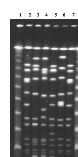
[image:4.612.54.291.92.402.2]implementa-tion of thespaAtyping technique, essentially according to the published procedure (22), was straightforward and easy to es-tablish in our laboratory, requiring only some expertise in PCR and DNA sequencing techniques, which are easily achieved with modern reagent kits, thermocyclers, and sequencers. To evaluate the reproducibility of thespaAtypes, four strains were typed twice, and in all cases the spaA types obtained were exactly the same. The stability ofspaAtypes was also evaluated for one strain, which was daily diluted in fresh medium over a 3-week period (approximately 15⫻109generations), and no changes inspaAtype were detected. In order to make a first evaluation of spaA typing, a small collection of 20 MRSA isolates representative of well-characterized and internation-ally spread clones, such as the Iberian (7, 21), Brazilian (24), FIG. 1. Representative gel of some subtypes of the most frequent SmaI-PFGE profiles and one sporadic clone. Strain codes indicate the period of isolation: HUSA, 1993 to 1994 (4); HU, 1994 to 1996; and HUR, 1997 to 1998. Samples are as follows: lanes 1 and 15, molecular size markers (lambda DNA ladder; New England Biolabs); lanes 2 and 14, reference strain NCTC8325; lanes 3 to 5, pattern A1 (HUSA67, HU1, and HUR36); lane 6, pattern A36 (HU221); lane 7, pattern A37 (HUR1); lane 8, pattern D1 (HU181); lane 9, pattern D2 (HU164); lane 10, pattern D3 (HU150); lane 11, pattern S1 (HUR9); lane 12, pattern S4 (HUR95); lane 13, pattern S5 (HUR94).
TABLE 2. Molecular typing of 53 MRSA clinical strains isolated from a single hospital in Hungary between 1997 and 1998 PFGE
pattern type isolationYr of isolatesNo. of Clonal typea Total no. of isolates(% of total)
A 1997 7 III::M1::A 9 (17.0)
1998 2
1997 1 III::B2::A 5 (9.4)
1998 4
1997 2 III::B1::A 6
1998 2 XI::B::A
1998 1 III⬘::B3::A 1998 1 III⬘::q::A
Total 20 (37.8)
D 1997 4 II::q::D 5
1997 1 II::q1::D
Total 5 (9.4)
S 1997 12 II::E1::S 15 (28.3)
1998 3
Varied 1997 2 III⬘::B2::B 13 (24.5) 1997 1 III::B2::B
1997 1 VIII::aa::M 1997 1 III::M4::T 1997 1 III::M1::Y 1998 1 III⬘::n::B
1998 2 I::NH::W
1998 1 III::B2::N 1998 1 VII::1::V
1998 1 V::W1::X
1998 1 III::1::Z
Total 13 (24.5)
aSee Table 1, footnotea.
VOL. 39, 2001 MRSA spaA TYPING 577
on May 15, 2020 by guest
http://jcm.asm.org/
and New York/Tokyo (1, 11) clones, was tested, and it was found thatspaA typing was able to discriminate among the different MRSA lineages: Iberian clone spaA type YHFG FMBQBLO; Brazilian clonespaAtype XKAOMQ; and New York/Tokyo clone spaA type TJMBMDMGMK (Oliveira et al., unpublished data).
Comparison betweenspaAtyping and other molecular
typ-ing techniques. In this study, spaA typing was excellent in
discriminating the clonal lineages more recently introduced in Hungarian hospitals (PFGE pattern D clones and clone II::E1::S), which were characterized by the specificspaAtypes WGKAQAQQ and TIMBMDMGMK/TIMBME, respectively.
However, the isolates belonging to the clonal lineage char-acterized by PFGE pattern A, circulating in Hungarian hospi-tals at least since 1993, were characterized by a cluster of four
spaA types with the KAOMQ motif, and two of these spaA
types were also found among strains belonging to the sporadic clones characterized by PFGE patterns B, T, and H. These findings suggest that these sporadic clones (III::B::B, III::B⬘::B, III::W::B, III::M4::T, and III::M⬘::H) may have evolved from clone III::B::A, so that a less discriminative technique like
spaAtyping might not be able to differentiate them. This hy-pothesis is supported by the fact that these sporadic clones show the same ClaI-mecA and ClaI-Tn554 types as PFGE pattern A-related clones. Moreover, another sporadic clone analyzed by spaA typing (clone I::NH::W), with nothing in common with clone III::B::A, displayed the unrelated and spe-cificspaAtype YHFGFMBQBLO, whereas the sporadic clone III::1::Z (sharingClaI-mecAtype III) was characterized by
spaA type WGKAOMO, with the KAOMQ motif. The vari-ability ofspaAtypes among the PFGE pattern A-related clones parallels the variability also detected by the other typing tech-niques, suggesting that the mutation rate of the spaA gene polymorphic region is comparable to the variability rate of
ClaI-mecApolymorphisms,ClaI-Tn554insertion patterns, and PFGE subtypes.
The application of spaA typing also provided interesting clonal relationships among MRSA. The sporadic clone I::NH::W found in Hungary in 1998 (Table 2) showed exactly the samespaAtype as the Iberian clone-related strains isolated since the mid-1980s and also strain DEN2125 isolated in Den-mark in 1964 (results not shown), confirming the previous
finding (4) that the Iberian clone, first described in Spain (7) and since then shown to be widely spread throughout Europe (12, 13, 21) and the United States (17, 18), may have in strain DEN2125 a evolutionary precursor. The I::NH::W clone may also be an Iberian clone derivative that has just been intro-duced in Hungary and will eventually disseminate among Hun-garian hospitals. Analysis of Fig. 2, in which the PFGE profiles of these strains are displayed, showed that these strains do not
[image:5.612.363.499.386.659.2]FIG. 2.SmaI-PFGE profiles of strains with the IberianspaAtype. Strains are as follows: lanes 1 and 8, molecular size markers (lambda DNA ladder; New England Biolabs); lanes 2 and 7, reference strain NCTC8325; lane 3, PER34 (Iberian clone representative strain [7]); lane 4, HUC191 (Iberian clone closely related strain [15]); lane 5, DEN2125 (archaic clone [1]); and lane 6, HUR97 (sporadic clone isolated in Hungary).
TABLE 3. spaAtyping of selected MRSA strains isolated in Hungary in 1994 to 1996 and 1997 to 1998
spaAtypea Clonal typeb(no. of strains)
WGKAKBAOKAOMQ...III::B1::A (2), IX::B::A (2), III::M::A (1)
WGKAKAOKAOMQ...III::B::A (5), III⬘::B::A (2), III::B2::A (1), III⬘::B2::A (2), XI::B::A (1), III::M::A (2), III::M1::A (6), III::W::A (2), III::B⬘::B (1), III::N::B (1), III::W::B (1)
WGKAOKAOMQ...III::B::A (1), III⬘::B3::A (1), III::M::A (1), III⬘::q::A (1), V::W1::A (1), III::B::B (1), III::M4::T (1), III::M⬘::H (1) XKAOKAOMQ...XI::B::A (2), III⬘::n::A (1)
WGKAQAQQ ...II::q::D (7), II::q1::D (1), II::D::D (3), II::A⬘::D (1) TIMBMDMGMK...II::E1::S (9)
TIMBME ...II::E1::S (1)
WGKAOMQ...III::1::Z (1) YHFGFMBQBLO ...I::NH::W (2)
aspaAtypes were assigned according to Shopsin and colleagues (22). Each letter corresponds to a different polymorphism of the 24-bp repeat; the sequence of letters correponds to the sequence of the 24-bp repeats within thespaAgene polymorphic X region.
bClonal types were defined on the basis ofClaI-mecApolymorphisms::ClaI-Tn554patterns::SmaI-PFGE restriction profiles.
on May 15, 2020 by guest
http://jcm.asm.org/
have the same PFGE pattern, although these patterns seem to be related (10 band differences). The stability in thespaAtype of Iberian clone-related strains over a span of at least three decades contrasts with the variability found in thespaAtype related to the Hungarian clone strains and was somehow un-expected, since the molecular basis forspaAtype is precisely the polymorphic X region within thespaAgene. These findings suggest that there are highly stable alleles of the X region, presumably very efficient in determining pathogenesis and/or adherence mechanisms.
As previously suggested by Shopsin and colleagues (22) and also confirmed by Tang and colleagues (23), DNA sequencing of the protein A gene polymorphic region as a typing technique seems to be a powerful technique for MRSA typing. This is especially true for MRSA isolates restricted to one location and to a short period of isolation (that is, for outbreak inves-tigations), as was shown in this study by the excellent discrim-ination of the clonal lineages more recently introduced in Hun-garian hospitals (clones related to PFGE patterns D and S).
spaAtyping also had a satisfactory capacity for discrimination in more diverse collections, as illustrated in this study with the PFGE pattern A clonal lineage. However, in these cases, other typing techniques and a more careful interpretation of the
spaAtypes may be needed. In our study, we foundspaAtypes to be stable and reproducible, and thespaAtyping technique was easy to implement and provided unambiguous results.
ACKNOWLEDGMENTS
We thank the late Anna Marton for the gift of some MRSA clinical isolates and L. Krippahl (Faculdade de Cieˆncias e Technologia, Uni-versidade Nova de Lisboa, Monte da Caparica, Portugal) for writing the GeneSearch computer program.
Partial support for this study was provided by projects PRAXIS XXI/2/2.2/SAU/1295/95 and PRAXIS XXI/P/SAU/14052/98 from Fundac¸a˜o para a Cieˆncia e Tecnologia, Lisbon, Portugal, and Project 31 CEM/NET from IBET, Oeiras, Portugal, awarded to H. de Len-castre. The 1997 to 1998 collection was recovered under Project RE-SIST, with a grant from Rhoˆne-Poulenc Rorer S.A. to A. Tomasz and H. de Lencastre. D. C. Oliveira and M. Aires-de-Sousa were supported by grants BD/4162/96 and BD/13731/97, respectively, from Fundac¸a˜o para a Cieˆncia e Tecnologia, Lisbon, Portugal, and C. R. Alves was supported by grant 001/99/BIC/P from ITQB, Oeiras, Portugal. P. Major was supported by Fundac¸a˜o Calouste Gulbenkian for his CEM/ NET Fellowship Project at ITQB/UNL, Oeiras, Portugal.
REFERENCES
1.Aires-de-Sousa, M., H. de Lencastre, I. Santos-Sanches, K. Kikuchi, K. Totsuka, and A. Tomasz.2000. Similarity of antibiotic resistance patterns and molecular typing properties of methicillin-resistantStaphylococcus au-reus(MRSA) isolates widely spread in New York City and in a hospital in Tokyo, Japan. Microb. Drug Resist.6:253–258.
2.Aires-de-Sousa, M., I. Santos-Sanches, A. van Belkum, W. van Leeuwen, H. Verbrugh, and H. de Lencastre.1996. Characterization of methicillin-resis-tantStaphylococcus aureusisolates from Portuguese hospitals by multiple genotyping methods. Microb. Drug Resist.2:331–341.
3.Chung, M., H. de Lencastre, P. Matthews, A. Tomasz, and the Multilab Project Collaborators: I. Adamsson, M. Aires-de-Sousa, T. Camou, C. Cocuzza, A. Corso, I. Couto, A. Dominguez, M. Gniadkowski, R. Goering, A. Gomes, K. Kikuchi, A. Marchese, R. Mato, O. Melter, D. Oliveira, R. Pala-cio, R. Sa´-Lea˜o, I. Santos Sanches, J.-H. Song, P. T. Tassios, and P. Villari.
2000. Molecular typing of methicillin resistantStaphylococcus aureusby pulsed field gel electrophoresis: Comparison of results obtained in a multi-laboratory effort using identical protocols and MRSA strains. Microb. Drug Resist.6:189–198.
4.de Lencastre, H., M. Chung, and H. Westh.2000. Archaic strains of methi-cillin-resistantStaphylococcus aureus: molecular and microbiological
prop-erties of isolates from the 1960s in Denmark. Microb. Drug. Resist.6:1–10. 5.de Lencastre, H., I. Couto, I. Santos, J. Melo-Cristino, A. Torres-Pereira, and A. Tomasz.1994. Methicillin-resistantStaphylococcus aureusdisease in a Portuguese hospital: characterization of clonal types by a combination of DNA typing methods. Eur. J. Clin. Microbiol. Infect. Dis.13:64–73. 6.de Lencastre, H., E. P. Severina, H. Milch, M. K. Thege, and A. Tomasz.
1997. Wide geographic distribution of a unique methicillin-resistant Staph-ylococcus aureusclone in Hungarian hospitals. Clin. Microbiol. Infect.3:289– 296.
7.Dominguez, M. A., H. de Lencastre, J. Linares, and A. Tomasz.1994. Spread and maintenance of a dominant methicillin-resistantStaphylococcus aureus
(MRSA) clone during an outbreak of MRSA disease in a Spanish hospital. J. Clin. Microbiol.32:2081–2087.
8.Enright, M. C., N. P. Day, C. E. Davies, S. J. Peacock, and B. G. Spratt.2000. Multilocus sequence typing for characterization of methicillin-resistant and methicillin-susceptible clones ofStaphylococcus aureus. J. Clin. Microbiol.
38:1008–1015.
9.Guss, B., M. Uhlen, B. Nilsson, M. Lindberg, J. Sjoquist, and J. Sjodahl.
1984. Region X, the cell-wall-attachment part of staphylococcal protein A. Eur. J. Biochem.138:413–420.
10. Kreiswirth, B., J. Kornblum, R. D. Arbeit, W. Eisner, J. N. Maslow, A. McGeer, D. E. Low, and R. P. Novick.1993. Evidence for a clonal origin of methicillin resistance inStaphylococcus aureus. Science259:227–230. 11. Kreiswirth, B. N., S. M. Lutwick, E. K. Chapnick, J. D. Gradon, L. I.
Lutwick, D. V. Sepkowitz, W. Eisner, and M. H. Levi.1995. Tracing the spread of methicillin-resistantStaphylococcus aureusby Southern blot hy-bridization using gene-specific probes ofmecand Tn554. Microb. Drug Resist.1:307–313.
12. Mato, R., I. S. Sanches, M. Venditti, D. J. Platt, A. Brown, and H. de Lencastre.1998. Spread of the multiresistant Iberian clone of methicillin-resistantStaphylococuus aureus(MRSA) to Italy and Scotland. Microb. Drug Resist.4:107–112.
13. Melter, O., I. Santos-Sanches, J. Schindler, M. Aires-de-Sousa, R. Mato, V. Kovarova, H. Zemlickova, and H. de Lencastre.1999. Methicillin-resistant
Staphylococcus aureusclonal types in the Czech Republic. J. Clin. Microbiol.
37:2798–2803.
14. National Committee for Clinical Laboratory Standards.1997. Performance standards for antimicrobial disk susceptibility tests. Approved standard M2– A6. National Committee for Clinical Laboratory Standards, Wayne, Pa. 15. Oliveira, D., I. Santos-Sanches, M. Tamayo, G. Ribeiro, R. Mato, D. Costa,
and H. de Lencastre.1998. Virtually all MRSA infections in the largest Portuguese hospital are caused by two internationally spread multiresistant strains: the “Iberian” and the “Brazilian” clones of MRSA. Clin. Microbiol. Infect.4:373–384.
16. Oliveira, D. C., S. W. Wu, and H. de Lencastre.2000. Genetic organization of the downstream region of themecAelement in methicillin-resistant Staph-ylococcus aureusisolates carrying different polymorphs of the antibiotic re-sistance gene. Antimicrob. Agents Chemother.44:1906–1910.
17. Roberts, R. B., A. de Lencastre, W. Eisner, E. P. Severina, B. Shopsin, B. N. Kreiswirth, and A. Tomasz.1998. Molecular epidemiology of methicillin-resistantStaphylococcus aureusin 12 New York hospitals. MRSA Collabo-rative Study Group. J. Infect. Dis.178:164–171.
18. Roberts, R. B., A. M. Tennenberg, W. Eisner, J. Hargrave, L. M. Drusin, R. Yurt, and B. N. Kreiswirth.1998. Outbreak in a New York City teaching hospital burn center caused by the Iberian epidemic clone of MRSA. Mi-crob. Drug Resist.4:175–183.
19. Ryffel, C., R. Bucher, F. H. Kayser, and B. Berger-Bachi.1991.The Staph-ylococcus aureus mecdeterminant comprises an unusual cluster of direct repeats and codes for a gene product similar to theEscherichia coli sn -glycerophosphoryl diester phosphodiesterase. J. Bacteriol.173:7416–7422. 20. Sa´-Lea˜o, R., I. Santos-Sanches, D. Dias, I. Peres, R. M. Barros, and H. de
Lencastre.1999. Detection of an archaic clone ofStaphylococcus aureuswith low-level resistance to methicillin in a pediatric hospital in Portugal and in international samples: relics of a formerly widely disseminated strain? J. Clin. Microbiol.37:1913–1920.
21. Sanches, I. S., M. Ramirez, H. Troni, M. Abecassis, M. Padua, A. Tomasz, and H. de Lencastre.1995. Evidence for the geographic spread of a methi-cillin-resistant Staphylococcus aureusclone between Portugal and Spain. J. Clin. Microbiol.33:1243–1246.
22. Shopsin, B., M. Gomez, S. O. Montgomery, D. H. Smith, M. Waddington, D. E. Dodge, D. A. Bost, M. Riehman, S. Naidich, and B. N. Kreiswirth.1999. Evaluation of protein A gene polymorphic region DNA sequencing for typing ofStaphylococcus aureusstrains. J. Clin. Microbiol.37:3556–3563. 23. Tang, Y. W., M. G. Waddington, D. H. Smith, J. M. Manahan, P. C. Kohner,
L. M. Highsmith, H. Li, F. R. Cockerill 3rd, R. L. Thompson, S. O. Mont-gomery, and D. H. Persing.2000. Comparison of protein A gene sequencing with pulsed-field gel electrophoresis and epidemiologic data for molecular typing of methicillin-resistantStaphylococcus aureus. J. Clin. Microbiol.38:
1347–1351.
24. Teixeira, L. A., C. A. Resende, L. R. Ormonde, R. Rosenbaum, A. M. Figueir-edo, H. de Lencastre, and A. Tomasz.1995. Geographic spread of epidemic
VOL. 39, 2001 MRSA spaA TYPING 579
on May 15, 2020 by guest
http://jcm.asm.org/
multiresistantStaphylococcus aureusclone in Brazil. J. Clin. Microbiol.33:
2400–2404.
25. Tenover, F. C., R. Arbeit, G. Archer, J. Biddle, S. Byrne, R. Goering, G. Hancock, G. A. Hebert, B. Hill, R. Hollis, et al.1994. Comparison of tradi-tional and molecular methods of typing isolates ofStaphylococcus aureus. J. Clin. Microbiol.32:407–415.
26. Tenover, F. C., R. D. Arbeit, R. V. Goering, P. A. Mickelsen, B. E. Murray,
D. H. Persing, and B. Swaminathan.1995. Interpreting chromosomal DNA restriction patterns produced by pulsed-field gel electrophoresis: criteria for bacterial strain typing. J. Clin. Microbiol.33:2233–2239.
27. Uhlen, M., B. Guss, B. Nilsson, S. Gatenbeck, L. Philipson, and M. Lind-berg.1984. Complete sequence of the staphylococcal gene encoding protein A, a gene evolved through multiple duplications. J. Biol. Chem.259:1695– 1702.
on May 15, 2020 by guest
http://jcm.asm.org/
ERRATA
Comparison of DNA Sequencing of the Protein A Gene
Polymorphic Region with Other Molecular Typing Techniques
for Typing Two Epidemiologically Diverse Collections of
Methicillin-Resistant
Staphylococcus aureus
DUARTE C. OLIVEIRA, INEˆS CRISO´ STOMO, ILDA SANTOS-SANCHES, PETER MAJOR, C. RUTE ALVES, MARTA AIRES-DE-SOUSA, MARIANNE K. THEGE,
ANDHERMI´NIADELENCASTRE
Instituto de Technologia Quı´mica e Biolo´gica, Universidade Nova de Lisboa, Oeiras, Portugal; The Rockefeller University, New York, New York 10021; Faculdade de Cieˆncias e Tecnologia, Universidade Nova de Lisboa, Monte da
Caparica, Portugal; and National Institute of Food Hygiene and Nutrition, Budapest, Hungary
Volume 39, no. 2, p. 574–580, 2001. Page 575, column 1, line 30: The sequence for SpaF1 should read “GAC GAT CCT TCG GTG AGC.”
Genetic Organization of
Pasteurella multocida cap
Loci
and Development of a Multiplex Capsular
PCR Typing System
KIRSTY M. TOWNSEND, JOHN D. BOYCE, JING Y. CHUNG, ALAN J. FROST,ANDBEN ADLER
Veterinary Pathology and Anatomy, School of Veterinary Science and Animal Production, The University of Queensland, Brisbane, QLD 4072, and Bacterial Pathogenesis
Research Group, Department of Microbiology, Monash University, VIC 3800, Australia