Epidemiology, Prevalence and Identification of
Citrobacter Species in Clinical Specimens in a Tertiary
Care Hospital in India
Ritu Nayar
*, Indu Shukla
**, Asfia Sultan
***
Department of Immunoassays & Infectious Serology, Dr. Lal Path Labs Pvt. Ltd., Delhi
**
Department of Microbiology, J. N. Medical College & Hospital, A.M.U.
Abstract- Citrobacter, a member of the family
Enterobacteriaceae, is usually present as intestinal commensal of man and animals. It is well known now that it has been associated with various nosocomial and community acquired infections in humans. This study was conducted in Department of Microbiology, J. N. Medical College and Hospital, Aligarh for a period of one and half years. Isolates were identified as
Citrobacter and then till species level using O’Hara scheme. They were isolated in an overall prevalence rate of 2.1% with
Citrobacter koseri and Citrobacter freundii as the most commonly isolated species in laboratory samples. Citrobacter
was most commonly isolated from pus & urine in both the sexes with significant predominance in male population. It usually affects people of all age groups including extremes of age predominantly in adolescent and middle age.
Index Terms- Citrobacter, Identification, Prevalence, Nosocomial Infections
I. INTRODUCTION
itrobacter species are considered to be environmental contaminants or harmless inhabitants in intestinal tracts of man and animals. These bacilli are commonly distributed in soil, sewage, water& food. The importance of this species lies in their association with serious nosocomial infections and high degree resistance to common antimicrobial agents used for the treatment of various infections [1,2,3]. Citrobacter freundii, Citrobacter koseri, Citrobacter amalonaticus, Citrobacter farmeri, Citrobacter sedlakii, Citrobacter braakii, Citrobacter werkmanii, Citrobacter youngae, Citrobacter rodenticum,Citrobacter gilleni
& Citrobacter murliniae. Citrobacter freundii and Citrobacter koseri have been isolated predominantly as superinfecting agents
and Serratia marcescens constituted 1-2% of nosocomial bloodstream, cardiovascular and ear, nose and throat infections [4]. Citrobacter species isolated from the nosocomial urinary tract infections are frequently seen in pure culture (60 to 75%) contrary to the sepsis which is often polymicrobial in nature [5]. Mortality rates are as high as 48 to 50%, death is more often associated with polymicrobic [5] than monomicrobic infections.
II. MATERIALANDMETHODS
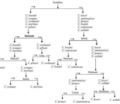
This study was conducted in the Department of Microbiology, J. N. Medical College Hospital, Aligarh, India for a period of one year and six months (2007-2008). It comprised of the samples that were received in the department from outpatient departments, different wards, nurseries, Intensive Care Units (ICUs) and emergency care unit. Various clinical specimens such as urine, pus, cerebrospinal fluid, blood, body fluids including peritoneal,pleural and bile fluids, respiratory tract specimens like bronchial aspirates, tracheal aspirates, bronchoalveolar lavage fluid and ear and nasal swabs, catheter tips and drain tips were received in leak proof sterile containers. Detailed clinical history was also obtained and recorded. Each sample was subjected to standard microbiological techniques for isolation and characterization of Citrobacter on the basis of cultural and biochemical characters [6].The genus Citrobacter was established if the isolate was motile, catalase positive, oxidase negative, lactose non fermenting, Methyl Red positive, VP negative, PPA negative, Citrate positive, ONPG positive Gram-negative rod.Further identification to species level was done by applying battery of tests as shown in Figure 1. A total of 105 randomly selected non repetitive bacterial isolates were included in the study.
Figure 1: Identification Key for Citrobacter Species Ornithine
C. freundii C. youngae C. werkmanii C. murlinae C. gillenii
C. koseri C. amalonaticus C. farmeri C. braakii C. rodenticum C. sedlakii
Malonate
C. freundii C. youngae C. murlinae
C. werkmanii C. gillenii
Malonate
Indole
C. braakii C. rodenticum
C. koseri C. amalonaticus C. farmeri C. sedlakii
C. braakii C. rodenticum
Malonate
Malonate
C. amalonaticus C. farmeri
C. koseri C. sedlakii
Citrate
C. farmeri C. amalonaticus
Melibiose
C. koseri C. sedlakii
Dulcitol
C. freundii C. youngae C. murlinae
Indole
C. youngae C. murlinae
Urea
C. gillenii C. werkmanii
−ve +ve
−ve
−ve
−ve
+ve
+ve
+ve −ve
−ve −ve
−ve
−ve +ve
+ve
+ve −ve +ve
III. RESULTSANDOBSERVATIONS
During the study period a total of 24,442 samples were received, among which 526 were identified as
[image:3.595.93.514.180.266.2]Citrobacter spp. The overall prevalence was 2.1%. Citrobacter spp were isolated in 5.8% of all isolates of pus followed by blood and urine (Table I).
Table I: Prevalence of Citrobacter in Various Clinical Specimens
Samples Total no. of isolates
Isolates identified as Citrobacter
Isolates under study
%age prevalence
Pus 6082 356 68 5.8
Urine 8023 134 20 1.7
Blood 8553 171 8 2.0
Others 1784 28 9 1.6
Total 24442 526 105 2.1
[image:3.595.162.436.341.489.2]Out of 105 randomly selected isolates 41.9% were identified as C. koseri, 19.0% were C. freundii, 11.4% were C. amalonaticus while 27.7% constituted other species of Citrobacter. (Table II)
Table II
:
Percentage Distribution of Various Citrobacter SpeciesSpecies % of strains
C. koseri 41.9
C. freundii 19.0
C. amalonaticus 11.4
C. braakii 9.5
C. sedlaaki 9.5
C. youngae 3.8
C. gilleni 2.8
C. werkmanii 0.95
C. murlinae 0.95
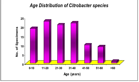
Maximum number of Citrobacter isolates were from 11-20 year age group(21.9%).Only one isolate (0.95%) was from the age group of >60 years (Figure 2). Citrobacter spp were isolated from 65.7% of males and 34.3% of female patients. The male to female ratio was 1.92:1. The maximum number of isolates were from
pus (64.76%) followed by urine and blood with 19.05% and 7.62% respectively (Table III).
20 25
n
s
[image:3.595.158.438.564.730.2]Table III: Samplewise Distribution of Citrobacter Isolates (N = 105)
SAMPLES NUMBER OF ISOLATES
(%age)
Pus 68 (64.7) Urine 20 (19.05) Blood 8 (7.6) Sputum 2 (1.9) Tracheal Swab 2 (1.9) Umbilical Tip 2 (1.9)
CSF 1 (0.95)
Pleural Fluid 1 (0.95) Endotracheal Tube 1 (0.95)
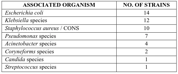
Citrobacter spp were found in mixed cultures in 40% of the isolates (Figure 3) of which maximum co-infections were recorded with Escherichia coli followed by Klebsiella spp. (Table IV)
DISTRBUTION OF ISOLATION OF CITROBACTER WITH OTHER MICRO-ORGANISMS
Monomicrobic Polymicrobic
Figure 3: Distribution of isolation of Citrobacter with other micro-organisms
Table IV: Organisms isolated in Mixed Cultures with Citrobacter Species
ASSOCIATED ORGANISM NO. OF STRAINS
Escherichia coli 14
Klebsiella species 12
Staphylococcus aureus / CONS 10
Pseudomonas species 7
Acinetobacter species 4
Coryneforms species 2
Candida species 1
[image:4.595.147.450.505.632.2]IV. DISCUSSION
Citrobacter spp. are uncommon causes of infections in neonates, young children, immunocompromised adults & older children [8, 9]. Neonates may acquire the organisms horizontally as nosocomial infections or vertically from the mother at the time of delivery. They are normal inhabitants of the gut and have been clubbed with the coliforms but when host defenses are weak or other factors favour their establishment in other tissues, serious infections may result. An association with virulence markers like the serum resistance, the cell surface hydrophobicity and the killing in the polymorphonuclear leucocytes, which had been studied in Escherichia coli, were found to exist in Citrobacter
spp. leading to its pathogenicity [10].
Citrobacter spp are one of the most misidentified genera in routine laboratory practice. It mimics many other bacteria of the family Enterobacteriaceae in colony morphology and biochemical properties hence confirmation and identification to species level is cumbersome. O’Hara scheme of identification is a simple scheme to identify Citrobacter upto species level utilising few biochemical tests. Ornithine decarboxylation remains the crucial step in establishment of species once the isolate has been identified to belong to the genus Citrobacter. It has been reported in various studies that the prevalence rate of
Citrobacter spp ranges from as low as 1.0% [11] to as high as 8.23% [12] which is consistent with our study where we report a prevalence rate of 2.1%. A prevalence rate of 2.18% and 1.8% among the uropathogens and in patients of peritonitis respectively was reported in other studies [13, 14] while in our study percentage prevalence in pus, urine and blood was 5.8, 1.7 and 2.0 percent respectively. The affected mean age of 35.5 years (range 3 days-87 years) was reported from an Indian study [15]. Comparable results were obtained in our study where we observed maximum isolation from 11-20 years and 31-40 years age range. There are other reports suggesting Citrobacter spp. to be an infective agent in extremes of age [16,17]. Male predominance was seen in our study which had been reported by other workers also [18]. In a study conducted by S. Mohanty [15], 67.3% Citrobacter isolates were from male patients while 32.7% were from female patients.
Our study emphasizes that wound infection, urinary tract infections and bacteremia are the commonest infections caused by Citrobacter. Similar observations had been made in a study who reported a maximum isolation of Citrobacter species from pus followed by urine [19]. On the contrary there are reports of their higher isolation from urine than pus [15]. In our study, limited association could be made with the site of pus collection
of the aforementioned study was from a tertiary care centre with referral from nearby areas. Acinetobacter spp as already known is a nosocomial pathogen hence was found to be more commonly associated in co-infection. Contrary to it more than half of our isolates were community acquired hence Escherichia coli took a lead.
V. CONCLUSIONS
The magnitude of Citrobacter infections have increased over time considering its potential to cause nosocomial infections and the growing numbers of immunocompromised patients in hospitals; C. koseri and C. freundii being the commonest species isolated. They are usually isolated from patients with wound infections, urinary tract infections and bacteremia. These are monomicrobial in more than half of the cases but polymicrobial infection can also be encountered. Citrobacter spp can cause infection in any age group with significant predilection in adolescent and middle age. Infection is seen in both sexes with significant proportion of infection in males. Identification should be done upto species level in all microbiology laboratories to quantitate and assess the real magnitude of these infections.
ACKNOWLEDGEMENTS
I am thankful to Late Dr. Fatima Shujatullah, Assistant Professor, Department of Microbiology, J. N. Medical College and Hospital, A.M.U., Aligarh who guided me throughout my study.
REFERENCES [1] Nada T, et al; Jpn J Infect Dis 2004;57:181-2.
[2] El Harrif-Heraud Z, et al; J Clin Microbiol 1997;35:2561-7. [3] Lin FY, et al; Pediatr Infect Dis J 1987;6:50-55.
[4] Richards, M. J., et al; Crit. Care Med. 1999;27: 887-892.
[5] Janda, J. M., and S. L. Abbott 1998. The Enterobacteria. Lippincott-Raven, Philadelphia, Pa.
[6] Collee JG, Miles RS, Watt B. Tests for identification of bacteria. In: Mackie and McCartney Practical Medical Microbiology, 14th ed. Collee JG, Fraser AG, Marmion BP, Simmons A, editors. (Churchil Livingstone: New York) 1996; 131-49.
[7] Caroline M. O’Hara, et al; J. Clin. Microbiol 1995; 242-45.
[8] Schwartz DA. Citrobacter infections. In: Connor DH, Chandler FW, Schwartz DA, Manz HJ, Lack EE, eds. Pathology of infectious diseases. Stanford, Connecticut: Appleton and Lange, 1997:513–6.
AUTHORS
First Author – Dr Ritu Nayar, MBBS, MD Microbiology,
Department of Immunoassays & Infectious Serology, Dr. Lal Path Labs Pvt Ltd, National Reference Laboratory, New Delhi, India., Email: dr.rits79@gmail.com
Second Author – Dr Indu Shukla, MBBS, MD Microbiology,
Department of Microbiology, J. N. Medical College and Hospital, A.M.U., Aligarh., Email: indushukla@hotmail.com
Third Author – Dr Asfia Sultan, MBBS, MD Microbiology,
Department of Microbiology, J. N. Medical College and Hospital, A.M.U., Aligarh, Email: drasfia@gmail.com
Corresponding Author – Dr Ritu Nayar, MBBS, MD