0095-1137/09/$08.00⫹0 doi:10.1128/JCM.01449-08
Copyright © 2009, American Society for Microbiology. All Rights Reserved.
Identification of Clinically Relevant Nonhemolytic Streptococci on the
Basis of Sequence Analysis of 16S-23S Intergenic Spacer Region
and Partial
gdh
Gene
䌤
Xiaohui Chen Nielsen,
1,2Ulrik Stenz Justesen,
1,3Rimtas Dargis,
1Michael Kemp,
1and Jens Jørgen Christensen
1*
Department of Bacteriology, Mycology and Parasitology, Statens Serum Institut, Copenhagen,1Department of Clinical Microbiology,
Rigshospitalet, Copenhagen,2and Department of Clinical Microbiology, Odense University Hospital, Odense,3Denmark
Received 29 July 2008/Returned for modification 30 July 2008/Accepted 23 January 2009
Nonhemolytic streptococci (NHS) cause serious infections, such as endocarditis and septicemia. Many conven-tional phenotypic methods are insufficient for the identification of bacteria in this group to the species level. Genetic analysis has revealed that single-gene analysis is insufficient for the identification of all species in this group of bacteria. The aim of the present study was to establish a method based on sequence analysis of the 16S-23S
intergenic spacer (ITS) region and the partial gdh gene to identify clinical relevant NHS to the species level.
Sequence analysis of the ITS region was performed with 57 NHS reference or clinical strains. Satisfactory identi-fication to the species level was achieved for 14/19 NHS species included in this study on the basis of sequence
analysis of the ITS region.Streptococcus salivariusandStreptococcus vestibularisobtained the expected taxon as the
best taxon match, but there was a short maximum score distance to the next best match (distance, <10).
Strepto-coccus mitis, Streptococcus oralis, andStreptococcus pneumoniaecould not be unambiguously discriminated by se-quence analysis of the ITS region, as was also proven by phylogenetic analysis. These five species could be identified
to the group level only by ITS sequence analysis. Partialgdhsequence analysis was applied to the 11S. oralisstrains,
the 11S. mitisstrains, and the 17S. pneumoniaestrains. All except one strain achieved a satisfactory identification
to the species level. A phylogenetic algorithm based on the analysis of partialgdhgene sequences revealed three
distinct clusters. We suggest that sequence analysis of the combination of the ITS region and the partialgdhgene
can be used in the reference laboratory for the species-level identification of NHS.
Streptococci are a heterogeneous group of bacteria consist-ing of more than 50 species. In addition to the traditional pathogenic pneumococci and hemolytic streptococci (HS), many species of non-HS (NHS), which are part of the com-mensal microbiota in the human body, are known to be op-portunistic pathogens that cause serious systemic and local infections. These infections include subacute infective endo-carditis (24), bacteremia in immunocompromised patients (19, 44), brain abscesses (34), meningitis (35), and pneumonia (5). There are reports describing associations between some spe-cific species and clinical manifestations.Streptococcus
gallolyti-cusandStreptococcus lutetiensisin the bovis group are reported
to have a strong association with colon cancer (31–33).
Strep-tococcus anginosusand Streptococcus intermediusin the
angi-nosus group are associated with abscess formation (9, 26), and
Streptococcus mitisand other normal oral commensals have a
strong association with infective endocarditis (6, 10, 22). The precise species-level identification of NHS from relevant clin-ical specimens is crucial to making the right diagnosis and understanding the pathogenesis of the infection.
The conventional phenotypic tests do not always allow ac-curate identification. The automated systems, such as the Rapid ID 32 Strep and the Vitek 2 GP systems (BioMerieux,
France), which are based on phenotypic tests, are widely ap-plied in the clinical microbiology laboratory. The large number of species relative to the number of biochemical traits exam-ined, the variability of several traits within species, the poor reproducibility of some tests, and the lack of sufficient pheno-typic data for more recently described species in the underlying databases often result in shortcomings with regard to the exact species designations (14, 23, 29, 41). In a study by Hoshino et al. (23) with 148 strains consisting of 115 clinical isolates and 33 reference strains, the rate of correct identification by commer-cial kits was below 50% but varied significantly between spe-cies. The most significant problems were observed withS. mitis,
S. oralis, and the 11Streptococcus species that have been
de-scribed since 1991. These inherent problems call for alternative means of identification.
Early and effective antimicrobial treatment can result in negative cultures with important clinical specimens, e.g., heart valve tissue or brain abscess material. This stresses the need for the possibility of performing non-culture-based molecular bi-ology examinations (43).
Gene sequence analysis has been applied in an attempt to make an accurate species-level identification of streptococci. The target sequences have included genes encoding functional
RNA (the 16S rRNA gene [3], rnpB [45]), protein-coding
genes (sodA [23],tuf[37],groESL[42],rpoB[11],gdhandddl
[12]), and noncoding spacer regions like the intergenic spacer (ITS) region (7). However, it seems that the analysis of no single gene is sufficient for this very heterogeneous group (23, 40).
* Corresponding author. Mailing address: Department of Bacteriol-ogy, Mycology and ParasitolBacteriol-ogy, Statens Serum Institut, Artillerivej 5, Copenhagen 2300, Denmark. Phone and fax: 45 32683268. E-mail: [email protected].
䌤Published ahead of print on 4 February 2009.
932
on May 16, 2020 by guest
http://jcm.asm.org/
Alternatively, the performance of multilocus sequence anal-ysis, as suggested by some studies (23), is not always realistic in the local clinical microbiology laboratory or even in reference laboratories.
The ITS region has been reported to be used for the strain typing of staphylococci andClostridium difficile(15, 16). It is also suggested to be a good candidate for species identification (17, 20, 36). According to Chen et al., the intraspecies similar-ity scores of 11 viridans group streptococcal species were high and ranged from 0.97 to 1.0, and pairwise comparison of two species of viridans group streptococci revealed a lower level of sequence similarity between their ITS regions than between their 16S rRNA gene sequences (7). These characteristics in-dicate that the ITS region might constitute a more discrimina-tive target sequence than the 16S rRNA gene for the differ-entiation of closely related species of NHS.
Thegdhgene, which is about 1,500 bp in length, encodes a 45-kDa glutamate-6-phosphate dehydrogenase. It is used to-gether with six other housekeeping genes for multilocus se-quence typing of pneumococci (23). Thegdhgene is reportedly highly conserved, as it exhibits an extremely small number of point mutations relative to the numbers in other genes (30). It
has been shown that partial gdhsequences could be used to
unambiguously differentiateS. pneumoniaefromS. mitisandS.
oralis(25, 30).
The purpose of this study was to establish a method based on ITS and partialgdhsequence analysis that is capable of unam-biguously identifying clinically relevant NHS to the species level. At the same time, the method should be able to be easily applied in a reference laboratory.
MATERIALS AND METHODS
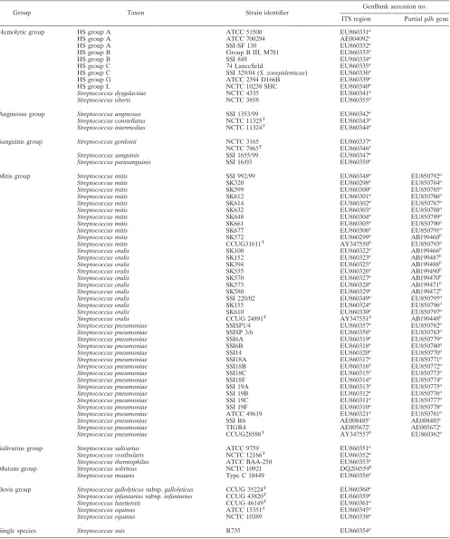
Bacterial strains.The 68 streptococcal strains used in our study are listed in Table 1. There were 57 strains representing 19 species of NHS and 11 strains representing 5 species of HS. Twenty-three of the strains were obtained from the American Type Culture Collection (ATCC; Manassas, VA), the National Col-lection of Type Strains (NCTC; London, United Kingdom), or the Culture Collection of the University of Go¨teborg (CCUG; Go¨teborg Sweden). The strains with SSI numbers were reference strains from theNeisseriaand Strepto-coccusReference Laboratory at the Statens Serum Institut (SSI; Copenhagen, Denmark). The strains with SK numbers were obtained from M. Kilian (Institute of Medical Microbiology and Immunology, University of Aarhus, Aarhus, Den-mark). All the strains used in the study were well characterized by conventional phenotypic methods, including microscopy; the evaluation of growth character-istics; performance of the catalase test; evaluation with the Rapid ID32 Strep system; determination of the production of pyrrolidonyl aminopeptidase, leucine aminopeptidase, -galactosidase, -N-acetylglucosaminidase, -glucosidase, ␣-galactosidase, alkaline phosphatase, arginine decarboxylase, urease, extracel-lular polysaccharide (dextran and levan), and esculetin from esculin; perfor-mance of the peroxide test; and the detection of acid production from inulin, salicin, raffinose, amygdalin, and glycogen. The reference strains from SSI, ATCC, NCTC, and CCUG were also identified by 16S rRNA gene sequence analysis at SSI (8). Most of the SK strains were also well identified by phyloge-netic analysis of the nucleotide sequences of four housekeeping genes,ddl,sodA,
gdh, andrpoB, at the Institute of Medical Microbiology and Immunology, Uni-versity of Aarhus (23).
DNA extraction.The genomic DNA of 35 strains was extracted from the cultures by using a QIAmp DNA minikit (Qiagen, Hilden, Germany), according to the manufacturer’s specifications. The genomic DNA of 33 strains was ex-tracted by boiling the culture: one to three colonies of each strain were boiled for 10 min at 95°C in 100l PCR-grade water.
PCR primers.To amplify the ITS region, we designed a forward primer, primer Strep16S-1471F (5⬘-GTG GGA TAG ATG ATT GGG GTG AAG T-3⬘), the 5⬘end of which is located at position 1471 of the 16S rRNA gene (Escherichia colinumbering). Reverse primer 6R-IGS (5⬘-GGG TTC CCC CAT TCG GAH
AT-3⬘) was adapted and improved from the reverse primer of Chen et al. (7). The 5⬘end of primer 6R-IGS is located at position 108 downstream of the 5⬘end of the 23S rRNA gene (E. colinumbering).
To amplify the partialgdhgene, we used two primers, primer Strep-gdhF (5⬘-ATGGACAAACCAGCNAGYTT-3⬘) and primer Strep-gdhR (5⬘-GCT TGA GGT CCC ATR CTN CC-3⬘), which amplify a 660-bp amplicon.
PCR analysis of ITS region and partialgdhgene.PCR of the ITS region and subsequent sequence analysis were performed with all 68 streptococcal strains. PCR of the partialgdhgene and subsequent sequence analysis were performed only with the 39 strains belonging to the mitis group (11S. oralisstrains, 11S. mitisstrains, and 17S. pneumoniaestrains).
The PCR was performed with 50-l reaction volumes consisting of 1⫻ Hot-StarTaq master mix (containing final concentrations of 2.5 U HotHot-StarTaq DNA polymerase, 1⫻ PCR buffer, 200 M each deoxynucleoside triphosphate; Qiagen) and 0.4M (final concentration) each primer. The conditions of the PCR with the primers for the ITS region (primers Strep16S-1471F and 6R-IGS) were as follows: 95°C for 15 min and 40 cycles of 94°C for 30 s, 61°C for 30 s, and 72°C for 30 s. The conditions of the PCR with the primers for the partialgdh
region were as follows: 95°C for 15 min and 35 cycles of 94°C for 30 s, 55°C for 30 s, and 72°C for 30 s. Both PCRs were performed with a DNA Engine Dyad cycler (Bio-Rad). The PCR products were analyzed by 2% gel electrophoresis after staining of the gels with ethidium bromide. The PCR products were purified with spin columns (Microcon YM-100 filter units; Millipore, Billerica, MA).
Sequencing of PCR products. (i) sequencing of ITS region and sequence editing.Both DNA strands of the amplicons were sequenced on an ABI Prism 3100 Avant genetic analyzer (Applied Biosystems, Foster City, CA) with a Big-Dye (version 3.1) kit (Applied Biosystems). Primers Strep16S-1471F and 6R-IGS were used as sequencing primers.
After sequencing of the PCR product, the sequence had to be edited, as described by Chen et al. (7). Portions of the 16S and 23S rRNA gene regions were removed to obtain the full sequences of the ITS region with CTAAGG at the 5⬘ends and AATAA at the 3⬘ends of the sequences of the ITS region.
(ii) Sequencing of partialgdhsequence and sequence editing.Primers Strep-gdhF and Strep-gdhR were used for the sequencing PCR. The sequences were manually edited as described by M. Kilian (Institute of Medical Microbiology and Immunology, University of Aarhus) so that the sequences were more com-parable in the NCBI BLAST search engine. For all the strains evaluated in this study, the 5⬘ends of thegdhsequences were TTTAAAAACCT, whereas the 3⬘ ends of thegdhsequences were cut just before the sequence AGA ACC ATA C, so that the 3⬘end of the edited sequence was TGC TTC/A TCC.
The edited sequences of the ITS region and the partialgdhgene were com-pared to sequences deposited in the NCBI database by using the BLAST search engine and by taking into consideration the percentage and number of identities, the maximum score, and E values for the best and the next best taxon matches. Phylogenetic analysis.DNA sequences were aligned by using the ClustalW program built into the MEGA (version 4.0) program package. Phylogenetic analysis on the basis of the sequences of the ITS region and the partialgdhgene for the 39 strains belonging to the mitis group were performed by the neighbor-joining and minimal evolution methods in the MEGA (version 4.0) program package (downloaded from http://www.megasoftware.net). The distance between the sequences was calculated by using the Kimura two-parameter model.
Nucleotide sequence accession numbers.The GenBank accession numbers of the sequences of the ITS region and the partialgdhgene obtained from our study are listed in Table 1.
RESULTS
PCR amplification and determination of sequences of ITS
region and partialgdhgene.PCR of the ITS region yielded a
single band for all strains, and the sizes of the bands varied from 550 bp to 650 bp. The DNA fragment encompassed a small portion of the 16S rRNA gene region, the ITS region, and a small portion of the 23S rRNA gene region.
PCR of the partialgdhgene yielded a single band at 660 bp, with the size being constant independent of the species.
Identification ofStreptococcusstrains (HS and NHS) on the
basis of sequence analysis of ITS region.Sequence analysis of
the ITS region was performed with all 68 streptococcal strains. The sizes of the edited ITS region sequences varied from 248 to 498 bp. All species except for theS. mitisandS. pneumoniae
on May 16, 2020 by guest
http://jcm.asm.org/
TABLE 1. The 68 strains of the genusStreptococcusused in the study
Group Taxon Strain identifier GenBank accession no.
ITS region Partialgdhgene
Hemolytic group HS group A ATCC 51500 EU860331a
HS group A ATCC 700294 AE004092c
HS group A SSI-SF 130 EU860332a
HS group B Group B III, M781 EU860333a
HS group B SSI 848 EU860334a
HS group C 74 Lancefield EU860335a
HS group C SSI 329/04 (S. zooepidemicus) EU860336a
HS group G ATCC 2394 D166B EU860339a
HS group L NCTC 10238 SHC EU860340a
Streptococcus dysgalactiae NCTC 4335 EU860341a
Streptococcus uberis NCTC 3858 EU860355a
Anginosus group Streptococcus anginosus SSI 1353/99 EU860342a
Streptococcus constellatus NCTC 11325T EU860343a
Streptococcus intermedius NCTC 11324T
EU860344a
Sanguinis group Streptococcus gordonii NCTC 3165 EU860337a
NCTC 7865T EU860346a
Streptococcus sanguinis SSI 1655/99 EU860347a
Streptococcus parasanguinis SSI 16/03 EU860350a
Mitis group Streptococcus mitis SSI 992/99 EU860348a
EU850792a
Streptococcus mitis SK320 EU860298a EU850784a
Streptococcus mitis SK599 EU860300a EU850785a
Streptococcus mitis SK612 EU860301a
EU850786a
Streptococcus mitis SK614 EU860302a
EU850787a
Streptococcus mitis SK632 EU860303a EU850788a
Streptococcus mitis SK648 EU860304a EU850789a
Streptococcus mitis SK661 EU860305a
EU850790a
Streptococcus mitis SK677 EU860306a
EU850791a
Streptococcus mitis SK572 EU860299a AB199460b
Streptococcus mitis CCUG31611T AY347550b EU850793a
Streptococcus oralis SK100 EU860322a
AB199466b
Streptococcus oralis SK152 EU860323a
AB199487b
Streptococcus oralis SK394 EU860325a AB199488b
Streptococcus oralis SK555 EU860326a AB199490b
Streptococcus oralis SK570 EU860327a
AB199470b
Streptococcus oralis SK573 EU860328a
AB199471b
Streptococcus oralis SK580 EU860329a AB199472b
Streptococcus oralis SSI 220/02 EU860349a EU850795a
Streptococcus oralis SK155 EU860324a
EU850796a
Streptococcus oralis SK610 EU860330a
EU850797a
Streptococcus oralis CCUG 24891T AY347551b AB199448b
Streptococcus pneumoniae SSISP1/4 EU860357a EU850782a
Streptococcus pneumoniae SSISP 3/6 EU860358a
EU850783a
Streptococcus pneumoniae SSI6A EU860319a
EU850779a
Streptococcus pneumoniae SSI6B EU860318a EU850780a
Streptococcus pneumoniae SSI14 EU860320a EU850770a
Streptococcus pneumoniae SSI18A EU860317a
EU850771a
Streptococcus pneumoniae SSI18B EU860316a
EU850772a
Streptococcus pneumoniae SSI18C EU860315a EU850773a
Streptococcus pneumoniae SSI18F EU860314a EU850774a
Streptococcus pneumoniae SSI 19A EU860313a
EU850775a
Streptococcus pneumoniae SSI 19B EU860312a
EU850776a
Streptococcus pneumoniae SSI 19C EU860311a EU850777a
Streptococcus pneumoniae SSI 19F EU860310a EU850778a
Streptococcus pneumoniae ATCC 49619 EU860321a
EU850781a
Streptococcus pneumoniae SSI R6 AE008485c
AE008485c
Streptococcus pneumoniae TIGR4 AE005672c AE005672c
Streptococcus pneumoniae CCUG28588T AY347557b EU860362a
Salivarius group Streptococcus salivarius ATCC 9759 EU860351a
Streptococcus vestibularis NCTC 12166T
EU860352a
Streptococcus thermophilus ATCC BAA-250 EU860353a
Mutans group Streptococcus sobrinus NCTC 10921 DQ204559b
Streptococcus mutans Type C 10449 EU860356a
Bovis group Streptococcus gallolyticussubsp.gallolyticus CCUG 35224T
EU860360a Streptococcus infantariussubsp.infantarius CCUG 43820T EU860359a
Streptococcus lutetiensis CCUG 46149T EU860361a
Streptococcus equinus ATCC 15351d
EU860345a
Streptococcus equinus NCTC 10389 EU860338a
Single species Streptococcus suis R735 EU860354a
a
Sequences obtained from our study.
b
Sequences already published in GenBank.
c
Published whole genome.
d
This strain was namedS. bovisearlier.
on May 16, 2020 by guest
http://jcm.asm.org/
strains achieved the expected taxon as the best taxon match.S.
anginosushad only a 96% sequence identity. Low maximum
score differences from the second best taxon match (difference,
⬍10) were obtained for the S. salivarius and S. vestibularis
strains, although the percent sequence identities were high (99 to 100%), and the next best taxon matches belonged to the salivarius group. Among the 39 strains in the mitis group, all 11
S. oralisstrains achieved the expected taxon as the best taxon
match, although maximum score differences to the next best taxon match (S. mitisorS. pneumoniae) were relatively low (differences, 7 to 17). Four of the 11 strains ofS. mitis(strains
SK612, SK614, SK648, and SK661) hadS. pneumoniaeas the
best taxon match andS. mitisas the second best taxon match. However, the maximum score differences between the two taxons were very low (differences, 5 to 6). Six of the 11S. mitis
strains and the 17 S. pneumoniae strains had the expected
taxon as the best taxon match, and the next best taxon matches
wereS. pneumoniaeandS. mitis, respectively. The maximum
score differences between the two best taxon matches were low (differences, 0 to 11) (Table 2).
Identification of strains belonging to the mitis group on the
basis of sequence analysis of partialgdhgene.Sequence
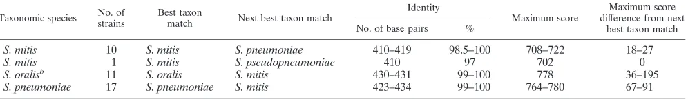
[image:4.585.47.539.81.494.2]anal-ysis of the partialgdhgene was performed with the 39 strains belonging to the mitis group. All of the edited sequences of the partialgdhgene were 431 bp in length (Table 3). Among these strains, 38 strains achieved satisfactory identification to the expected taxon with a long maximum score distance to the next best taxon (difference, 18 to 195). Only one S. mitis strain (strain SK611) could not be allocated to a single species on the basis of partialgdhgene sequence analysis, as the maximum scores forS. mitisandS. pseudopneumoniaein both cases were 710. Three other S. mitisstrains (strains SK612, SK614, and SK648) that could not be allocated to the expected taxon on the basis of sequence analysis of the ITS region achieved the correct identification on the basis of the subsequent sequence analysis of the partialgdhgene.
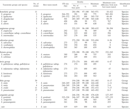
TABLE 2. Identification of HS and NHS strains on the basis of ITS sequence dataa
Taxonomic groups and species No. of
strains Best taxon match
ITS size (bp)
Identity
Maximum score
Maximum score difference from next
best taxon match
Identification level No. of
base pairs %
Hemolytic group 11 284–496 97–100 563–971 18–422 Species
S. pyogenes 3 S. pyogenes 364–391 391 100 775 365–381 Species
S. agalactiae 2 S. agalactiae 295–372 295–372 100 585–735 48–49 Species
S. dysgalactiae 4 S. dysgalactiae 301 285–369 97–100 565–648 58–79 Species
S. equi 1 S. equi 498 496 99 971 767 Species
S. uberis 1 S. uberis 431 331 99 630 422 Species
Anginosus group 3 222–319 96–100 389–632 38–394 Species
S. anginosus 1 S. anginosus 292 222 96 389 38 Species
S. constellatussubsp.constellatus 1 S. constellatus 396 319 100 632 394 Species
S. intermedius 1 S. intermedius 347 270 100 535 143 Species
Salivarius group 3 272–350 99–100 533–694 8–36 Species
S. salivarius 1 S. salivarius 273 272 99 533 8 Group
S. vestibularis 1 S. vestibularis 350 350 100 694 8 Group
S. thermophilus 1 S. thermophilus 365 365 100 659 38 Species
Mutans group 2 229–388 99–100 454–755 232–543 Species
S. mutans 1 S. mutans 389 388 99 755 543 Species
S. sobrinus 1 S. sobrinus 407 229 100 454 232 Species
Bovis group 5 273–274 100 493–495 6–47 Species
S. gallolyticussubsp.gallolyticus 1 S. gallolyticussubsp.
gallolyticus
274 274 100 495 6 Species
S. infantariussubsp.infantarius 1 S. infantariussubsp.
infantarius
273 273 100 493 47 Species
S. lutetiensis 1 S. lutetiensis 273 273 100 493 14 Species
S. equinus 2 S. equinus 364 284 100 563 18 Species
Mitis group 39 194–248 99–100 351–492 0–17 Group
S. mitis 7 S. mitis 248–249 248–249 99–100 444–448 1–11 Group
S. mitis 4 S. pneumoniae 248–249 239–248 100 432–448 5–6
S. oralis 11 S. oralis 246 194–246 99–100 351–472 7–17 Group
S. pneumoniae 17 S. pneumoniae 248 247–248 99–100 443–492 0–5 Group
Sanguinis group 4 246–336 98–100 476–628 127–397 Species
S. gordonii 2 S. gordonii 323–324 246–247 99–100 476–488 127–133 Species
S. sanguinis 1 S. sanguinis 341 294 100 583 397 Species
S. parasanguinis 1 S. parasanguinis 341 336 98 628 253 Species
S. suis 1 S. suis 419 419 100 831 617 Species
aData are compiled for strains belonging to the same species.
on May 16, 2020 by guest
http://jcm.asm.org/
No misidentification was observed on the basis of sequence analysis of the ITS region and the partialgdhgene.
Phylogenetic analysis on the basis of the sequences of the
ITS region and partialgdhgene.The phylogenetic relationship
ofS. mitis, S. oralis, andS. pneumoniaederived from the
se-quences of the ITS region is presented in Fig. 1. The 39 strains failed to form any distinct clusters according to the taxon. The evolutionary distances for 38 strains were within 0.002. The dis-tance between the lastS. oralisstrain (strain SK152) to the other strains was only 0.005. Therefore, sequence analysis of the ITS region was insufficient for discrimination of the three speciesS.
oralis,S. pneumoniae, andS. mitisfrom each other.
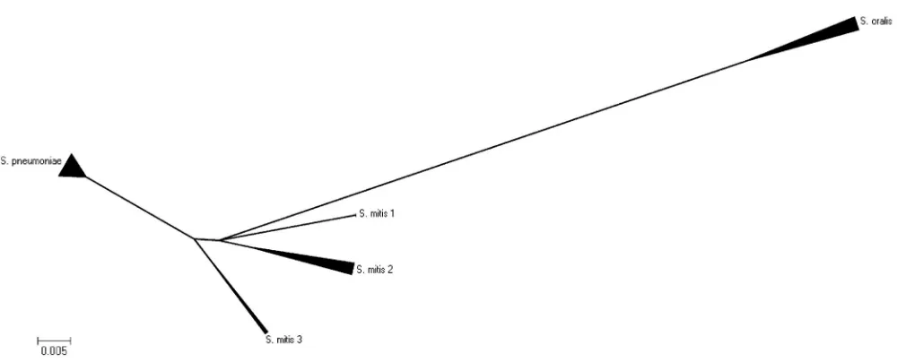
The phylogenetic relationship of S. mitis, S. oralis, andS.
pneumoniaeon the basis of the sequences of the partial gdh
genes of the 39 strains are presented in Fig. 2.S. oralis, S.
pneumoniae, andS. mitiswere separated as three distinct
clus-ters, with the type strains represented in each of the distinct clusters. The S. pneumoniae and the S. mitis clusters had a shorter distance to each other than to theS. oraliscluster. The
S. mitis cluster was much more scattered than the S.
pneu-moniaecluster. The 11S. mitisstrains formed three subclusters
within theS. mitiscluster.
DISCUSSION
We describe a method for species-level identification by combining sequence analysis of the ITS region and sequence analysis of the partialgdhgene that is capable of identifying 24 clinically relevant streptococcal species (19 NHS, 5 HS).
In our study, sequence analysis of the ITS region was used as a first-line tool for the identification of species in the genus
Streptococcus. All 11 strains (100%) belonging to the five HS
species achieved the correct species as the best taxon match on the basis of sequence analysis of the ITS region. The maximum score differences from the next best taxon match varied from 18 to 422. These differences are large enough for the differen-tiation of species. Of the strains belonging to the 19 NHS species, 53/57 strains (94%) achieved the correct species des-ignation as the best taxon match.
Members of the mutans, sanguinis, and anginosus groups achieved unambiguous identifications with high identity scores, and the differences in the maximum scores from the best to the next best taxon match were significant (232 to 543, 127 to 397, and 38 to 394, respectively).
A high degree of heterogeneity within theS. anginosus spe-cies has been reported previously (27). It was suggested that the species contains several subspecies or new species. In our
study, we included only one strain ofS. anginosus. Therefore, the low percent identity (96%) probably reflects the heteroge-neity in this species. Further molecular taxonomic studies are needed to explore this heterogeneity within strains belonging to this species.
The type strain ofS. gallolyticussubsp. gallolyticus achieved the expected taxon as the best taxon match, although the
max-imum score difference from the next best taxon match, S.
macedonicus, was only 6. The taxonomy of the bovis group has
undergone dynamic changes in the last two decades. Recent studies based on DNA-DNA hybridization, 16S rRNA gene
sequencing, and sodA gene sequencing revealed that the S.
bovis/S.equinuscomplex consists of five clusters.S. gallolyticus
and S. macedonicus belong to one cluster. The DNA-DNA
hybridization data from the same study revealed that the
ge-nomes of S. macedonicus and S. gallolyticus display ⬎70%
homology, which supports the hypothesis thatS. macedonicus
and S. gallolyticus are a single species (38). The four other
strains belonging to the bovis group included in this study,S.
infantarius subsp. infantarius, S. lutetiensis, and S. equinus,
achieved unambiguous species identification on the basis of sequence analysis of the ITS region. Strain ATCC 15351 was previously named S. bovis. The high degrees of similarity by both DNA-DNA hybridization and 16S rRNA gene sequenc-ing brought the conclusion that the speciesS. equinusandS.
bovis belong to a single species. The name S. equinus has
nomenclatural priority. Therefore,S. bovisis no longer a rec-ognized taxon (39).
Among the members of the salivarius group, onlyS. saliva-riusis commonly identified from a variety of human infections (4, 13, 21).S. thermophilushas been isolated only from dairy products.S. vestibularis was identified from the human oral cavity, and its association with human infections has not been confirmed. In our study, bothS. salivarius and S. vestibularis
achieved the correct taxon as the best taxon match, although the maximum score distance to the next taxon was only 8. The second best taxon forS. salivariuswasS. vestibularisand vice versa. The failure ofS. salivariusto produce extracellular poly-saccharides on sucrose-containing agar is helpful in securing a correct distinction between these two species.
[image:5.585.45.543.83.155.2]The differences in maximum scores between the best and the next best taxon matches were very small for the strains belong-ing to the mitis group (range of differences, 0 to 17), often making it impossible to allocate the strain examined to a spe-cific species. Four strains ofS. mitishadS. pneumoniaeas the best taxon match, followed very closely byS. mitis(maximum score differences, 5 to 6). The phylogenetic analysis of the
TABLE 3. Identifications of the 39 strains ofS. mitis,S. pneumoniae, andS. oralison the basis of the partialgdhsequencesa
Taxonomic species No. of strains
Best taxon
match Next best taxon match
Identity
Maximum score
Maximum score difference from next
best taxon match No. of base pairs %
S. mitis 10 S. mitis S. pneumoniae 410–419 98.5–100 708–722 18–27
S. mitis 1 S. mitis S. pseudopneumoniae 410 97 702 0
S. oralisb 11 S. oralis S. mitis 430–431 99–100 778 36–195
S. pneumoniae 17 S. pneumoniae S. mitis 423–434 99–100 764–780 67–91
aData are compiled for strains belonging to the same species. The partialgdhgene sequences are all 431 bp in length.
bThegdhsequences of strains SSI 220/202, SK155, and SK610 and type strain CCUG24891 were achieved in our laboratory. Thegdhsequences of strains SK100,
SK152, SK555, SK394, SK570, SK573, and SK580 were downloaded from M. Kilian’s website (www.immi.au.dk/service/download/kilian).
on May 16, 2020 by guest
http://jcm.asm.org/
results of sequence analysis of the ITS region revealed thatS.
oralisdid not form a distinct cluster in relation toS. mitisand
S. pneumoniae. It is well known that members of the mitis
group are also closely related on the basis of their 16S rRNA gene sequences (18, 28).
On the basis of these results, we concluded that the ITS region can be used for the species-level identification of strains belonging to the hemolytic, anginosus, mutans, and sanguinis groups. Strains belonging to the salivarius group and the mitis group can be identified only to the group level.
Sequence analysis of the partialgdhgene proved to be useful in separatingS. mitis,S. oralis, andS. pneumoniae, which are otherwise hard to differentiate from each other. In this study, 17/17 S. pneumoniae strains (100%), 11/11 S. oralis strains (100%), and 10/11S. mitisstrain (90.9%) were correctly iden-tified. Only oneS. mitisstrain could not be discriminated from
S. pseudopneumoniae. The maximum score difference between
these two taxons was 0. This is probably because of the taxo-nomic changes that have been made in recent years.S.
pseu-dopneumoniaeis a relatively new taxon that was first described
in 2004 and that is closely related toS. mitisandS. pneumoniae
(1). Phylogenetic analysis based on thesodA gene sequence
showed that the strains assigned to the speciesS.
pseudopneu-moniaewere more closely aligned withS. mitis than with S.
pneumoniae(1). The phylogenetic analysis based on
concate-nated partial sequences of theddl,gdh,rpoB, andsodAgenes showed thatS. pseudopneumoniaeis included within the pneu-moniae-mitis-pseudopneumoniae cluster (29). This species was, however, not included in our study.
The three distinct clusters ofS. mitis,S. pneumoniae, andS.
oralis in the phylogenetic algorithm based on partialgdh
se-quences proved thatgdhsequence analysis is capable of dis-criminating these three genetically closely related species from each other. As a result, we have a tool for the species-level identification of these species.
The phylogenetic algorithm also gave us information about the genetic relationship between the three species. There was a much longer distance from theS. oraliscluster to theS. mitis
andS. pneumoniae clusters than between theS. mitisand S.
pneumoniaeclusters. This suggests thatS. mitisand S.
pneu-moniaeare genetically more closely related to each other than
they are to S. oralis. The S. mitis cluster was much more
scattered than the clusters of the other two species, and it formed several subclusters. This suggests thatS. mitisis genet-ically more heterogeneous. This is in accordance with the re-cent observations of Bek-Thomsen et al., who observed that the range of interstrain gdh sequence distances was signifi-cantly larger for S. mitis than for what was found amongS.
pneumoniaestrains (2).
In this study, we present a reliable method for the identifi-cation of clinically relevant streptococci, with a focus on NHS, to the species level. The method is easy to perform in a labo-ratory that has sequencing facilities. By sequence analysis of
FIG. 1. Phylogenetic tree determined on the basis of the sequences of the ITS regions of 11.S. mitis(Smit) strains, 11S. oralis(Soral) strains, and 17S. pneumoniae(Spneu) strains obtained by the unrooted neighbor-joining method in the MEGA (version 4.0) program pack-age. The scale bar indicates the evolutionary distance between the
sequences determined by calculation of the percent sequence diver-gence. It clearly demonstrates thatS. mitis,S. pneumoniae, andS. oralis
are genetically closely related species and cannot be discriminated from each other on the basis of the sequences of their ITS regions.
on May 16, 2020 by guest
http://jcm.asm.org/
the ITS region, all HS strains and most NHS strains could be identified to the species level.S. mitis,S. pneumoniae, andS.
oralis could not be unambiguously discriminated from each
other by sequence analysis of the ITS region. A second se-quence analysis based on the partial gdh gene distinguished these three species from each other. Only oneS. mitisstrain
could not be unambiguously discriminated from S.
pseudo-pneumoniae, which is probably because of the new
nomencla-ture change. The phylogenetic tree based on the gdh gene
sequences clearly shows that S. oralis, S. mitis, and S.
pneu-moniaeform three distinct clusters. If colonies are available,
sequencing of the ITS region and the partialgdhgene can both be completed within 24 to 72 h.
On the basis of the results from this study, we conclude that the combination of sequence analysis of the ITS region and sequence analysis of the partialgdhgene is a potential tool for the identification of clinically relevant streptococci in a clinical microbiology reference laboratory.
ACKNOWLEDGMENT
We thank Mogens Kilian, Institute of Medical Microbiology and Immunology, University of Aarhus, for his kind advice in choosing the housekeeping genegdhfor sequencing and for supplying us with ref-erence and clinical strains.
REFERENCES
1.Arbique, J. C., C. Poyart, P. Trieu-Cuot, G. Quesne, M. G. Carvalho, A. G. Steigerwalt, R. E. Morey, D. Jackson, R. J. Davidson, and R. R. Facklam. 2004. Accuracy of phenotypic and genotypic testing for identification of
Streptococcus pneumoniae and description of Streptococcus pseudopneu-moniaesp. nov. J. Clin. Microbiol.42:4686–4696.
2.Bek-Thomsen, M., H. Tettelin, I. Hance, K. E. Nelson, and M. Kilian.2008. Population diversity and dynamics ofStreptococcus mitis,Streptococcus oralis, andStreptococcus infantisin the upper respiratory tracts of adults, deter-mined by a nonculture strategy. Infect. Immun.76:1889–1896.
3.Bosshard, P. P., S. Abels, M. Altwegg, E. C. Bottger, and R. Zbinden.2004. Comparison of conventional and molecular methods for identification of aerobic catalase-negative gram-positive cocci in the clinical laboratory. J. Clin. Microbiol.42:2065–2073.
4.Carley, N. H.1992.Streptococcus salivariusbacteremia and meningitis
fol-lowing upper gastrointestinal endoscopy and cauterization for gastric bleed-ing. Clin. Infect. Dis.14:947–948.
5.Carratala, J., B. Roson, A. Fernandez-Sevilla, F. Alcaide, and F. Gudiol. 1998. Bacteremic pneumonia in neutropenic patients with cancer: causes, empirical antibiotic therapy, and outcome. Arch. Intern. Med.158:868–872. 6.Catto, B. A., M. R. Jacobs, and D. M. Shlaes.1987.Streptococcus mitis. A
cause of serious infection in adults. Arch. Intern. Med.147:885–888. 7.Chen, C. C., L. J. Teng, and T. C. Chang.2004. Identification of clinically
relevant viridans group streptococci by sequence analysis of the 16S-23S ribosomal DNA spacer region. J. Clin. Microbiol.42:2651–2657. 8.Christensen, J. J., R. Dargis, M. S. Kaltoft, K. Andresen, and M. Kemp.
2006. Ribosomal DNA sequencing of streptococci: usefulness in species identification? Int. Congr. Ser.1289:155–158.
9.Claridge, J. E., III, S. Attorri, D. M. Musher, J. Hebert, and S. Dunbar.2001.
Streptococcus intermedius,Streptococcus constellatus, andStreptococcus angi-nosus(“Streptococcus millerigroup”) are of different clinical importance and are not equally associated with abscess. Clin. Infect. Dis.32:1511–1515. 10.Douglas, C. W., J. Heath, K. K. Hampton, and F. E. Preston.1993. Identity
of viridans streptococci isolated from cases of infective endocarditis. J. Med. Microbiol.39:179–182.
11.Drancourt, M., V. Roux, P. E. Fournier, and D. Raoult.2004.rpoBgene sequence-based identification of aerobic gram-positive cocci of the genera
Streptococcus, Enterococcus, Gemella, Abiotrophia, and Granulicatella. J. Clin. Microbiol.42:497–504.
12.Garnier, F., G. Gerbaud, P. Courvalin, and M. Galimand.1997. Identifica-tion of clinically relevant viridans group streptococci to the species level by PCR. J. Clin. Microbiol.35:2337–2341.
13.Gautam, M., K. B. Chopra, D. D. Douglas, R. A. Stewart, and S. Kusne. 2007.Streptococcus salivariusbacteremia and spontaneous bacterial perito-nitis in liver transplantation candidates. Liver Transplant.13:1582–1588. 14.Gorm, J. T., K. H. Bossen, and B. Bruun.1999. Evaluation of the Rapid ID
32 Strep system. Clin. Microbiol. Infect.5:417–423.
15.Gurtler, V.1993. Typing ofClostridium difficilestrains by PCR-amplification of variable length 16S-23S rDNA spacer regions. J. Gen. Microbiol.139: 3089–3097.
16.Gurtler, V., and H. D. Barrie.1995. Typing ofStaphylococcus aureusstrains by PCR-amplification of variable-length 16S-23S rDNA spacer regions: char-acterization of spacer sequences. Microbiology141(Pt 5):1255–1265. 17.Gurtler, V., and V. A. Stanisich.1996. New approaches to typing and
iden-tification of bacteria using the 16S-23S rDNA spacer region. Microbiology 142(Pt 1):3–16.
18.Haanpera, M., J. Jalava, P. Huovinen, O. Meurman, and K. Rantakokko-Jalava.2007. Identification of alpha-hemolytic streptococci by pyrosequenc-ing the 16S rRNA gene and by use of VITEK 2. J. Clin. Microbiol.45:762– 770.
19.Han, X. Y., M. Kamana, and K. V. Rolston.2006. Viridans streptococci isolated by culture from blood of cancer patients: clinical and microbiologic analysis of 50 cases. J. Clin. Microbiol.44:160–165.
[image:7.585.43.548.61.263.2]20.Henry, T., P. C. Iwen, and S. H. Hinrichs.2000. Identification ofAspergillus
FIG. 2. Minimal evolution algorithm (suppressed) obtained by using the MEGA (version 4.0) program and based on the partialgdhgene sequences of 11S. oralis strains, 17S. pneumoniaestrains, and 13S. mitisstrains. It shows that the three species form three distinct clusters. The
S. oraliscluster has a longer distance to the two other clusters, indicating thatS. pneumoniaeandS. mitisare genetically more closely related on the basis ofgdhgene evolution. There are three subclusters within theS. mitiscluster, indicating that the speciesS. mitiscontains a heterogeneous group of strains.
on May 16, 2020 by guest
http://jcm.asm.org/
species using internal transcribed spacer regions 1 and 2. J. Clin. Microbiol. 38:1510–1515.
21.Hoecker, J. L., L. K. Pickering, D. Groschel, and S. Kohl.1978.Streptococcus salivariussepsis in children with malignancies. J. Pediatr.92:337–338. 22.Horaud, T., and F. Delbos.1984. Viridans streptococci in infective
endocar-ditis: species distribution and susceptibility to antibiotics. Eur. Heart J. 5(Suppl. C):39–44.
23.Hoshino, T., T. Fujiwara, and M. Kilian.2005. Use of phylogenetic and phenotypic analyses to identify nonhemolytic streptococci isolated from bac-teremic patients. J. Clin. Microbiol.43:6073–6085.
24.Hsu, R. B., and F. Y. Lin.2006. Effect of penicillin resistance on presentation and outcome of nonenterococcal streptococcal infective endocarditis. Car-diology105:234–239.
25.Ip, M., F. Chi, S. S. Chau, M. Hui, J. Tang, and P. K. Chan.2006. Use of the housekeeping genes,gdh(zwf) andgki, in multilocus sequence typing to differentiateStreptococcus pneumoniaefromStreptococcus mitisand Strepto-coccus oralis. Diagn. Microbiol. Infect. Dis.56:321–324.
26.Jacobs, J. A., H. G. Pietersen, E. E. Stobberingh, and P. B. Soeters.1995.
Streptococcus anginosus,Streptococcus constellatusandStreptococcus interme-dius. Clinical relevance, hemolytic and serologic characteristics. Am. J. Clin. Pathol.104:547–553.
27.Jacobs, J. A., C. S. Schot, and L. M. Schouls.2000. TheStreptococcus anginosusspecies comprises five 16S rRNA ribogroups with different phe-notypic characteristics and clinical relevance. Int. J. Syst. Evol. Microbiol. 50(Pt 3):1073–1079.
28.Kawamura, Y., X. G. Hou, F. Sultana, H. Miura, and T. Ezaki.1995. De-termination of 16S rRNA sequences ofStreptococcus mitisandStreptococcus gordoniiand phylogenetic relationships among members of the genus Strep-tococcus. Int. J. Syst. Bacteriol.45:406–408.
29.Kilian, M., K. Poulsen, T. Blomqvist, L. S. Havarstein, M. Bek-Thomsen, H. Tettelin, and U. B. Sorensen.2008. Evolution ofStreptococcus pneumoniae
and its close commensal relatives. PLoS One3:e2683.
30.Kiratisin, P., L. Li, P. R. Murray, and S. H. Fischer.2005. Use of house-keeping gene sequencing for species identification of viridans streptococci. Diagn. Microbiol. Infect. Dis.51:297–301.
31.Klein, R. S., M. T. Catalano, S. C. Edberg, J. I. Casey, and N. H. Steigbigel. 1979.Streptococcus bovissepticemia and carcinoma of the colon. Ann. In-tern. Med.91:560–562.
32.Klein, R. S., R. A. Recco, M. T. Catalano, S. C. Edberg, J. I. Casey, and N. H. Steigbigel.1977. Association ofStreptococcus boviswith carcinoma of the colon. N. Engl. J. Med.297:800–802.
33.Kok, H., R. Jureen, C. Y. Soon, and B. H. Tey.2007. Colon cancer presenting asStreptococcus gallolyticusinfective endocarditis. Singapore Med. J.48:e43– e45.
34.Lu, C. H., W. N. Chang, Y. C. Lin, N. W. Tsai, P. C. Liliang, T. M. Su, C. S. Rau, Y. D. Tsai, C. L. Liang, C. J. Chang, P. Y. Lee, H. W. Chang, and J. J.
Wu.2002. Bacterial brain abscess: microbiological features, epidemiological trends and therapeutic outcomes. Q. J. Med.95:501–509.
35.Moller, K., E. H. Frederiksen, J. H. Wandall, and P. Skinhoj.1999. Menin-gitis caused by streptococci other thanStreptococcus pneumoniae: a retro-spective clinical study. Scand. J. Infect. Dis.31:375–381.
36.Olsen, R. J., P. A. Cernoch, C. M. Austin, E. A. Graviss, D. H. Farkas, and G. A. Land.2007. Validation of the MycoAlign system forMycobacterium
spp. identification. Diagn. Microbiol. Infect. Dis.59:105–108.
37.Picard, F. J., D. Ke, D. K. Boudreau, M. Boissinot, A. Huletsky, D. Richard, M. Ouellette, P. H. Roy, and M. G. Bergeron.2004. Use oftufsequences for genus-specific PCR detection and phylogenetic analysis of 28 streptococcal species. J. Clin. Microbiol.42:3686–3695.
38.Poyart, C., G. Quesne, and P. Trieu-Cuot.2002. Taxonomic dissection of the
Streptococcus bovisgroup by analysis of manganese-dependent superoxide dismutase gene (sodA) sequences: reclassification of ‘Streptococcus infan-tariussubsp.coli’ asStreptococcus lutetiensissp. nov. and ofStreptococcus bovisbiotype 11.2 asStreptococcus pasteurianussp. nov. Int. J. Syst. Evol. Microbiol.52:1247–1255.
39.Schlegel, L., F. Grimont, E. Ageron, P. A. Grimont, and A. Bouvet.2003. Reappraisal of the taxonomy of theStreptococcus bovis/Streptococcus equinus
complex and related species: description ofStreptococcus gallolyticussubsp.
gallolyticussubsp. nov.,S. gallolyticussubsp.macedonicussubsp. nov. andS. gallolyticussubsp.pasteurianussubsp. nov. Int. J. Syst. Evol. Microbiol.53: 631–645.
40.Simmon, K. E., L. Hall, C. W. Woods, F. Marco, J. M. Miro, C. Cabell, B. Hoen, M. Marin, R. Utili, E. Giannitsioti, T. Doco-Lecompte, S. Bradley, S. Mirrett, A. Tambic, S. Ryan, D. Gordon, P. Jones, T. Korman, D. Wray, L. B. Reller, M. F. Tripodi, P. Plesiat, A. J. Morris, S. Lang, D. R. Murdoch, and C. A. Petti.2008. Phylogenetic analysis of viridans group streptococci causing endocarditis. J. Clin. Microbiol.46:3087–3090.
41.Spelleberg, B., and C. Brandt.2007.Streptococcus, p. 412–429. InP. R. Murray, E. J. Baron, J. H. Jorgensen, M. L. Landry, and M. A. Pfaller (ed.), Manual of clinical microbiology, 9th ed. ASM Press, Washington, DC. 42.Teng, L. J., P. R. Hsueh, J. C. Tsai, P. W. Chen, J. C. Hsu, H. C. Lai, C. N.
Lee, and S. W. Ho.2002.groESL sequence determination, phylogenetic analysis, and species differentiation for viridans group streptococci. J. Clin. Microbiol.40:3172–3178.
43.Tsai, J. C., L. J. Teng, and P. R. Hsueh.2008. Direct detection of bacterial pathogens in brain abscesses by polymerase chain reaction amplification and sequencing of partial 16S ribosomal deoxyribonucleic acid fragments. Neu-rosurgery62(Suppl. 2):547–555.
44.Tunkel, A. R., and K. A. Sepkowitz.2002. Infections caused by viridans streptococci in patients with neutropenia. Clin. Infect. Dis.34:1524–1529. 45.Westling, K., I. Julander, P. Ljungman, M. Vondracek, B. Wretlind, and S.
Jalal.2008. Identification of species of viridans group streptococci in clinical blood culture isolates by sequence analysis of the RNase P RNA gene,rnpB. J. Infect.56:204–210.