Introduction
Stem cells
Stem cell (SC) therapy is not a new concept. In the after-math of the bombings of Hiroshima and Nagasaki in 1945, researchers discovered that bone marrow (BM) trans planted into irradiated mice produced hemato-poiesis [1]. Hematopoietic stem cells (HSCs) were fi rst identifi ed in 1961, and their ability to migrate and diff erentiate into multiple cell types was documented [2].
Distinct SC types have been established from embryos and identifi ed in fetal tissues and umbilical cord blood (UCB) as well as in specifi c niches in many adult mammalian tissues and organs such as BM, brain, skin, eyes, heart, kidneys, lungs, gastrointestinal tract, pancreas, liver, breast, ovaries, prostate, and testis [3]. All SCs are
undiff erentiated cells that exhibit unlimited self-renewal and can generate multiple cell lineages or more restricted progenitor populations that can contribute to tissue homeostasis by replenishing the cells or to tissue regeneration after injury [4,5].
A progenitor cell is a biological cell that, like an SC, has a tendency to diff erentiate into a specifi c type of cell but is already more specifi c than an SC and is pushed to diff erentiate into its ‘target’ cell. Th e most important diff erence between SCs and progenitor cells is that SCs can replicate indefi nitely, whereas progenitor cells can divide only a limited number of times. Controversy about the exact defi nition remains and the concept is still evolving.
Several investigations [5-7] have been carried out with isolated embryonic, fetal, and adult SCs in a well-defi ned culture microenvironment to defi ne the sequential steps and intracellular pathways that are involved in their diff erentiation into the specifi c cell lineages. More particularly, diff erent methods, including the use of cell feeder layers, cell-free conditions, and extracellular matrix molecules such as collagen, gelatin, and laminin and diverse growth factors and cytokines, have been developed for the in vitro culture of SCs [3,5].
Retinal diseases
Age-related macular degeneration (AMD), glaucoma, and diabetic retinopathy are the three most common causes of visual impairment and legal blindness in developed countries [8-10]. One common denominator of these conditions is progressive loss of the neural cells of the eye – photoreceptors, interneurons, and retinal gang lion cells, or RGCs – and essential supporting cells such as the retinal pigment epithelium (RPE). Retinal dys trophies – retinitis pigmentosa (RP) (Figure 1), Stargardt disease, Best disease, Leber congenital amaurosis, and so on – all evolve with early loss of photoreceptors and subsequent loss of RGC. Recent years have seen enormous progress in the treatment options that stop the progression of AMD from a neovascular state to fi brosis, that slow down the progression of glaucoma by reducing intraocular pressure, and that prevent progression of diabetic retinopathy by optimizing glycemic control and treat retinal neovascularization early [11-14]. However, irreversible visual loss still occurs in a signifi cant
Abstract
Distinct stem cell types have been established from embryos and identifi ed in the fetal tissues and umbilical cord blood as well as in specifi c niches in many adult mammalian tissues and organs such as bone marrow, brain, skin, eyes, heart, kidneys, lungs, gastrointestinal tract, pancreas, liver, breast, ovaries, and prostate. All stem cells are undiff erentiated cells that exhibit unlimited self-renewal and can generate multiple cell lineages or more restricted progenitor populations that can contribute to tissue homeostasis by replenishing the cells or to tissue regeneration after injury. The remarkable progress of regenerative medicine in the last few years indicates promise for the use of stem cells in the treatment of ophthalmic disorders. Experimental and human studies with intravitreal bone marrow-derived stem cells have begun. This paper reviews recent advances and potential sources of stem cells for cell therapy in retinal diseases.
© 2010 BioMed Central Ltd
Stem cell therapy for retinal diseases: update
Rubens Camargo Siqueira*
1-3R E V I E W
*Correspondence: [email protected]
2Rubens Siqueira Research Center, São José do Rio Preto, Rua Saldanha Marinho 2815 conj 42, São Paulo 15010-100, Brazil
Full list of author information is available at the end of the article
pro portion of cases. Research is aimed at developing novel treatments using neuroprotective and regenerative strategies.
SCs can potentially be used for both neuroprotection and cell replacement. Intravitreal delivery of neuro-trophic factors slows down photoreceptor degeneration in rodent models of RP, RGC loss in glaucoma models, and optic nerve and optic tract trauma, but the eff ect may be temporary. Slow-release preparations and gene therapy approaches used to induce retinal cells to secrete neurotrophic factors are two ways to induce longer-term eff ects. A third option is to use SCs as long-term delivery agents, possibly encapsulated in a device, because many SCs either secrete neurotrophins naturally or can be genetically engineered to do so [15-17]. SCs can be injected into the eye through the intravitreal or subretinal technique [18-21].
Progress has also been made in the fi eld of photo-receptor, RPE, and RGC replacement by SCs and progenitor cells, although long-term restoration of visual function has notbeen confi rmed. Th e recent discoveries that human fi broblasts can be ‘reprogrammed’ to behave like embryonic SCs and that adult eyes harbor retinal progenitor cells (RPCs) also increase the potential availa-bility of SCs for trans plantation, including autolo gous transplantation, and stimulate intrinsic ‘self-regeneration’, which could poten tially overcome a lot of the problems
associated with non-autologous transplan tation in
humans [15].
Potential sources of stem cells for cell therapy in retinal diseases
Bone marrow-derived stem cells
BM-derived SCs have been proposed as a potential source of cells for regenerative medicine [9,22]. Th is is based on the assumption that HSCs isolated from BM are plastic and are able to ‘transdiff erentiate’ into tissue-committed SCs for other organs (for example, heart, liver, or brain). Unfortunately, the concept of SC plasticity was
not confi rmed in recent studies, and previously encour-ag ing data demonstrating this phenomenon in vitro could be explained by a phenomenon of cell fusion or, as believed by our group, by the presence of heterogeneous populations of SCs in BM [23]. Th e identifi cation of very small, embryonic-like SCs in BM supports the notion that this tissue contains a population of primitive SCs, which, if transplanted together with HSCs, would be able to regenerate damaged tissues in certain experimental settings. Cells from BM are easily and safely aspirated. After administration of local anesthesia, about 10 mL of the BM is aspirated from the iliac crest by means of a sterile BM aspiration needle; subsequently, mononuclear BM-derived SCs are separated by using the Ficoll density separation method [3,18].
SC-based therapy has been tested in animal models for several diseases, including neurodegenerative disorders such as Parkinson disease, spinal cord injury, and multiple sclerosis. Th e replacement of lost neurons that are not physiologically replaced is pivotal for therapeutic success. In the eye, degeneration of neural cells in the retina is a hallmark of widespread ocular diseases such as AMD and RP. In these cases, the photoreceptor loss that occurs as a primary event (as in RP) or secondary to loss of RPE (as in AMD) leads to blindness [3,9].
BM is an ideal tissue for studying SCs because of its accessibility and because proliferative dose responses of BM-derived SCs can be readily investigated. Further-more, there are a number of well-defi ned mouse models and cell surface markers that allow eff ective studies of hematopoiesis in healthy and injured mice. Because of these characteristics and the experience of BM trans-plantation in the treatment of hematological cancers, BM-derived SCs have also become an important tool in regenerative medicine. Th e BM harbors at least two distinct SC populations: HSCs and mesen chymal stem cells (MSCs) [24,25].
Hematopoietic stem cells
HSCs are multipotent SCs that give rise to all of the blood cell types, including myeloid lineages (monocytes, macrophages, neutrophils, basophils, eosinophils, eryth ro-cytes, megakaryocytes/platelets, and dendritic cells) and lymphoid lineages (T cells, B cells, and natural killer cells). HSCs are found in adult BM in, for example, femurs, hips, ribs, the sternum, and other bones. Cells can be obtained directly from the hip by using a needle and syringe (Figure 1) or from the blood after pre treatment with cytokines, such as G-CSFs (granulocyte colony-stimulating factors), that induce cells to be released from the BM compartment. Other sources for clinical and scientifi c use include UCB and placenta [23,24].
population) by vital dyes such as rhodamine 123 (rhodamine-dull, also called rholo) or Hoechst 33342, and presence of various surface antigenic markers, many of which belong to the cluster of diff erentiation (CD) series (CD34, CD38, CD90, CD133, CD105, and CD45) and also c-kit and SC factor receptor [26-31]. Otani and colleagues [16] demonstrated that, whenever a fraction of mouse or human adult BM-derived SCs (lineage-negative hematopoietic stem cells, or Lin-HSCs) containing endo-thelial precursors stabilizes and rescues retinal blood vessels that would ordinarily completely degenerate, a dramatic neurotrophic rescue eff ect is also observed. Retinal nuclear layers are preserved in two mouse models of retinal degeneration, rd1 and rd10, and detectable, albeit severely abnormal, electroretinogram recordings are observed in rescued mice at times when they are never observed in control-treated or untreated eyes. Th e normal mouse retina consists predominantly of rods, but the rescued cells after treatment with Lin-HSCs are nearly all cones. Microarray analysis of rescued retinas demonstrates signifi cant upregulation of many anti-apoptotic genes, including small heat shock proteins and transcription factors.
Some reports have demonstrated the clinical feasibility of the intravitreal administration of autologous bone marrow-derived mononuclear cells (ABMCs) in patients with advanced degenerative retinopathies [32,33]. More recently, our group conducted a prospective, phase I, non-randomized, open-label study that included three patients with RP and two patients with cone-rod dys-trophy and an Early Treatment Diabetic Retinopathy Study best-corrected visual acuity of 20/200 or worse [34]. Evaluations such as best-corrected visual acuity, full-fi eld electroretinography, kinetic visual full-fi eld (Goldman), fl uorescein and indo cyanine green angiography, and optical coherence tomography were performed at base-line and 1, 7, 13, 18, 22, and 40 weeks after intravitreal injection of 10 × 106 ABMCs (0.1 mL) into one study eye
of each patient. No adverse event associated with the injection was observed. A 1-line improvement in best-corrected visual acuity was measured in four patients 1 week after injection and was maintained throughout follow-up. Th ree patients showed undetectable electro-retinography responses at all study visits, whereas one
patient demonstrated residual res ponses for
dark-adapted standard fl ash stimulus (a wave amplitude of approximately 35 mV), which remained recordable throughout follow-up, and one patient showed a small response (a wave amplitude of approximately 20 mV) recordable only at weeks 7, 13, 22, and 40. Visual fi elds showed no reduction (with a Goldman Standard V5e stimulus) for any patient at any visit. No other changes were observed on optical coherence tomography or fl uorescein and indocyanine green angiograms. We
conclude that intravitreal injection of ABMCs in eyes with advanced RP or cone-rod dystrophy was associated with no detectable structural or functional toxicity over a period of 10 months [34].
Mesenchymal stem cells
MSCs are progenitors of all connective tissue cells. In adults of multiple vertebrate species, MSCs have been isolated from BM and other tissues, expanded in culture, and diff erentiated into several tissue-forming cells such as bone, cartilage, fat, muscle, tendon, liver, kidney, heart, and even brain cells.
According to the International Society for Cellular Th erapy [35], there are three minimum requirements for a population of cells to be classifi ed as MSCs. Th e fi rst is that MSCs, unlike BM-derived hematopoietic cells, are isolated from a population of mononuclear cells on the basis of their selective adherence to the surface of the plastic of culture dishes; a disadvantage of this method of identifi cation is the possible contamination by hemato-poietic cells and cellular heterogeneity with respect to the potential for diff erentiation. Second, CD105, CD73, and CD90 must be present and CD34, CD45, CD14 or CD11b, CD79, or CD19 and HLA-DR must not be expressed in more than 95% of the cells in culture. Finally, the cells can be diff erentiated into bone, fat, and cartilage [36].
A number of studies have shown that BM-derived MSCs can diff erentiate into cells expressing photo-receptor proteins when injected into the subretinal space [37,38]. Interestingly, it has been suggested that rat MSCs can be made to express photopigment (rhodopsin) in vitro simply by adding epidermal growth factor to the culture media [39]. Additionally, although other retina-relevant cell types have been engineered, a number of studies have shown that BM- or adipose tissue-derived MSCs are converted to RPE cells [39-41]. As with work on other neuronal phenotypes, however, there has now been a reassessment of the ability of MSCs to diff erentiate into functionally useful retinal cells. Some studies have shown that transplanted BM-derived MSCs do not diff erentiate into neural retinal cells [42]. In an in vitro rat retina-explant model, untreated MSCs seemed to transdiff erentiate into microglia109 in a way reminiscent of earlier work on MSC transplants in other neurological tissue [43]. Some limited improvement was seen with pre-treatment with brain-derived neurotrophic factor (BDNF), nerve growth factor, and basic fi broblast growth factor (bFGF) in terms of morphological diff er entiation into retinal neurons and expression of NF200, GFAP, PKC-alpha, and recoverin, but these cells did not express rhodopsin [44].
expression of neuron-specifi c enolase and neurofi lament) and secrete ciliary neurotrophic factor (CNTF), bFGF, and BDNF for at least 4 weeks [45]. Animal studies have also demonstrated that subretinal transplantation of MSCs delays retinal degeneration and preserves retinal function through a trophic response [46]. UCB-derived MSCs have also been shown to be neuroprotective of rat ganglion cells [47]. Very recently, the intravenous
adminis tration of BM-derived MSCs was shown to
prevent photoreceptor loss and preserve visual function in the Royal College of Surgeons (RCS) rat model of RP [48].
A role for genetically modifi ed MSCs may emerge in the treatment of subretinal neovascularization. It has been shown that BM-derived MSCs accumulate around sub-retinal membranes induced by sub-retinal laser burns. Intra-venous injection of mouse BM-derived MSCs genetically engineered to secrete pigment epithelium-derived factor resulted in smaller neovascular complexes [49].
Induced pluripotent stem cells
Current methods of producing SCs from adult somatic cells off er an alternative cell source for transplantation. Induced pluripotent stem cells (iPSCs) are morpho-logically identical to embryonic SCs, display similar gene expression profi les and epigenetic status, and have the potential to form any cell in the body [45,50]. Th ese cells have been employed to generate cells for the treatment of various diseases, including diabetes, cardio vascular disease, sickle cell anemia, Parkinson disease, and hemophilia [51-55]. Meyer and colleagues [56] recently showed that iPSCs can diff erentiate into retinal cell types, whereas a paper by Buchholz and colleagues [57] showed that human iPSCs can be diff erentiated into retinal pigment epithelial cells that display functionality in vitro.
Carr and colleagues [58] demonstrated that iPSCs can be diff erentiated into functional iPSC RPE cells and that transplantation of these cells can facilitate the short-term maintenance of photoreceptors through phagocytosis of photoreceptor outer segments. Long-term visual func-tion is maintained in this model of retinal disease even though the xenografted cells are eventually lost, suggest-ing a secondary protective host cellular response. Zhao and colleagues [59] showed that abnormal gene expres-sion in some cells diff erentiated from iPSCs can induce T cell-dependent immune response in syngeneic recipients. Th erefore, the immunogenicity of therapeu-tically valuable cells derived from patient-specifi c iPSCs should be evaluated before any clinical application of these autologous cells into the patients.
Owing to viral insertions of pluripotency genes, this particular line of iPSC RPE cells cannot be used as a direct therapy, but recent advances in iPSC cell re pro-gramming technology, including the use of small
molecules [60], piggyBac transposition [61,62], non-integrating episomal vectors [63], and manipulation of endogenous transcription factors [64], should eliminate the risks associated with the integration of SC genes into the genome. Furthermore, the fi nding that blood cells can be used to derive iPSCs [65] may remove the need for the invasive biopsies required to collect somatic cells and accelerate the ethical production of SC-derived tissue for therapeutic use.
Human embryonic stem cells
Th e human embryonic stem cell (hESC) is defi ned as a cell that can both renew itself by repeated division and diff erentiate into any one of the 200 or more adult cell types in the human body. An hESC arises from the eight-cell stage morula. Outside of normal development, hESCs have been diff erentiated in vitro into neural cell types and even pigmented epithelium, although control-ling their diff erentiation has proven challenging. Several hESC lines exist and are supported by public research funds. Th e use of hESCs has signifi cant limitations, including ethical issues, and a risk of teratoma formation, but the chief problem is that we are still struggling to understand the developmental cues that diff erentiate hESCs into the specifi c adult cell types required to repair damaged tissues [66].
Nistor and colleagues [67] showed, for the fi rst time, that three-dimensional early retinal progenitor tissue constructs can be derived from hESCs. Th ree-dimensional tissue constructs were developed by culturing hESC-derived neural retinal progenitors in a matrix on top of hESC-derived RPE cells in a cell culture insert. An osmolarity gradient maintained the nutrition of the three-dimensional cell constructs. Cross-sections through hESC-derived tissue constructs were character ized by immunohistochemistry for various transcription factors and cell markers. Tissue constructs derived from hESCs expressed transcription factors characteristic of retinal development, such as pax6, Otx2, Chx10, retinal RAX, Brn3b (necessary for diff erentiation of RGCs), and crx and nrl (which have a role in photoreceptor develop-ment). Many cells expressed neuronal markers, including nestin, beta-tubulin, and microtubule-associated protein.
Stargardt disease, respectively. Good manufacturing practice-compliant hESC RPE cells survived subretinal transplantation in RCS rats for prolonged periods (>220 days). Th e cells sustained visual function and photo-receptor integrity in a dose-dependent fashion without teratoma formation or untoward pathological reactions.
Near-normal functional measurements were recorded at survival of greater than 60 days in RCS rats. To further address safety concerns, a good laboratory practice-compliant study was carried out in the NIH III immune-defi cient mouse model. Long-term data (spanning the life of the animals) showed no gross or microscopic evidence of teratoma/tumor formation after subretinal hESC RPE cell transplantation. Th ese results suggest that hESCs could serve as a potentially safe and inexhaustible source of RPE cells for the effi cacious treatment of a range of retinal degenerative diseases.
In 2010, the US Food and Drug Administration granted orphan drug designation to RPE cells for Advanced Cell Technology, Inc. (Santa Monica, CA, USA) to initiate its phase 1/2 clinical trials using RPE cells derived from hESCs to treat patients with Stargardt macular dystrophy. Moreover, in 2011, the company received a positive opinion from the Committee for Orphan Medicinal Products of the European Medicines Agency for the designation of this product as an orphan medicinal product for the treatment of Stargardt disease [69,70].
Retinal progenitor cells
RPCs, considered the active cellular component of fetal retinal transplants, were purifi ed from green fl uorescent
protein transgenic mice and trans planted into the
degenerating retina of a mature mouse model. Th e
transplanted RPCs developed into neurons, including presumptive photoreceptor cells expres sing rhodopsin,
opsin, and recoverin. Th e host showed rescue of the outer retina layer cells with integration of donor cells occurring in multiple retinal layers. Th e greatest concentration of integration in the outer retina and, most importantly, recipient mice, demonstrated an improved response to light [71,72].
In adults, the neural retina and RPE have overlapping regenerative capacity after injury (for example, in amphibians, injury can activate both retinal and RPE progenitor cells to mediate regeneration and repair).
Table 1. Mechanisms of the paracrine eff ect
Paracrine eff ect Mechanisms
Increased angiogenesis Stem cells produce local signaling molecules that may improve perfusion and enhance angiogenesis to chronically ischemic tissue. Although the particular growth factors contributing to this neovascular eff ect remain to be defi ned, the list includes vascular endothelial growth factor, hepatocyte growth factor, and basic fi broblast growth factor 2 [84,85]. Decreased infl ammation Stem cells appear to attenuate infarct size and injury by modulating local infl ammation. When transplanted into injured
tissue, the stem cell faces a hostile, nutrient-defi cient, infl ammatory environment and may release substances that limit local infl ammation in order to enhance its survival. Recent studies implicate the release of the anti-infl ammatory cytokine interleukin-10 as playing an integral role in modulating the activity of innate and adaptive immune cells, such as dendritic cells, T cells, and B cells [3,83,86].
Anti-apoptotic and Stem cells in a third pathway promote salvage of tenuous or malfunctioning cell types at the infarct border zone. Injection chemotactic signaling of mesenchymal stem cells (MSCs) into a cryo-induced infarct reduces myocardial scar width 10 weeks
later. MSCs appear to activate an anti-apoptosis signaling system at the infarct border zone and this eff ectively protects ischemia-threatened cell types from apoptosis [3,39,83].
Benefi cial remodeling of the Stem cell transplantation alters the extracellular matrix, resulting in a more favorable post-infarct remodeling, extracellular matrix strengthening of the infarct scar, and prevention of deterioration in organ function [3,83,87].
Activation of neighboring Exogenous stem cell transplantation may activate neighboring resident tissue stem cells. These resident stem cells may resident stem cells possess growth factor receptors that can be activated to induce their migration and proliferation and promote both the
restoration of dead tissue and the improved function in damaged tissue [3,26].
Limited damage of amphibian retina activates progenitor cells within the neural retina, presumably retina stem cells (RSCs), to mediate regeneration and repair, whereas extensive damage that destroys most of the neural retina results in the activation of a progenitor cell population within the RPE layer, presumably retinal pigment epithelial SCs, to regenerate both neural retina and RPE [72,73]. RSCs have been isolated from the edge of the peripheral regionof the neurosensory mammalian retina, although questions regarding the true origin of
these cells and the ability to self-renew have been raised [74,75].
More recently, RSCs that demonstrate self-renewal and diff erentiated progeny have been isolated from the posterior neural retina. Of the multiple types of RSCs and RPCs reported, retinal glial Müller cells are the most studied late progenitors that retain competency to produce neuronal lineages, including photoreceptor cells. Müller glial cells actively regenerate damaged retina in lower animals and can be similarly activated in mice by
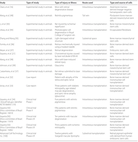
Table 2. Clinical and experimental studies using cell therapy for retinal diseases
Study Type of study Type of injury or illness Route used Type and source of cells
Otani, et al. [16] Experimental study in animals Mice with retinal degenerative disease
Intravitreous transplantation Adult bone marrow-derived lineage-negative hematopoietic stem cells Wang, et al. [48] Experimental study in animals Retinitis pigmentosa Tail vein Pluripotent bone
marrow-derived mesenchymal stem cells
Li, et al. [93] Experimental study in animals Rat injured by ischemia/
reperfusion
Intravitreous transplantation Bone marrow mesenchymal stem cells
Uteza, et al. [94] Experimental study in animals Photoreceptor cell degeneration in Royal College of Surgeon rats
Intravitreous transplantation Encapsulated fi broblasts
Zhang and Wang [95] Experimental study in animals Light-damaged retinal structure
Subretinal space Bone marrow mesenchymal stem cells
Tomita, et al. [96] Experimental study in animals Retinas mechanically injured using a hooked needle
Intravitreous transplantation Bone marrow-derived stem cells
Meyer, et al. [97] Experimental study in animals Retinal degeneration Intravitreous transplantation Embryonic stem cells Siqueira, et al. [21] Experimental study in animals Chorioretinal injuries caused
by laser red diode 670N-M
Intravitreous transplantation Bone marrow-derived stem cells
Wang, et al. [98] Experimental study in animals Mice with laser-induced retinal injury
Intravitreous transplantation Bone marrow-derived stem cells
Johnson, et al. [99] Experimental study in animals Glaucoma Intravitreous transplantation Bone marrow-derived mesenchymal stem cell Castanheira, et al. [37] Experimental study in animals Rat retinas submitted to laser
damage
Intravitreous transplantation Bone marrow-derived mesenchymal stem cell Jonas, et al. [32] Case report Patient with atrophy of the
retina and optic nerve
Intravitreous transplantation Bone marrow-derived mononuclear cell transplantation Jonas, et al. [33] Case report Three patients with diabetic
retinopathy, age-related macular degeneration, and optic nerve atrophy (glaucoma)
Intravitreous transplantation Bone marrow-derived mononuclear cell transplantation
Siqueira, et al. [34] ClinicalTrials.gov identifi er NCT01068561 [19]
Clinical trial Phase I
Five patients with retinitis pigmentosa
Intravitreous transplantation Bone marrow-derived mononuclear cell transplantation Siqueira [92]
Ethics committee of Brazil Register: 16018
Clinical trial Phase II
Fifty patients with retinitis pigmentosa
Intravitreous transplantation Bone marrow-derived mononuclear cell transplantation Siqueira [92]
Ethics committee of Brazil Register: 15978
Clinical trial Phases I/II
Ten patients with macular degeneration
Intravitreous transplantation Bone marrow-derived mononuclear cell transplantation Siqueira [92]
Ethics Committee of Brazil Register: 16062
Clinical trial Phases I/II
Thirty patients with ischemic retinopathy
Intravitreous transplantation Bone marrow-derived mononuclear cell transplantation Advanced Cell Technology
Inc. [100]
Clinical trial Phases I/II
Twelve patients with Stargardt macular dystrophy
application of growth factors. Although the Müller cells reside in the retina and have properties of RSCs, they also produce all major neural lineages, in which there are multipotent neural SCs [72,76-79].
Endothelial progenitor cells
Endothelial progenitor cells (EPCs) are a minor popu-lation of mononuclear cells circulating in peripheral blood. Th ough rare in comparison with other blood cells, EPCs are capable of facilitating vascular repair in diff erent ischemic tissues; therefore, EPCs have been regarded as promising candidates for inducing thera-peutic angiogenesis in multiple diseases such as acute myocardial infarction, unstable angina, stroke, diabetic microvasculopathies, pulmonary arterial hypertension, atherosclerosis, and ischemic retinopathies [80,81].
Numerous studies suggest that EPCs re-vascularize ischemic tissues, and recent clinical trials have high-lighted the feasibility, safety, and potential therapeutic benefi t of an EPC-based cytotherapy for myocardial infarction. However, there are still discrepancies about the extent to which these cells incorporate into the vascu-lature and the level to which they restore functionality to damaged tissue. Th ese controversies are caused by an imprecise EPC defi nition as many diff erent cell popu-lations are collectively referred to as EPCs. Recently, Medina and colleagues [82] described a distinct EPC population named outgrowth endothelial cells (OECs). OECs were isolated from adult human peripheral blood and grown on collagen substrate. OECs were able to closely interact with mature endothelial cells through adherens and tight junctions. OECs demon strated de novo tubulogenic potential and fully incor porate into a mature vascular network. Th is is also demon strated in vivo, where OECs directly incorporate into resident ischemic vasculature and signifi cantly decreased avascular areas (P <0.001) when compared with vehicle-injected retinas. Th e authors concluded that OECs have great potential as an alternative treatment for ischemic retinopathies [82].
Possible mechanisms of retinal function recovery with the use of cell therapy (paracrine eff ect)
SCs may be able to restore the functioning of the retina through two paths: cell replacement (described above with the diff erent types of SC) and rescue therapy (the paracrine eff ect). Th e paracrine eff ect is defi ned as an action exerted by a substance secreted by a cell on local cellular environments. Some cell-to-cell communication requires direct cell-to-cell contact. Some cells can form gap junctions that connect their cytoplasm to the cytoplasm of adjacent cells. In cardiac muscle, gap junctions between adjacent cells allow action potential propagation from the cardiac pacemaker region of the
heart to spread and coordinately cause contraction of the heart.
SCs transplanted into injured tissue express paracrine signaling factors, including cytokines, chemokines, and growth factors, which are involved in orchestrating the SC-driven repair process. However, our understanding of the mechanistic basis for SC-mediated paracrine enhance ment of end-organ function remains incomplete. Th e paracrine eff ect can be divided into several mecha-nisms and may benefi t the treatment of retinal diseases, helping to improve the functioning of cells, as described in Table 1 [3,26,39,83-87].
Routes of stem cell transplantation for retinal diseases
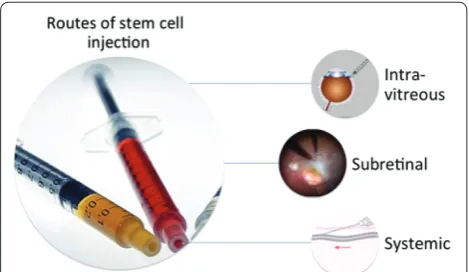
Routes that were tested for cell therapy for retinal diseases are systemic administration (intravenous), intra-vitreal injection, and subretinal injection (Figure 1). Figure 2 shows the two subretinal injection techniques: (A) injection of cell suspensions and (B)injection of cells adhered to a matrix (scaff old) [20,88-90].
Conclusions
SCs maintain the balance between somatic cell popu-lations in various tissues and are responsible for organ regeneration. Th e remarkable progress of regenerative medicine in the last few years indicates promise for the use of SCs in the treatment of ophthalmic disorders. Based on the abovementioned mechanisms, experimental and human studies with intravitreal BM-derived SCs have begun (Table 2) [91,92]. History is starting to be written in this very promising therapeutic fi eld.
Abbreviations
ABMC, autologous bone marrow-derived mononuclear cell; AMD, age-related macular degeneration; BDNF, brain-derived neurotrophic factor; bFGF, basic fi broblast growth factor; BM, bone marrow; EPC, endothelial progenitor cell; hESC, human embryonic stem cell; HSC, hematopoietic stem cell; iPSC, induced pluripotent stem cell; Lin-HSC, lineage-negative hematopoietic stem cell; MSC, mesenchymal stem cell; OEC, outgrowth endothelial cell; RCS, Royal College of Surgeons; RGC, retinal ganglion cell; RP, retinitis pigmentosa; RPC, retinal progenitor cell; RPE,retinal pigment epithelium; RSC, retina stem cell; SC, stem cell; UCB, umbilical cord blood.
Competing interests
The author declares that he has no competing interests.
Acknowledgments
I thank Júlio Cesar Voltarelli, André Marcio Vieira Messias, and Rodrigo Jorge, who are medical researchers at the University of São Paulo (Ribeirão Preto, São Paulo, Brazil) and who are participating in the clinical study of stem cells for treatment of retinitis pigmentosa.
Author details
1São Paulo University, Ribeirão Preto, Rua Saldanha Marinho 2815 conj 42, São Paulo 15010-100, Brazil. 2Rubens Siqueira Research Center, Sao Jose do Rio Preto, Rua Saldanha Marinho 2815 conj 42, São Paulo 15010-100, Brazil. 3Retina Cell, Sao Jose do Rio Preto, Rua Saldanha Marinho 2815 conj 42, São Paulo 15010-100, Brazil.
References
1. Lorenz E, Congdon C, Uphoff D: Modifi cation of acute irradiation injury in
mice and guinea-pigs by bone marrow injections.Radiology 1951,
58:863-877.
2. Till JE, McCulloch EA: A direct measurement of the radiation sensitivity of
normal mouse bone marrow cells.Radiat Res 1961, 14:213-222.
3. Siqueira RC, Voltarelli JC, Messias AM, Jorge R: Possible mechanisms of retinal function recovery with the use of cell therapy with bone
marrow-derived stem cells.Arq Bras Oftalmol 2010, 73:474-479.
4. Lanza R, Rosenthal N: The stem cell challenge.Sci Am 2004, 290:93-99. 5. Mimeault M, Batra SK: Concise review: recent advances on the signifi cance
of stem cells in tissue regeneration and cancer therapies.Stem Cells 2006,
24:2319-2345.
6. Ortiz-Gonzalez XR, Keene CD, Verfaillie C, Low WC: Neural induction of adult
bone marrow and umbilical cord stem cells.Curr Neurovasc Res 2004,
1:207-213.
7. Trounson A: The production and directed diff erentiation of human
embryonic stem cells.Endocr Rev 2006, 27:208-219. Review.
8. Kolb H: Simple anatomy of the retina. In Webvision: The Organization of the Retina and Visual System [Internet]. Edited by Kolb H, Fernandez E, Nelson R. Salt Lake City, UT: University of Utah Health Sciences Center; 1995 to 1 May 2005 [updated 1 May 2007].
9. Machalinska A, Baumert B, Kuprjanowicz L, Wiszniewska B, Karczewicz D, Machalinski B: Potential application of adult stem cells in retinal repair –
challenge for regenerative medicine.Curr Eye Res 2009, 34:748-760. Review.
10. Bunce C, Wormald R: Leading causes of certifi cation for blindness and
partial sight in England & Wales.BMC Public Health 2006, 6:58.
11. Chakravarthy U, Evans J, Rosenfeld PJ: Age related macular degeneration.
BMJ 2010, 340:c981.
12. Maier PC, Funk J, Schwarzer G, Antes G, Falck-Ytter YT: Treatment of ocular hypertension and open angle glaucoma: meta-analysis of randomised
controlled trials.BMJ 2005, 331:134.
13. O’Doherty M, Dooley I, Hickey-Dwyer M: Interventions for diabetic macular
oedema: a systematic review of the literature.Br J Ophthalmol 2008,
92:1581-1590.
14. Mohamed Q, Gillies MC, Wong TY: Management of diabetic retinopathy:
a systematic review.JAMA 2007, 298:902-916.
15. Dahlmann-Noor A, Vijay S, Jayaram H, Limb A, Khaw PT: Current approaches and future prospects for stem cell rescue and regeneration of the retina
and optic nerve. Can J Ophthalmol 2010, 45:333-341.
16. Otani A, Dorrell MI, Kinder K, Moreno SK, Nusinowitz S, Banin E, Heckenlively J, Friedlander M: Rescue of retinal degeneration by intravitreally injected adult bone marrow-derived lineagenegative hematopoietic stem cells.
J Clin Invest 2004, 114:765-774. Comment in: J Clin Invest 2004, 114:755-757. 17. Otani A, Kinder K, Ewalt K, Otero FJ, Schimmel P, Friedlander M: Bone
marrow-derived stem cells target retinal astrocytes and can promote or
inhibit retinal angiogenesis.Nat Med 2002, 8:1004-1010. Comment in:
Nat Med 2002, 8:932-934.
18. Siqueira RC, Messias A, Voltarelli JC, Scott I, Jorge R: Autologous bone marrow-derived stem cells transplantation for retinitis pigmentosa.
Cytotherapy 2010, 12 Suppl 1:58.
19. Autologous Bone Marrow-Derived Stem Cells Transplantation for Retinitis
Pigmentosa. ClinicalTrials.gov identifi er: NCT01068561 [http://clinicaltrials.
gov/ct2/show/NCT01068561].
20. Siqueira RC: Autologous transplantation of retinal pigment epithelium in
age related macular degeneration.Arq Bras Oftalmol 2009, 72:123-130.
21. Siqueira RC, Abad L, Benson G, Sami M: Behaviour of stem cells in eyes of
rabbits with chorioretinal injuries caused by laser red diode 670N-M. In:
Annual Meeting of the Association for Research in Vision and Ophthalmology (ARVO), 2008, Fort Lauderdale. Invest Ophthalmol Vis Sci 2008, 49:536. 22. Enzmann V, Yolcu E, Kaplan HJ, Ildstad ST: Stem cells as tools in regenerative
therapy for retinal degeneration.Arch Ophthalmol 2009, 127:563-571.
23. Müller-Sieburg CE, Cho RH, Thoman M, Adkins B, Sieburg HB: Deterministic regulation of hematopoietic stem cell self-renewal and diff erentiation.
Blood 2002, 100:1302-1309.
24. Ratajczak MZ, Kucia M, Reca R, Majka M, Janowska-Wieczorek A, Ratajczak J: Stem cell plasticity revisited: CXCR4-positive cells expressing mRNA for
early muscle, liver and neural cells ‘hide out’ in the bone marrow.Leukemia
2004, 18:29-40.
25. Spangrude GJ, Heimfeld S, Weissman IL: Purifi cation and characterization of
mouse hematopoietic stem cells.Science 1988, 241:58-62. Erratum in:
Science 1989, 244:1030.
26. Cheng AS, Yau TM: Paracrine eff ects of cell transplantation: strategies to
augment the effi cacy of cell therapies.Semin Thorac Cardiovasc Surg 2008,
20:94-101.
27. Nielsen JS, McNagny KM: CD34 is a key regulator of hematopoietic stem
cell traffi cking to bone marrow and mast cell progenitor traffi cking in the
periphery.Microcirculation 2009, 16:487-496.
28. Kuçi S, Kuçi Z, Latifi -Pupovci H, Niethammer D, Handgretinger R, Schumm M, Bruchelt G, Bader P, Klingebiel T: Adult stem cells as an alternative source of
multipotential (pluripotential) cells inregenerative medicine.Curr Stem Cell
Res Ther 2009, 4:107-117.
29. Challen GA, Boles N, Lin KK, Goodell MA: Mouse hematopoietic stem cell
identifi cation and analysis.Cytometry A 2009, 75:14-24. Review.
30. Voltarelli JC, Ouyang J: Hematopoietic stem cell transplantation for autoimmune diseases in developing countries: current status and future
perspectives.Bone Marrow Transplant 2003, 32 Suppl 1:S69-71.
31. Voltarelli JC: Applications of fl ow cytometry to hematopoietic stem cell
transplantation.Mem Inst Oswaldo Cruz 2000, 95:403-414.
32. Jonas JB, Witzens-Harig M, Arseniev L, Ho AD: Intravitreal autologous bone marrow-derived mononuclear cell transplantation: a feasibility report.
Acta Ophthalmol 2008, 86:225-226.
33. Jonas JB, Witzens-Harig M, Arseniev L, Ho AD: Intravitreal autologous bone
marrow-derived mononuclear cell transplantation.Acta Ophthalmol 2010,
88:e131-132.
34. Siqueira RC, Messias A, Voltarelli JC, Scott IU, Jorge R: Intravitreal injection of autologous bone marrow-derived mononuclear cells for hereditary retinal
dystrophy: a phase I trial.Retina 2011, 31:1207-1214.
35. Horwitz EM, Le Blanc K, Dominici M, Mueller I, Slaper-Cortenbach I, Marini FC, Deans RJ, Krause DS, Keating A; International Society for Cellular Therapy: Clarifi cation of the nomenclature for MSC: The International Society for
Cellular Therapy position statement.Cytotherapy 2005, 7:393-395.
36. Phinney DG, Prockop DJ: Concise review: mesenchymal stem/multipotent stromal cells: the state of transdiff erentiation and modes of tissue
repair-current views.Stem Cells 2007, 25:2896-2902.
37. Castanheira P, Torquetti L, Nehemy MB, Goes AM: Retinal incorporation and diff erentiation of mesenchymal stem cells intravitreally injected in the
injured retina of rats.Arq Bras Oftalmol 2008, 71:644-650.
38. Gong L, Wu Q, Song B, Lu B, Zhang Y: Diff erentiation of rat mesenchymal stem cells transplanted into the subretinal space of sodium
iodate-injected rats.Clin Experiment Ophthalmol 2008, 36:666-671.
39. Zhang P, Li J, Liu Y, Chen X, Kang Q, Zhao J, Li W: Human neural stem cell transplantation attenuates apoptosis and improves neurological
functions after cerebral ischemia in rats.Acta Anaesthesiol Scand 2009,
53:1184-1191.
40. Vossmerbaeumer U, Ohnesorge S, Kuehl S, Haapalahti M, Kluter H, Jonas JB, Thierse HJ, Bieback K: Retinal pigment epithelial phenotype induced in
human adipose tissue-derived mesenchymal stromal cells.Cytotherapy
2009, 11:177-188.
41. Arnhold S, Heiduschka P, Klein H, Absenger Y, Basnaoglu S, Kreppel F, Henke-Fahle S, Kochanek S, Bartz-Schmidt KU, Addicks K, Schraermeyer U: Adenovirally transduced bone marrow stromal cells diff erentiate into
pigment epithelial cells and induce rescue eff ects in RCS rats.Invest
Ophthalmol Vis Sci 2006, 47:4121-4129.
42. Yu J, Vodyanik MA, Smuga-Otto K, Antosiewicz-Bourget J, Frane JL, Tian S, Nie J, Jonsdottir GA, Ruotti V, Stewart R, Slukvin II, Thomson JA: Induced
pluripotent stem cell lines derived from human somatic cells.Science 2007,
318:1917-1920.
43. Azizi SA, Stokes D, Augelli BJ, DiGirolamo C, Prockop DJ: Engraftment and migration of human bone marrow stromal cells implanted in the brains of
albino rats—Similarities to astrocyte grafts.Proc Natl Acad Sci U S A 1998,
95:3908-3913.
44. Erices A, Conget P, Minguell JJ: Mesenchymal progenitor cells in human
umbilical cord blood.Br J Haematol 2000, 109:235-242.
45. Li W, Zhou H, Abujarour R, Zhu S, Young Joo J, Lin T, Hao E, Schöler HR, Hayek A, Ding S: Generation of human induced pluripotent stem cells in the
absence of exogenous Sox2.Stem Cells 2009, 27:2992-3000.
46. Inoue Y, Iriyama A, Ueno S, Takahashi H, Kondo M, Tamaki Y, Araie M, Yanagi Y: Subretinal transplantation of bone marrow mesenchymal stem cells delays retinal degeneration in the RCS rat model of retinal degeneration.
Exp Eye Res 2007, 85:234-241.
Navarrete R, Navarrete C, Jen LS: Umbilical cord blood mesenchymal stromal cells are neuroprotective and promote regeneration in a rat optic
tract model.Exp Neurol 2009, 216:439-448.
48. Wang S, Lu B, Girman S, Duan J, McFarland T, Zhang QS, Grompe M, Adamus G, Appukuttan B, Lund R: Non-invasive stem cell therapy in a rat model for
retinal degeneration and vascular pathology. PLoS ONE 2010, 5:e9200.
49. Hou HY, Liang HL, Wang YS, Zhang ZX, Wang BR, Shi YY, Dong X, Cai Y: A therapeutic strategy for choroidal neovascularization based on
recruitment of mesenchymal stem cells to the sites of lesions.Mol Ther
2010, 18:1837-1845.
50. Takahashi K, Tanabe K, Ohnuki M, Narita M, Ichisaka T, Tomoda K, Yamanaka S: Induction of pluripotent stem cells from adult human fi broblasts by
defi ned factors.Cell 2007, 131:861-872.
51. Zhang D, Jiang W, Liu M, Sui X, Yin X, Chen S, Shi Y, Deng H: Highly effi cient diff erentiation of human ES cells and iPS cells into mature pancreatic
insulin-producing cells.Cell Res 2009, 52:615-621.
52. Zhang J, Wilson GF, Soerens AG, Koonce CH, Yu J, Palecek SP, Thomson JA, Kamp TJ: Functional cardiomyocytes derived from human induced
pluripotent stem cells.Circ Res 2009, 104:e30-41.
53. Hanna J, Wernig M, Markoulaki S, Sun CW, Meissner A, Cassady JP, Beard C, Brambrink T, Wu LC, Townes TM, Jaenisch R: Treatment of sickle cell anemia
mouse model with iPS cells generated from autologous skin.Science 2007,
318:1920-1923.
54. Xu D, Alipio Z, Fink LM, Adcock DM, Yang J, Ward DC, Ma Y: Phenotypic
correction of murine hemophilia A using an iPS cell-based therapy.Proc
Natl Acad Sci U S A 2009, 106:80.
55. Wernig M, Zhao JP, Pruszak J, Hedlund E, Fu D, Soldner F, Broccoli V, Constantine-Paton M, Isacson O, Jaenisch R: Neurons derived from reprogrammed fi broblasts functionally integrate into the fetal brain and
improve symptoms of rats with Parkinson’s disease.Proc Natl Acad Sci U S A
2008, 105:5856-5861.
56. Meyer JS, Shearer RL, Capowski EE, Wright LS, Wallace KA, McMillan EL, Zhang SC, Gamm DM: Modeling early retinal development with human
embryonic and induced pluripotent stem cells.Proc Natl Acad Sci U S A
2009, 106:16698-16703.
57. Buchholz DE, Hikita ST, Rowland TJ, Friedrich AM, Hinman CR, Johnson LV, Clegg DO: Derivation of functional retinal pigmented epithelium from
induced pluripotent stem cells.Stem Cells 2009, 27:2427-2434.
58. Carr AJ, Vugler AA, Hikita ST, Lawrence JM, Gias C, Chen LL, Buchholz DE, Ahmado A, Semo M, Smart MJ, Hasan S, da Cruz L, Johnson LV, Clegg DO, Coff ey PJ: Protective eff ects of human iPS-derived retinal pigment
epithelium cell transplantation in the retinal dystrophic rat. PLoS One 2009,
4:e8152.
59. Zhao T, Zhang ZN, Rong Z, Xu Y:Immunogenicity of induced pluripotent
stem cells.Nature 2011, 474:212-215.
60. Shi Y, Desponts C, Do JT, Hahm HS, Schöler HR, Ding S: Induction of pluripotent stem cells from mouse embryonic fi broblasts by Oct4 and Klf4
with small-molecule compounds.Cell Stem Cell 2008, 3:568-574.
61. Woltjen K, Michael IP, Mohseni P, Desai R, Mileikovsky M, Hämäläinen R, Cowling R, Wang W, Liu P, Gertsenstein M, Kaji K, Sung HK, Nagy A: piggyBac transposition reprograms fi broblasts to induced pluripotent stem cells.
Nature 2009, 458:766-770.
62. Kaji K, Norrby K, Paca A, Mileikovsky M, Mohseni P, Woltjen K: Virus-free induction of pluripotency and subsequent excision of reprogramming
factors.Nature 2009, 458:771-775.
63. Yu J, Hu K, Smuga-Otto K, Tian S, Stewart R, Slukvin II, Thomson JA: Human induced pluripotent stem cells free of vector and transgene sequences. Science 2009, 324:797-801.
64. Balasubramanian S, Babai N, Chaudhuri A, Qiu F, Bhattacharya S, Dave BJ, Parameswaran S, Carson SD, Thoreson WB, Sharp JG, Rao M, Ahmad I: Non cell-autonomous reprogramming of adult ocular progenitors: generation
of pluripotent stem cells without exogenous transcription factors. Stem
Cells 2009, 27:3053-3062.
65. Loh YH, Agarwal S, Park IH, Urbach A, Huo H, Heff ner GC, Kim K, Miller JD, Ng K, Daley GQ: Generation of induced pluripotent stem cells from human
blood. Blood 2009, 113:5476-5479.
66. MacLaren RE, Pearson RA: Stem cell therapy and the retina.Eye 2007, 21:1352-1359.
67. Nistor G, Seiler MJ, Yan F, Ferguson D, Keirstead HS: Three-dimensional early retinal progenitor 3D tissue constructs derived from human embryonic
stem cells.J Neurosci Methods 2010, 190:63-70.
68. Lu B, Malcuit C, Wang S, Girman S, Francis P, Lemieux L, Lanza R, Lund R: Long-term safety and function of RPE from human embryonic stem cells
in preclinical models of macular degeneration.Stem Cells 2009,
27:2126-2135.
69. Idelson M, Alper R, Obolensky A, Ben-Shushan E, Hemo I, Yachimovich-Cohen N, Khaner H, Smith Y, Wiser O, Gropp M, Cohen MA, Even-Ram S, Berman-Zaken Y, Matzrafi L, Rechavi G, Banin E, Reubinoff B: Directed diff erentiation of human embryonic stem cells into functional retinal pigment
epithelium cells.Cell Stem Cell 2009, 5:396-408.
70. Vugler A, Lawrence J, Walsh J, Carr A, Gias C, Semo M, Ahmado A, da Cruz L, Andrews P, Coff ey P: Embryonic stem cells and retinal repair.Mech Dev
2007, 124:807-829.
71. Klassen HJ, Ng TF, Kurimoto Y, Kirov I, Shatos M, Coff ey P, Young MJ: Multipotent retinal progenitors express developmental markers, diff erentiate into retinal neurons, and preserve light-mediated behavior.
Invest Ophthalmol Vis Sci 2004, 45:4167-4173.
72. Stern JH, Temple S: Stem cells for retinal replacement therapy.
Neurotherapeutics 2011, 8:736-743.
73. Stone LS: Neural retina degeneration followed by regeneration from
surviving retinal pigment cells in grafted adult salamander eyes.Anat Rec
1950, 106:89-109.
74. Tropepe V, Coles BL, Chiasson BJ, Horsford DJ, Elia AJ, McInnes RR, van der Kooy D: Retinal stem cells in the adult mammalian eye.Science 2000, 287:2032-2036.
75. Ahmad I, Tang L, Pham H: Identifi cation of neural progenitors in the adult
mammalian eye.Biochem Biophys Res Commun 2000, 270:517-521.
76. Cicero SA, Johnson D, Reyntjens S, Frase S, Connell S, Chow LM, Baker SJ, Sorrentino BP, Dyer MA: Cells previously identifi ed as retinal stem cells are
pigmented ciliary epithelial cells. Proc Natl Acad Sci U S A 2009,
106:6685-6690.
77. Moshiri A, Reh TA: Persistent progenitors at the retinal margin of ptc+/−
mice.J Neurosci 2004, 24:229-237.
78. Karl MO, Hayes S, Nelson BR, Tan K, Buckingham B, Reh TA: Stimulation of
neural regeneration in the mouse retina.Proc Natl Acad Sci U S A 2008,
105:19508-19513.
79. Das AV, Mallya KB, Zhao X, Ahmad F, Bhattacharya S, Thoreson WB, Hegde GV, Ahmad I: Neural stem cell properties of Muller glia in the mammalian
retina: regulation by Notch and Wnt signaling.Dev Biol 2006, 299:283-302.
80. Rafi i S, Lyden D: Therapeutic stem and progenitor cell transplantation for
organ vascularization and regeneration.Nat Med 2003, 9:702-712.
81. Medina RJ, O’Neill CL, Sweeney M, Guduric-Fuchs J, Gardiner TA, Simpson DA, Stitt AW: Molecular analysis of endothelial progenitor cell (EPC) subtypes
reveals two distinct cell populations with diff erent identities.BMC Med
Genomics 2010, 3:18.
82. Medina RJ, O’Neill CL, O’Doherty TM, Guduric-Fuchs J, Gardiner TA, Stitt AW: Endothelial progenitor cells contribute to vascular repair in the ischaemic
retina.Heart 2011, 97:e7.
83. Crisostomo PR, Markel TA, Wang Y, Meldrum DR: Surgically relevant aspects
of stem cell paracrine eff ects.Surgery 2008, 143:577-581.
84. Markel TA, Wang Y, Herrmann JL, Crisostomo PR, Wang M, Novotny NM, Herring CM, Tan J, Lahm T, Meldrum DR: VEGF is critical for stem cell-mediated cardioprotection and a crucial paracrine factor for defi ning the
age threshold in adult and neonatal stem cell function.Am J Physiol Heart
Circ Physiol 2008, 295:H2308-2314.
85. Markel TA, Crisostomo PR, Wang M, Herring CM, Meldrum DR: Activation of individual tumor necrosis factor receptors diff erentially aff ects stem cell
growth factor and cytokine production.Am J Physiol Gastrointest Liver
Physiol 2007, 293:G657-662.
86. Oh JY, Kim MK, Shin MS, Lee HJ, Ko JH, Wee WR, Lee JH: The anti-infl ammatory and anti-angiogenic role of mesenchymal stem cells in
corneal wound healing following chemical injury.Stem Cells 2008,
26:1047-1055.
87. Koontongkaew S, Amornphimoltham P, Yapong B: Tumor-stroma interactions infl uence cytokine expression and matrix metalloproteinase
activities in paired primary and metastatic head and neck cancer cells.Cell
Biol Int 2009, 33:165-173.
88. Binder S, Stanzel BV, Krebs I, Glittenberg C: Transplantation of the RPE in AMD.Prog Retin Eye Res 2007, 26:516-515.
clinical study.Br J Ophthalmol 2011, 95:370-375.
90. Singh MS, Maclaren RE: Stem cells as a therapeutic tool for the blind:
biology and future prospects.Proc Biol Sci 2011, 278:3009-3016.
91. Ballios BG, van der Kooy D: Biology and therapeutic potential of adult
retinal stem cells.Can J Ophthalmol 2010, 45:342-351.
92. Siqueira RC: Stem-cell therapy for retinal diseases.In Embryonic Stem Cells - Diff erentiation and Pluripotent Alternatives. Edited by Kallos MS. Rijeka, Croatia: InTech; 2011 [http://www.intechopen.com/articles/show/title/ stem-cell-therapy-for-retinal-diseases].
93. Li N, Li XR, Yuan JQ: Eff ects of bone-marrow mesenchymal stem cells transplanted into vitreous cavity of rat injured by ischemia/reperfusion.
Graefes Arch Clin Exp Ophthalmol 2009, 247:503-514.
94. Uteza Y, Rouillot JS, Kobetz A, Marchant D, Pecqueur S, Arnaud E, Prats H, Honiger J, Dufi er JL, Abitbol M, Neuner-Jehle M: Intravitreous transplantation of encapsulated fi broblasts secreting the human fi broblast growth factor 2 delays photoreceptor cell degeneration in Royal
College of Surgeons rats. Proc Natl Acad Sci U S A 1999, 96:3126-3131.
95. Zhang Y, Wang W: Eff ects of bone marrow mesenchymal stem cell
transplantation on light-damaged retina. Invest Ophthalmol Vis Sci 2010,
51:3742-3748.
96. Tomita M, Adachi Y, Yamada H, Takahashi K, Kiuchi K, Oyaizu H, Ikebukuro K,
Kaneda H, Matsumura M, Ikehara S: Bone marrow-derived stem cells can
diff erentiate into retinal cells in injured rat retina. Stem Cells 2002,
20:279-283.
97. Meyer JS, Katz ML, Maruniak JA, Kirk MD: Embryonic stem cell-derived neural progenitors incorporate into degenerating retina and enhance
survival of host photoreceptors.Stem Cells 2006, 24:274-283.
98. Wang HC, Brown J, Alayon H, Stuck BE: Transplantation of quantum dot-labelled bone marrow-derived stem cells into the vitreous of mice with
laser-induced retinal injury: survival, integration and diff erentiation.Vision
Res 2010, 50:665-673.
99. Johnson TV, Bull ND, Hunt DP, Marina N, Tomarev SI, Martin KR: Neuroprotective eff ects of intravitreal mesenchymal stem cell
transplantation in experimental glaucoma.Invest Ophthalmol Vis Sci 2010,
51:2051-2059.
100. Advanced Cell Technology, Inc. homepage [http://www.advancedcell.com].
doi:10.1186/scrt91
Cite this article as: Siqueira RC: Stem cell therapy for retinal diseases: