R E S E A R C H
Open Access
miR-205-5p inhibits human endometriosis
progression by targeting ANGPT2 in
endometrial stromal cells
Chen-Fei Zhou
1†, Min-Juan Liu
2†, Wei Wang
1, Sha Wu
3, Yu-Xin Huang
2, Guo-Bin Chen
4, Li-Min Liu
4,
Dong-Xian Peng
2, Xue-Feng Wang
2, Xu-Zi Cai
2, Xiao-Xuan Li
2, Wan-Qin Feng
2and Ying Ma
2*Abstract
Background:miRNA expression profiles in ectopic endometrium (EC) serving as pathophysiologic genetic fingerprints contribute to determining endometriosis progression; however, the underlying molecular mechanisms remain unknown.
Methods:miRNA microarray analysis was used to determine the expression profiling of EC fresh tissues. qRT-PCR was performed to screen miR-205-5p expression in EC tissues. The roles of miR-205-5p and its candidate target gene, angiopoietin-2 (ANGPT2), in endometriosis progression were confirmed on the basis of both in vitro and in vivo systems. miR-205-5p and ANGPT2 expression were measured by in situ hybridization and immunochemistry, and their clinical significance was statistically analysed.
Results:miR-205-5p was screened as a novel suppressor of endometriosis through primary ectopic endometrial stromal cell migration, invasion, and apoptosis assay in vitro, along with endometrial-like xenograft growth and apoptosis in vivo. In addition, ANGPT2 was identified as a direct target of miR-205-5p through bioinformatic target prediction and luciferase reporter assay. Re-expression and knockdown of ANGPT2 could respectively rescue and simulate the effects induced by miR-205-5p. Importantly, the miR-205-5p-ANGPT2 axis was found to activate the ERK/ AKT pathway in endometriosis. Finally, miR-205-5p and ANGPT2 expression were closely correlated with the
endometriosis severity.
Conclusion:The newly identified miR-205-5p-ANGPT2-AKT/ERK axis illustrates the molecular mechanism of endometriosis progression and may represent a novel diagnostic biomarker and therapeutic target for disease treatment.
Keywords: Endometriosis, miR-205-5p, Endometrial stromal cells, ANGPT2
Background
Endometriosis is a common and multifactorial gynaecologic condition characterised by the presence of endometrial-like tissue in aberrant locations outside the uterus. Endometriosis affects an estimated 10 to 15% of women of reproductive age worldwide [1]. The disease has multiple manifestations, such as dysmenorrhoea, chronic pelvic pain, infertility, and cancer-ous lesions, and it can severely affect the quality of patients’
life [2, 3]. The current gold standard for the diagnosis of endometriosis is laparoscopic surgery [4]. It is unlikely that reproductive-age women would undergo such an invasive surgery when they can choose temporarily diminish pain symptoms by other treatments. However, none of the cur-rently available techniques, including imaging methods or la-boratory-developed platforms, is a suitable replacement for laparoscopy [5, 6]. Therefore, it is urgent to better under-stand the molecular mechanism underlying the endometri-osis progression and to identify novel and more efficient diagnostic predictors for accurate identification of the dis-ease, so that the optimal therapeutic strategies can be found.
© The Author(s). 2019Open AccessThis article is distributed under the terms of the Creative Commons Attribution 4.0 International License (http://creativecommons.org/licenses/by/4.0/), which permits unrestricted use, distribution, and reproduction in any medium, provided you give appropriate credit to the original author(s) and the source, provide a link to the Creative Commons license, and indicate if changes were made. The Creative Commons Public Domain Dedication waiver (http://creativecommons.org/publicdomain/zero/1.0/) applies to the data made available in this article, unless otherwise stated. * Correspondence:mayingwuzhuoyi@126.com
†Chen-Fei Zhou and Min-Juan Liu contributed equally to this work.
2Department of Obstetrics and Gynecology, Zhujiang Hospital of Southern
Medical University, No.253, Middle Gongyeda Road, Haizhu District, Guangzhou 510280, China
miRNAs are small non-coding RNAs, approximately 20 to 24 nucleotides in length, that regulate target gene ex-pression at the posttranscriptional level by pairing to 3′ untranslated regions (UTRs) of mRNAs and triggering RNA degradation or translational suppression [7]. These miRNAs are involved in multiple biological processes, such as cellular migration, invasion, and apoptosis [8, 9]. As a transcriptional regulator, aberrant expression of miR-NAs usually leads to dysregulated expression of target genes that are involved in initiation and progression of various diseases including endometriosis, suggesting that they are excellent candidate biomarkers and potential therapeutic targets [10–12]. However, miRNA profiling of ectopic endometrium reflecting endometriosis progression is still unclear.
To address this problem, we performed the current study to investigate the underlying molecular mecha-nisms for differentially expressed miRNAs of ectopic endometrium in endometriosis progression, as well as its clinical relevance, to explore the potential clinical appli-cations in diagnosis and therapy.
Materials and methods Clinical specimens
Ectopic endometrial tissues and serum were collected from patients with endometriosis, and normal endometrial tissues and serum from hysterectomy in patients with grade II–III cervical intraepithelial neoplasia or uterine leiomyoma. The pathological diagnosis was performed preoperatively and confirmed postoperatively. All of the clinical specimens were obtained from the Department of Gynecology of Zhujiang Hospital (Guangzhou, People’s Republic of China) between 2006 and 2012 according to the ethical and legal standards. Patients receiving hor-mone treatment or those with concurrent malignancies were excluded. The characteristics of the patients with endometriosis are described in Additional file1: Table S1.
miRNA microarray
Agilent miRNA microarray 21.0 for normal endomet-rium (n= 3) and ectopic endometrium (n= 3) was estab-lished by Guangdong Longsee Biomedical Corporation and analysed by GeneSpringGX software 11.0 (Agilent).
Isolation of primary endometrial stromal cells and cell culture
Primary normal and ectopic endometrial stromal cells were isolated as described previously [13]. Briefly, ectopic endometrial tissues were minced and digested in phosphate-buffered saline (PBS) containing collagenase (1 mg/mL, 15 U/mg) and 1% penicillin/streptomycin for 60 min in an orbital shaker at 37 °C. The obtained homogenous cell suspension were plated in T25 cell cul-ture flasks (Corning) and culcul-tured in Dulbecco’s
modified Eagle medium (DMEM)/Ham’s F12 (1:1) with 10% foetal bovine serum (FBS, Gibco) and bio-antibiotics (100 IU/mL penicillin and 100μg/mL streptomycin). Cel-lular purities were examined by immunofluorescence staining for vimentin (ab8978, Abcam) and cytokeratin (ab76126, Abcam) antibodies. Primary endometrial stro-mal cells were used between passages 3 and 5.
Stable transfection with lentivirus
Lentivirus containing an miR-205-5p overexpression se-quence and its negative control were all purchased from GeneChem Inc. Primary ectopic endometrial stromal cells were transfected with lenti-miR-205, and polyclonal cells expressing green fluorescent protein signals were selected for further experiments by flow cytometer.
Transient transfection with oligonucleotides and plasmids The 205-5p mimic and its negative control, or miR-205-5p inhibitor and its negative control, were designed and cloned by RiboBio Inc. The ANGPT2 coding sequence (without 3′-UTR) was cloned into pCDNA3.1(+)-Vector (Invitrogen). The empty vector was used as a blank control. siVASH1 and its negative control siRNA were designed and synthesised by GenePharma Inc. Lipofectamine 2000 Re-agent (Invitrogen) was then used to transfect miR-205-5p mimic and inhibitor, plasmid-ANGPT2, and siANGPT2 ac-cording to the manufacturer’s protocol. For RNA extraction and western blot and in vitro functional assays, cells were used 48 h after transfection. The sequence of siANGPT2 and siRNA are shown in Additional file1: Table S2.
RNA extraction and quantitative reverse transcriptase polymerase chain reaction
RNA was extracted from clinical specimens, serum sam-ples, primary cells, and xenografted lesions by the miR-Neasy Mini kit (Qiagen, 217004). qRT-PCR was performed according to the protocol of our previous study [14]. Spe-cific primer sets for miRNAs and U6 were purchased from RiboBio Inc. The expression of miRNAs and mRNAs was normalised to U6 and glyceraldehy3-phosphate de-hydrogenase (GAPDH), respectively. The primer sequences are shown in Additional file1: Table S2.
Immunohistochemistry
In situ hybridisation
In situ hybridisation (ISH) was performed as described previously [16]. Briefly, after incubation with H2O2, the
tissue sections were treated with pepsin at 37 °C for 2 min, washed, and prehybridised for 4 h at 37 °C. Hybrid-isation with digoxygenin-labelled miR-205-5p LNA probes (Exiqon) occurred at 37 °C overnight. The sec-tions were then washed at 37 °C and incubated with bio-tinylated mouse anti-digoxin for 1 h at 37 °C. Staining was visualised by adding 3,3-diaminobenzidine and counterstaining with haematoxylin.
Staining assessment
The IHC- and ISH-stained tissue sections were reviewed and scored separately by two independent pathologists. For semi-quantitative evaluation of ANGPT2, Annexin V, and miR-205-5p expression in tissue sections, the German Immuno-Reactive Score was applied as previ-ously described [17]. The immunohistochemical score was calculated by combining the proportion of positively stained cells and the intensity of staining. The staining intensity was rated on a scale of 1 to 4, with 1 represent-ing no stainrepresent-ing, 2 weak stainrepresent-ing, 3 moderate stainrepresent-ing, and 4 strong staining. No staining was scored as 0; stain-ing of 1 to 10% as 1; 11 to 50% as 2; 51 to 80% as 3; and 81 to 100% as 4. The raw data were converted to IHS by multiplying the quantity score and the intensity score. Tissues with an IHS of 4 or greater were defined as high expression; tissues with a IHS of less than 4 were defined as low expression.
Animal models
Female nude mice (6 weeks old) were purchased from the Experimental Animal Center, Southern Medical University (Guangzhou, People’s Republic of China). The studies were approved by the Institutional Animal Research Ethics Com-mittee of Southern Medical University. The endometriosis mouse model was established in the Experimental Animal Center of Southern Medical University as described previ-ously [10]. Briefly, 1 × 10⁷ectopic endometrial stromal cells mixed with gland cells (1:1) were subcutaneously injected into the flanks of each nude mice (n= 6, per group). Lesion size (mm3) was measured every 4 days and calculated by the following formula: volume = (width)2× length/2. The mice were euthanised 30 days after the injection of human ectopic endometrial cells to evaluate the extent of endometriosis.
Western blot assay
Western blot assay was performed as in our previous study [14]. The detailed antibody information is pro-vided in Additional file1: Table S3.
Other methods
Luciferase reporter assay, wound healing assay, ELISA assay, Transwell invasion assay, and apoptosis assay were performed according to the manufacturers’ protocols. Detail information is described in Additional file2: Sup-plemental materials and methods.
Statistical analysis
SPSS V.13.0 software was used for statistical analysis. Data are expressed as the mean ± standard deviation (SD). A two-tailed Student ttest or one-way analysis of variance (ANOVA) was used for comparisons among the groups. The Fisher or chi-square test was applied for categorical variables. Partial correlations were applied in the multi-variate correlation analysis. The Cox regression model was used for the univariate and multivariate analysis. Dif-ferences were considered to be statistically significant whenP< 0.05.
Results
Identification of miR-205-5p as a negatively pathologic miRNA in endometriosis
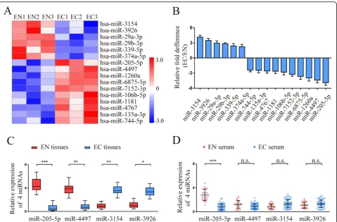
To identify the differences in miRNAs for endometriosis, miRNA expression profiles of normal endometrium (EN) (n= 3) and ectopic endometrium (EC) (n= 3) were ana-lysed by Agilent miRNA microarray 21.0. Using a 2-fold change and P< 0.05 as the threshold cut-off, we found that 16 miRNAs were significantly different between EN and EC (Fig. 1a). The differential expression of 16 miR-NAs was verified by qRT-PCR in the same tissues used for microarray analysis. The results showed that a 4.89 ± 0.51-fold lower level of miR-205-5p, 4.37 ± 0.53-0.51-fold lower level of miR-4497, 4.13 ± 0.38-fold higher level of miR-3154, and 3.46 ± 0.39-fold higher level of miR-3926 were exam-ined in EC compared with EN (Fig. 1b). miR-205-5p showed the greatest downregulation among the 4 repre-sentative miRNAs, suggesting that miR-205-5p may be the best candidate for further study.
To further confirm the statistical significance of the 4 representative miRNAs (205-5p, 4497, miR-3154, miR-3926), additional tissues and serum sample from the EN (n= 23) and EC (n= 68) groups were ana-lysed by qRT-PCR. Compared with the EN group, a sig-nificantly lower level of miR-205 was detected in both tissues and serum samples of the EC group (Fig.1c, d). Taken together, these results suggested that low levels of miR-205-5p expression may be associated with endo-metriosis progression.
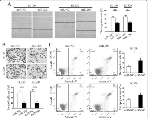
miR-205-5p suppressed migration and invasion but promoted apoptosis of endometriosis-derived endometrial stromal cells in vitro
endometrial stromal cells, EC109 and EC520, and two primary normal endometrial stromal cells, EN211 and EN307. The immunofluorescence staining showed that both vimentin and cytokeratin were expressed in EC and EN stromal cells (Additional file 3: Figure S1A). Add-itionally, in the presence of E2 plus medroxyprogester-one acetate (MPA), the cells transformed from a spindle-shaped appearance to large polygonal-shaped cells and significantly increased PRL secretion (Add-itional file 3: Figure S1B & C), indicating that the in-duced endometrial stromal cells were successfully differentiated in vitro. Collectively, these results were consistent with previous studies [18,19], suggesting that the isolated EC109, EC520, EN211, and EN307 cells were endometrial stromal cells.
To further explore the suppressive role of miR-205-5p in endometriosis in vitro, we constructed a lentiviral vec-tor expressing miR-205-5p and established miR-205-5p-overexpressing EC109 and EC520 cells after lentivector transfection (Additional file 4: Figure S2A). More than
500-fold increase in miR-205-5p expression was exam-ined in Lenti-miR-205-5p-transfected EC109 and EC520 cells compared with the negative control (NC) group (Additional file 4: Figure S2B). Despite very low en-dogenous expression of miR-205-5p in EC109 and EC520 cells (Additional file 4: Figure S2C), miR-205-5p inhibitors were transiently transfected into two cells. However, no significant difference in the expression of miR-205-5p was observed between the miR-205-5p inhibitors group and the NC group by qRT-PCR (Add-itional file4: Figure S2D).
knockdown in normal endometrial stromal cells showed similar functional phenotypes as endometriosis-derived endometrial stromal cells (Additional file5: Figure S3). More importantly, western blot analysis showed that miR-205-5p overexpression significantly upregulated Bax and E-Cad protein expression but downregulated Bcl-2 and Vimentin protein expression (Additional file6: Figure S4). Taken to-gether, these results showed that miR-205-5p suppressed migration and invasion but promoted apoptosis of endomet-riosis-derived endometrial stromal cells.
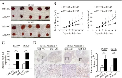
miR-205-5p suppressed endometriosis progression in vivo To better evaluate the biologic function of miR-205-5p in vivo, we performed an in vivo endometriosis study by
receptors 1 and 2 (sTNFR-1 and 2), and high-sensitivity C-reactive protein (hs-CRP) (Additional file 7: Figure S5). These results demonstrated that miR-205-5p could exert a significant inhibitory effect on endometriosis of endometrial stromal cells in vivo.
ANGPT2 is the direct target of miR-205-5p
Multiple algorithms (miRWalk, TargetScan, miRDB) were used to identify the candidate targets of miR-205-5p. As a re-sult, 31 target genes were predicted to be regulated by miR-205-5p (Fig.4a). Among these candidates, CDH11, ANGPT2, PLCB1, KPNA1, and HIF1AN lowly expressed in normal endometrial stromal cells confirmed by The Human Protein Atlas (THPA) database and were chosen for the next study (Fig.4b). To further verify this prediction, a qRT-PCR analysis was performed and the results showed that the CDH11, ANGPT2, PLCB1, KPNA1, and HIF1AN expression in cells and xenografts associated with miR-205-5p-overexpressed EC109 and EC520 cells were all downregulated at mRNA levels (Fig.4c; Additional file 8: Figure S6A). Among them, ANGPT2 was the most significantly downregulated gene.
To examine miR-205-5p regulation of the putative target ANGPT2, the predicted miR-205-5p binding site in the 3′ -UTR of ANGPT2 (wild type) or the mutated sequence (mu-tant type) was cloned into luciferase reporter plasmids and assessed for their response to miR-205-5p in EC109 and EC520 cells. The results showed that the expression of the reporter gene followed by a wild-type 3′-UTR of ANGPT2 was significantly reduced by the co-transfected miR-205-5p mimic, whereas reporter gene expression had no change if followed by the 3′-UTR of the ANGPT2 gene with a mu-tated putative target site of miR-205-5p (Fig. 4d; Add-itional file 8: Figure S6B). Thus, we concluded that ANGPT2 is a direct target of miR-205-5p.
overexpressed EC109 and EC520 cells (Fig. 4e). Overall, miR-205-5p negatively regulated ANGPT2 expression in vitro and in vivo.
ANGPT2 was a critical downstream mediator of miR-205-5p-suppressive effects in endometriosis
To explore whether the role of miR-205-5p in endomet-riosis was mediated through suppressing ANGPT2, plas-mids expressing ANGPT2 without 3′-UTR and specific
detected by qRT-PCR and western blot (Fig. 5a, b). As the results showed, re-expression of ANGPT2 rescued, whereas knockdown of ANGPT2 simulated, the suppres-sion of ANGPT2 and AKT/ERK pathway activation me-diated by miR-205-5p.
Further in vitro studies confirmed that re-expression of ANGPT2 dramatically attenuated the effects induced by miR-205-5p. Meanwhile, the knockdown of ANGPT2 gen-erated similar functional phenotypes associated with miR-205-5p overexpression in ectopic endometrial stromal cells, including suppressing the abilities of migration and invasion (Fig.5c, d; Additional file9: Figure S7A & B), as well as pro-moting cellular apoptosis (Fig.5e; Additional file9: Figure
S7C). Collectively, these results indicated that ANGPT2 was a critical downstream mediator of miR-205-5p suppressive effects in endometriosis.
Clinical associations of miR-205-5p with ANGPT2 expression in human endometriosis tissues
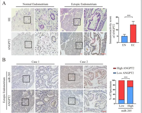
We first used the antibody that specifically recognised ANGPT2 to examine the expression pattern in the aforementioned 68 EN and 23 EC clinical specimens. As shown in Fig. 6a, the expression of ANGPT2 protein in EC tissues was significantly higher than that in EN tis-sues. We further investigated whether there was an asso-ciation between the expression of ANGPT2 and
205-5p in human endometriosis tissues. The results showed that the expression of ANGPT2 protein was negatively correlated with miR-205-5p levels in the EC tissues (Fig. 6b; Additional file 1: Table S4), suggesting that miR-205-5p suppressed ANGPT2 expression in clinical endometriosis progression.
Moreover, we analysed the relationship between clini-copathological characteristics and miR-205 expression in patients with endometriosis. The dysmenorrhoea score, CA125 level, and endometriosis score were dramatically correlated with downregulation of miR-205-5p and up-regulation of ANGPT2 (Table 1). More importantly, multivariate analysis confirmed that downregulation of miR-205-5p and upregulation of ANGPT2 score were also markedly correlated with endometriosis score (Table2). Taken together, these data indicated that
miR-205-5p and ANGPT2 were promising biomarkers for reflecting the severity of endometriosis.
Discussion
personalised diagnostic and therapeutic strategies for patients with confirmatory diagnosis. Recently, miRNAs have been recognised to be promising biomarkers because of their tis-sue-specific expression profiles and minimally invasive diag-nostic means [27–29]. In our study, miR-205-5p was identified as a novel pathologic suppressor of endometriosis using tissue microarray analysis. We further confirmed that miR-205-5p could directly target angiopoietin-2 (ANGPT2) by binding to its 3′-UTR and be involved in the procession of endometriosis via regulating the ANGPT2 pathway. More importantly, our clinical data showed that both miR-205-5p and ANGPT2 are valuable factors for reflecting the severity of endometriosis. These results provided us with enough rea-son to further explore the roles of the miR-205-5p-ANGPT2 axis in the progression of endometriosis.
Profiles of miRNA expression in normal endometrium exhibit dynamic changes across the menstrual cycle [30], where their implication into physiologic systems can be used to identify pathologic phenotypes. Endometriosis can be typically categorised into two types: asymptomatic and symptomatic, depending on the arrival location of endo-metrial-like tissues [31–33]. As expected, the miRNA pro-files of ectopic endometrium are more representable than those of eutopic endometrium, which provides more use-ful pathophysiologic fingerprints to confirm a diagnosis of symptomatic endometriosis. Here, we found that miR-205-5p levels in both tissues and serum from ectopic endometrium patients were significant downregulation compared with those from normal endometrium patients. Moreover, our study data confirmed the suppressive effect
Table 1Correlation between the clinicopathological factors and expression of miR-205-5p and ANGPT2 in endometriosis
miR-205-5p expression ANGPT2 expression
High Low P High Low P
Patients (n) 19 49 45 23
Age (years) 31.26 ± 5.98 31.98 ± 6.36 0.67 32.18 ± 6.43 31.00 ± 5.85 0.464
Haemoglobin (g/L) 113.89 ± 15.08 114.80 ± 18.00 0.85 113.98 ± 18.07 115.65 ± 15.44 0.706
Endometrial phase (%)
Proliferative phase 78.95% 83.67% 82.22% 82.61%
Secretive phase 21.05% 16.33% 0.917 17.78% 17.39% 0.97
Dysmenorrhoea pain score (%)
Less than 4 points 57.89% 30.61% 28.89% 56.52%
More than 4 points 42.11% 69.39% 0.038 71.11% 43.48% < 0.001
Chronic pelvic pain score (%)
Less than 4 points 42.11% 36.69% 31.11% 47.83%
More than 4 points 57.89% 65.31% 0.57 68.89% 52.17% 0.18
E2 (pmol/L) 369.58 ± 355.63 296.55 ± 301.26 0.245 276.76 ± 279.00 395.61 ± 373.30 0.054
CA-125 (kU/L) 34.31 ± 21.27 109.55 ± 140.91 0.001 116.67 ± 144.86 33.47 ± 21.40 < 0.001
Endometriosis score (%)
Less than 16 points 68.42% 38.78% 35.56% 69.57%
More than 16 points 31.58% 61.22% 0.028 64.44% 30.43% 0.008
Table 2Summary of univariate and multivariate Cox regression analysis of the severity of endometriosis
Univariate analysis Multivariate analysis
SE B P SE B P
Age (years) 0.132 −0.517 < 0.001 0.003 −0.031 < 0.001
Haemoglobin (g/L) 0.025 −0.001 0.971 0.059 0.001 0.552
Endometrial phase 1.034 −0.382 0.712 3.012 0.096 0.139
Dysmenorrhoea pain score 0.646 2.066 0.001 1.669 0.217 < 0.001
Chronic pelvic pain score 0.644 2.128 0.001 1.577 0.103 0.051
E2 (pmol/L) 0.017 0.011 0.313 0.12 0.001 0.121
CA-125 (kU/L) 0.054 0.157 0.004 0.08 0.001 0.001
miR-205-5p expression 0.573 2.228 < 0.001 1.688 1.628 < 0.001
of miR-205-5p through primary ectopic endometrial stro-mal cell migration, invasion, and apoptosis assay in vitro, along with endometrial-like xenograft growth and apop-tosis in vivo. Although endometriosis cells can hardly transform into cancer cells, the cancerous behaviour, such as invasion into adjacent organs and spreading to distant organs, frequently appears in endometriosis [34, 35]. Interestingly, previous studies supported our data that the overexpression of miR-205-5p in multiple cancer types in-duced similar in vitro and in vivo phenotypes to our study
[36–38], indicating that miR-205-5p could be an ideal
therapeutic target that contributes to suppressing the can-cerous behaviour.
To further explore the molecular mechanisms underlying the involvement of miR-205-5p in human endometriosis, we combined three typical miRNA prediction algorithms and the THPA database to identify the potential targets of miR-205-5p. THPA is an online platform for simultaneously identifying and quantifying protein of potential targets in both human physiologic and pathologic tissues [39]. Compared with the conventional qRT-PCR analysis for targets of miRNAs, THPA can be used as a better identification and quantification tool for primarily screening target protein due to its large and accurate data in human tissues. Then, we integrated the prediction results of bioinformatic methods and luciferase reporter assay. Consequently, ANGPT2 was identified as a novel dir-ect target of miR-205-5p. Various genes have been identified as the direct targets of miR-205-5p. For ex-ample, miR-205-5p directly targeted ERBB2 and p63, leading to resistance to standard therapy for Her2-positive breast cancer [40]. A lipid metabolism-related gene called acetyl-CoA carboxylase β (ACACβ) was targeted by miR-205-5p in hepatic lipid metabolism [41]. A recent study by Di Carlo et al. found that angiopoietin-2 (ANGPT2) was gradually downregu-lated in normal endometrium, eutopic endometrium, and ectopic endometrium using immunohistochemical staining [42]. However, up to now, the underlying mechanism for ANGPT2 affecting the pathogenesis of endometriosis is still unclear. ANGPT2, a well-recog-nised vascular destabilising factor, is a biomarker of poor outcome in many human diseases [43, 44]. Our data here suggested that overexpression of miR-205-5p could cause the significant downregulation of ANGPT2 at both mRNA and protein levels in ectopic endometrial stromal cells and xenografted lesions. Meanwhile, the re-expression and knockdown of ANGPT2 could respectively rescue and simulate the effects induced by miR-205-5p. Moreover, activation of the AKT and ERK pathways involved in endomet-riosis progression was responsible for the downregula-tion of miR-205-5p and upreguladownregula-tion of ANGPT2.
Although decades of research have gone into develop-ing a set of diagnostic biomarkers for evaluatdevelop-ing the severity of endometriosis, the effective biomarkers for differentiating different types and degrees of endometri-osis are still lacking. For example, CA-125 is a clinical biomarker for the prediction of endometriosis severity [45]. However, the correlation of CA-125 levels with dis-ease progression is not high [46]. Several other schemes that are being implemented to detect inflammatory bio-markers such as IL-8 and IL-6 are still unsatisfactory
[47, 48]. Based on the widely accepted theory on the
retrograde movement of sloughed menstruation [49], the menstrual endometrial cells are the source of ectopic endometriotic foci. Therefore, using the direct source of the disease, including peripheral blood or even urine, is perhaps reasonable and logical in our quest to identify biomarkers for endometriosis. In our study, ISH and IHC analyses in human ectopic endometrium serial sec-tions also showed an adverse relasec-tionship between miR-205-5p and ANGPT2. More importantly, we showed that clinical endometriosis severity scores are also closely correlated with miR-205-5p and ANGPT2 expression, and predicting endometriosis severity was also deter-mined according to the multivariate regression model.
Conclusion
Taken together, our data provided evidence that miR-205-5p may function as an ectopic endometriotic suppressor when evaluating the disease severity in human endometri-osis. Decreased miR-205-5p may contribute to apoptosis reduction and promoting migration and invasion by regu-lating the ANGPT2-AKT/ERK pathway. The newly identi-fied miR-205-5p-ANGPT2-AKT/ERK signalling axis illustrated a critical molecular mechanism of endometri-osis progression and provided a novel diagnostic and therapeutic target for endometriosis treatment.
Additional files
Additional file 1:Table S1.Descriptive characteristics of patients with endometriosis.Table S2.Detailed primer sequences in the study.Table S3.The antibodies used in western blot.Table S4. Expression of miR-205-5p and ANGPT2 in endometriosis, related to Figure6. (DOCX 20 kb)
Additional file 2:Supplemental materials and methods. (DOCX 16 kb)
Additional file 3:Figure S1.Identification of endometrial stromal cells, related to Fig.1. a. Representative fluorescent images of vimentin and cytokeratin expression in EC109, EC520, EN211, and EN307 cells. b. Representative morphological changes of EC109, EC520, EN211, and EN307 cells induced with 10-8M E2 + 10-7M MPA for 14 d. c. The PRL protein levels in supernatant of
E2+MPA-induced cells was detected by ELISA. Scale bar, 20μm. Error bars represent the mean ± SD of three independent experiments. ***,P<0.001. (TIF 4359 kb)
stably transfected with miR-205-5p (miR-205) or negative control (miR-NC) lentivectors were detected by qRT-PCR. c. miR-205-5p levels in EN, EC109 and EC520 were detected by qRT-PCR. d. miR-205-5p levels in EC109 and EC520 transiently transfected with miR-205-5p inhibitors (anti-205) or negative control (miR-NC) were detected by qRT-PCR. EN, normal endometrium. miR-205, miR-205-5p. anti-205, anti-205-5p. Error bars represent the mean ± SD of three independent experiments. ***,P< 0.001. (TIF 4478 kb)
Additional file 5:Figure S3.miR-205-5p knockdown promoted migration and invasion but suppressed apoptosis of EN211 and EN307 in vitro, related to Fig.2. a. miR-205-5p levels in EN211 and EN307 stably transfected with anti-205-5p (anti-205) or negative control (anti-NC) lentivectors were detected by qRT-PCR. b. Representative micrographs of wound healing assay in miR-205-5p-knockdown EN211 and EN307 compared with NC were shown. Images were acquired at 0 (white dotted line) and 48 h (black dotted line). Average migration rate per field was calculated. Scale bar, 20μm. c. Representative micrographs of Transwell invasion assay in miR-205-5p- knockdown EN211 and EN307 compared with NC were shown. Average invasive cells per field were calculated. Scale bar, 50μm. d. Representative micrographs of apoptosis assay in miR-205-5p-knockdown EN211 and EN307 compared with NC were shown. Average apoptosis rate per time was analysed. anti-205, anti-miR-205-5p. Error bars represent the mean ± SD of three independent experiments. **,P<0.01; ***,P<0.001. (TIF 4383 kb)
Additional file 6Figure S4.The protein levels associated with migration, invasion and apoptosis in EC109 and EC520 cells transfected by lentivectors were detected by the western blot analysis, related to Fig.2. 205, miR-205-5p. (TIF 937 kb)
Additional file 7:Figure S5.The protein levels of interleukin-1 beta (IL-1β), interleukin-6 (IL-6), soluble tumor necrosis factorαreceptors 1 and 2 (sTNFR-1 and sTNFR-2), and high-sensitivity C-reactive protein (hs-CRP) in peripheral blood from animal model of endometriosis were detected by ELISA, related to Fig.3. miR-205, miR-205-5p. Error bars represent the mean ± SD of three independent experiments. ***,P<0.001. (TIF 509 kb)
Additional file 8:Figure S6.miR-205-5p directly inhibited ANGPT2 expression via its 3’-UTR, related to Fig.4. a. The RNA levels of CDH11, ANGPT2, PLCB1, KPNA1 and HIF1AN in cells and xenografts associated with miR-205-5p-overexpressed EC520 cells were analysed by qRT-PCR. b. The effect of miR-NC and miR-205-5p on the activity of the luciferase reporter containing either wild type (WT) or mutant type (MT) in EC520 were tested by dual-luciferase reporter assay. miR-205, miR-205-5p. Error bars represent the mean ± SD of three independent experiments. **,P< 0.01. (TIF 483 kb)
Additional file 9:Figure S7.The re-expression of ANGPT2 could rescue the suppressive effects of miR-205-5p in endometriosis, related to Fig.5. a. Representative images of wound healing assay in EC109 and EC520 treated as indicated were shown. Images were acquired at 0 (white dotted line) and 48 h (black dotted line). Scale bar, 20μm. b. Representative images of Transwell invasion assay in EC109 and EC520 treated as indicated were shown. Scale bar, 50μm. c. Representative images of apoptosis assay in EC109 and EC520 treated as indicated were shown. (TIF 9701 kb)
Abbreviations
ANGPT2:Angiopoietin-2; UTRs: Untranslated regions; PBS: Phosphate-buffered saline; qRT-PCR: Quantitative reverse transcriptase polymerase chain reaction; GAPDH: Glyceraldehyde-3-phosphate dehydrogenase;
IHC: Immunohistochemistry; ISH: In situ hybridization; SD: Standard deviation; EN: Normal endometrium; EC: Ectopic endometrium; THPA: The Human Protein Atlas; WT: Wild type; MT: Mutant type; E2: Estradiol;
MPA: Medroxyprogesterone acetate; PRL: Prolactin; IL-1β: Interleukin-1 beta; IL-6: Interleukin-6; sTNFR-1: Soluble tumour necrosis factorαreceptor 1; sTNFR-2: Soluble tumour necrosis factorαreceptor 2; hs-CRP: High-sensitivity C-reactive protein
Acknowledgements Not applicable.
Authors’contributions
MY and ZCF designed the experiments and wrote the manuscript. ZCF, LMJ, and HYX conducted the experiments. WW, WS, and WXF provided experimental guidance. ZCF, CGB, and PDX analysed the data. MY, LMJ, WS, and LXX interpreted the experiment results. WW, LLM, CXZ, and FWQ provided clinical specimens. All of the authors gave final approval to the submitted version of the manuscript.
Funding
This work was supported by the National Natural Science Foundation of China [grant number: 81701418] and the Science and Technology Planning Project of Guangdong province [grant number: 2014A020212667].
Availability of data and materials
The raw data of miRNA microarray have been submitted to GEO (accession number: GSE124010).
Ethics approval and consent to participate
The human study was approved by the World Medical Association Declaration of Helsinki and Institutional Research Ethics Committee at the Ministry of Public Health of People’s Republic of China. Written informed consent was obtained from all patients. All animal studies were approved by the Institutional Animal Research Ethics Committee of Southern Medical University.
Consent for publication Not applicable.
Competing interests
The authors declare that they have no competing interests.
Author details
1Department of Obstetrics and Gynecology, The First Affiliated Hospital of
Guangzhou Medical University, Guangzhou 510120, China.2Department of Obstetrics and Gynecology, Zhujiang Hospital of Southern Medical University, No.253, Middle Gongyeda Road, Haizhu District, Guangzhou 510280, China.
3Department of Immunology/Guangdong Provincial Key Laboratory of
Proteomics, School of Basic Medical Sciences, Southern Medical University, Guangzhou 510515, China.4Department of Obstetrics and Gynecology,
Shenzhen Maternal and Child Healthcare Hospital of Southern Medical University, Shenzhen 518028, China.
Received: 12 April 2019 Revised: 21 July 2019 Accepted: 16 August 2019
References
1. Giudice LC. Clinical practice. Endometriosis. N Engl J Med. 2010;362(25): 2389–98.
2. Dunselman GA, Vermeulen N, Becker C, Calhaz-Jorge C, D’Hooghe T, De Bie B, et al. ESHRE guideline: management of women with endometriosis. Human Reprod. 2014;29(3):400–12.
3. Nnoaham KE, Hummelshoj L, Webster P, d’Hooghe T, de Cicco NF, de Cicco NC, et al. Impact of endometriosis on quality of life and work productivity: a multicenter study across ten countries. Fertil Steril. 2011;96(2):366–73. 4. Giudice LC, Kao LC. Endometriosis. Lancet. 2004;364(9447):1789–99. 5. Nisenblat V, Bossuyt PM, Shaikh R, Farquhar C, Jordan V, Scheffers CS, et al.
Blood biomarkers for the non-invasive diagnosis of endometriosis. Cochrane Database Syst Rev. 2016;5:CD012179.
6. Gupta D, Hull ML, Fraser I, Miller L, Bossuyt PM, Johnson N, et al. Endometrial biomarkers for the non-invasive diagnosis of endometriosis. Cochrane Database Syst Rev. 2016;4:CD012165.
7. Lee YS, Nakahara K, Pham JW, Kim K, He Z, Sontheimer EJ, et al. Distinct roles for Drosophila Dicer-1 and Dicer-2 in the siRNA/miRNA silencing pathways. Cell. 2004;117(1):69–81.
8. Ambros V. The functions of animal microRNAs. Nature. 2004;431(7006):350–5. 9. Pitzler L, Auler M, Probst K, Frie C, Bergmeier V, Holzer T, et al. miR-126-3p promotes matrix-dependent perivascular cell attachment, migration and intercellular interaction. Stem Cells. 2016;34(5):1297–309.
11. Hirakawa T, Nasu K, Abe W, Aoyagi Y, Okamoto M, Kai K, et al. miR-503, a microRNA epigenetically repressed in endometriosis, induces apoptosis and cell-cycle arrest and inhibits cell proliferation, angiogenesis, and contractility of human ovarian endometriotic stromal cells. Human Reprod. 2016;31(11):2587–97. 12. Zhou M, Fu J, Xiao L, Yang S, Song Y, Zhang X, et al. miR-196a
overexpression activates the MEK/ERK signal and represses the progesterone receptor and decidualization in eutopic endometrium from women with endometriosis. Human Reprod. 2016;31(11):2598–608. 13. Cho S, Mutlu L, Zhou Y, Taylor HS. Aromatase inhibitor regulates let-7
expression and let-7f-induced cell migration in endometrial cells from women with endometriosis. Fertil Steril. 2016;106(3):673–80.
14. Wu L, Han L, Zhou C, Wei W, Chen X, Yi H, et al. TGF-beta1-induced CK17 enhances cancer stem cell-like properties rather than EMT in promoting cervical cancer metastasis via the ERK1/2-MZF1 signaling pathway. FEBS J. 2017;284(18):3000–17.
15. Liu D, Li L, Zhang XX, Wan DY, Xi BX, Hu Z, et al. SIX1 promotes tumor lymphangiogenesis by coordinating TGFbeta signals that increase expression of VEGF-C. Cancer Res. 2014;74(19):5597–607.
16. Que T, Song Y, Liu Z, Zheng S, Long H, Li Z, et al. Decreased miRNA-637 is an unfavorable prognosis marker and promotes glioma cell growth, migration and invasion via direct targeting Akt1. Oncogene. 2015;34(38):4952–63. 17. Remmele W, Schicketanz KH. Immunohistochemical determination of
estrogen and progesterone receptor content in human breast cancer. Computer-assisted image analysis (QIC score) vs. subjective grading (IRS). Pathol Res Pract. 1993;189(8):862–6.
18. Cai Y, Jin H, Cao L, Gao Q, Tao J. Overexpression of TAFI promotes epithelial mesenchymal transition in endometriosis. Eur Rev Med Pharmacol Sci. 2017; 21(24):5527–33.
19. Shuya L, Menkhorst E, Yap J, Li P, Lane N, Dimitriadis E. Leukemia inhibitory factor enhances endometrial stromal cell decidualization in humans and mice. PLoS One. 2011;6(9):e25288.
20. Leconte M, Nicco C, Ngô C, Chéreau C, Chouzenoux S, Marut W, et al. The mTOR/AKT inhibitor temsirolimus prevents deep infiltrating endometriosis in mice. Am J Pathol. 2011;179(2):880–9.
21. Matsuzaki S, Darcha C. Co-operation between the AKT and ERK signaling pathways may support growth of deep endometriosis in a fibrotic microenvironment in vitro. Human Reprod. 2015;30(7):1606–16. 22. McKinnon BD, Kocbek V, Nirgianakis K, Bersinger NA, Mueller MD. Kinase
signalling pathways in endometriosis: potential targets for non-hormonal therapeutics. Hum Reprod Update. 2016;22(3):382–403.
23. Rogers PA, D’Hooghe TM, Fazleabas A, Giudice LC, Montgomery GW, Petraglia F, et al. Defining future directions for endometriosis research: workshop report from the 2011 World Congress of Endometriosis in Montpellier, France. Reproductive Sci. 2013;20(5):483–99.
24. Greene R, Stratton P, Cleary SD, Ballweg ML, Sinaii N. Diagnostic experience among 4,334 women reporting surgically diagnosed endometriosis. Fertil Steril. 2009;91(1):32–9.
25. Eisenberg VH, Weil C, Chodick G, Shalev V. Epidemiology of endometriosis: a large population-based database study from a healthcare provider with 2 million members. BJOG. 2018;125(1):55–62.
26. Ballweg ML. Impact of endometriosis on women’s health: comparative historical data show that the earlier the onset, the more severe the disease. Best Pract Res Clin Obstet Gynaecol. 2004;18(2):201–18.
27. Wei WF, Zhou CF, Wu XG, He LN, Wu LF, Chen XJ, et al. MicroRNA-221-3p, a TWIST2 target, promotes cervical cancer metastasis by directly targeting THBS2. Cell Death Dis. 2017;8(12):3220.
28. Liang Z, Chen Y, Zhao Y, Xu C, Zhang A, Zhang Q, et al. miR-200c suppresses endometriosis by targeting MALAT1 in vitro and in vivo. Cancer Biol Therapy. 2017;8(1):251.
29. Zhou Y, Huang T, Zhang J, Wong CC, Zhang B, Dong Y, et al. TEAD1/4 exerts oncogenic role and is negatively regulated by miR-4269 in gastric tumorigenesis. Oncogene. 2017;36(47):6518–30.
30. Rekker K, Saare M, Roost AM, Salumets A, Peters M. Circulating microRNA profile throughout the menstrual cycle. PLoS One. 2013;8(11):e81166. 31. Cheung KT, Trevisan J, Kelly JG, Ashton KM, Stringfellow HF, Taylor SE, et al.
Fourier-transform infrared spectroscopy discriminates a spectral signature of endometriosis independent of inter-individual variation. Analyst. 2011; 136(10):2047–55.
32. Kurotsuchi S, Iwase A, Goto M, Hariyama Y, Kikkawa F. Scar endometriosis after a laparotomy for uterine perforation as a complication of dilatation and curettage. Arch Gynecol Obstet. 2009;279(6):941–3.
33. Fuldeore MJ, Soliman AM. Prevalence and symptomatic burden of diagnosed endometriosis in the United States: national estimates from a cross-sectional survey of 59,411 women. Gynecol Obstet Investig. 2017;82(5): 453–61.
34. Anglesio MS, Papadopoulos N, Ayhan A, Nazeran TM, Noë M, Horlings HM, et al. Cancer-associated mutations in endometriosis without cancer. N Engl J Med. 2017;376(19):1835–48.
35. Bloski T, Pierson R. Endometriosis and chronic pelvic pain: unraveling the mystery behind this complex condition. Nursing Women’s Health. 2008; 12(5):382–95.
36. De Cola A, Lamolinara A, Lanuti P, Rossi C, Iezzi M, Marchisio M, et al. MiR-205-5p inhibition by locked nucleic acids impairs metastatic potential of breast cancer cells. Cell Death Dis. 2018;9(8):821.
37. Gulei D, Magdo L, Jurj A, Raduly L, Cojocneanu-Petric R, Moldovan A, et al. The silent healer: miR-205-5p up-regulation inhibits epithelial to mesenchymal transition in colon cancer cells by indirectly up-regulating E-cadherin expression. Cell Death Dis. 2018;9(2):66.
38. Vosgha H, Ariana A, Smith RA, Lam AK. miR-205 targets angiogenesis and EMT concurrently in anaplastic thyroid carcinoma. Endocr Relat Cancer. 2018;25(3):323–37.
39. Uhlen M, Oksvold P, Fagerberg L, Lundberg E, Jonasson K, Forsberg M, et al. Towards a knowledge-based Human Protein Atlas. Nat Biotechnol. 2010; 28(12):1248–50.
40. De Cola A, Volpe S, Budani MC, Ferracin M, Lattanzio R, Turdo A, et al. miR-205-5p-mediated downregulation of ErbB/HER receptors in breast cancer stem cells results in targeted therapy resistance. Cell Death Dis. 2015;6: e1823.
41. Tao YF, Qiang J, Bao JW, Li HX, Yin GJ, Xu P, et al. miR-205-5p negatively regulates hepatic acetyl-CoA carboxylase beta mRNA in lipid metabolism of Oreochromis niloticus. Gene. 2018;660:1–7.
42. Di Carlo C, Bonifacio M, Tommaselli GA, Bifulco G, Guerra G, Nappi C. Metalloproteinases, vascular endothelial growth factor, and angiopoietin 1 and 2 in eutopic and ectopic endometrium. Fertil Steril. 2009;91(6): 2315–23.
43. Lee SJ, Lee CK, Kang S, Park I, Kim YH, Kim SK, et al. Angiopoietin-2 exacerbates cardiac hypoxia and inflammation after myocardial infarction. J Clin Invest. 2018;128(11):5018–33.
44. Keskin D, Kim J, Cooke VG, Wu CC, Sugimoto H, Gu C, et al. Targeting vascular pericytes in hypoxic tumors increases lung metastasis via angiopoietin-2. Cell Rep. 2015;10(7):1066–81.
45. Shen A, Xu S, Ma Y, Guo H, Li C, Yang C, et al. Diagnostic value of serum CA125, CA19-9 and CA15-3 in endometriosis: a meta-analysis. J Int Med Res. 2015;43(5):599–609.
46. Maiorana A, Cicerone C, Niceta M, Alio L. Evaluation of serum CA 125 levels in patients with pelvic pain related to endometriosis. Int J Biol Markers. 2007;22(3):200–2.
47. Scholl B, Bersinger NA, Kuhn A, Mueller MD. Correlation between symptoms of pain and peritoneal fluid inflammatory cytokine concentrations in endometriosis. Gynecol Endocrinol. 2009;25(11):701–6.
48. Vodolazkaia A, El-Aalamat Y, Popovic D, Mihalyi A, Bossuyt X, Kyama CM, et al. Evaluation of a panel of 28 biomarkers for the non-invasive diagnosis of endometriosis. Human Reprod. 2012;27(9):2698–711.
49. Sampson JA. Metastatic or embolic endometriosis, due to the menstrual dissemination of endometrial tissue into the venous circulation. Am J Pathol. 1927;3(2):93–110.43.
Publisher’s Note