Open Access
Research article
Evaluation of developmental phenotypes produced by morpholino
antisense targeting of a sea urchin Runx gene
James A Coffman*, Carrie Dickey-Sims, Jeffrey S Haug, John J McCarthy and
Anthony J Robertson
Address: Stowers Institute for Medical Research, 1000 E. 50th Street, Kansas City, MO 64110, USA
Email: James A Coffman* - [email protected]; Carrie Dickey-Sims - [email protected]; Jeffrey S Haug - [email protected]; John J McCarthy - [email protected]; Anthony J Robertson - [email protected]
* Corresponding author
Abstract
Background: Runx transcription factors are important regulators of metazoan development. The sea urchin Runx gene SpRunt was previously identified as a trans-activator of the CyIIIa actin gene, a differentiation marker of larval aboral ectoderm. Here we extend the functional analysis of SpRunt, using morpholino antisense oligonucleotides (morpholinos) to interfere with SpRunt expression in the embryo.
Results: The developmental effects of four different SpRunt-specific morpholinos were evaluated. The two morpholinos most effective at knocking down SpRunt produce an identical mitotic catastrophe phenotype at late cleavage stage that is an artifact of coincidental mis-targeting to histone mRNA, providing a cautionary example of the insufficiency of two different morpholinos as a control for specificity. The other two morpholinos produce gastrula stage proliferation and differentiation defects that are rescued by exogenous SpRunt mRNA. The expression of 22 genes involved in cell proliferation and differentiation was analyzed in the latter embryos by quantitative polymerase chain reaction. Knockdown of SpRunt was found to perturb the expression of differentiation markers in all of the major tissue territories as well as the expression of cell cycle control genes, including cyclin B and cyclin D.
Conclusions: SpRunt is essential for embryonic development, and is required globally to coordinate cell proliferation and differentiation.
Background
The Runt domain (Runx) is a highly conserved, 128 amino acid sequence that defines a small family of het-erodimeric transcription factors that are key regulators of animal development (reviewed in [1]). Most develop-mental studies of Runx gene function have been carried out in Drosophila and in mice, each of which has multiple Runx genes. The genome of Drosophila melanogaster con-tains four Runx genes, including the well-studied genes
runt and lozenge, as well as two genes (genomic loci CG1379 and CG15455) that have not been well-charac-terized [2]. Runt is a primary pair rule gene involved in segmentation, sex determination and neurogenesis, whereas lozenge is a key regulator of patterning in the eye (reviewed in [3]). Mammalian genomes contain three Runx genes, each of which is essential for development of a major organ system: Runx1 is required for hematopoie-sis, Runx2 is required for osteogenesis, and Runx3 is
Published: 07 May 2004
BMC Biology 2004, 2:6
Received: 16 January 2004 Accepted: 07 May 2004
This article is available from: http://www.biomedcentral.com/1741-7007/2/6
sal root ganglia and for normal stomach development (reviewed in [1]). All three mammalian Runx genes are associated with cancer, and RUNX1 is a frequently mutated gene in acute leukemia [4]. The multiplicity of Runx genes in insects and vertebrates reflects independent duplication events within the arthropod and chordate lin-eages, and the primitive condition within bilaterians appears to be possession of a single Runx gene [2].
The sea urchin Strongylocentrotus purpuratus has a single Runx gene, SpRunt [5,6]. SpRunt was discovered biochem-ically through its specific regulatory interaction with the
CyIIIa actin gene [7,8]. The Runx target sequence in the
CyIIIa cis-regulatory domain is required for transcriptional activation, particularly after gastrulation when high levels of CyIIIa expression accompany terminal differentiation of the aboral ectoderm [8]. However, in the gastrula-stage embryo the highest levels of SpRunt mRNA are found not in aboral ectoderm, but rather in oral ectoderm and endomesoderm, a spatial pattern that is essentially iso-morphic with that of continued growth and cell prolifera-tion [5]. SpRunt mRNA is absent in unfertilized eggs, and accumulates zygotically to an initial steady-state level of ~700 molecules per embryo by morula stage, after which it accumulates further to a 10-fold higher level between mid-blastula and early gastrula stage [8]. In adults, SpRunt
expression has been shown to be an early response of the immune system to bacterial challenge [9].
The current study was undertaken to further define the role of SpRunt in sea urchin embryogenesis.
Results and discussion
Morpholino antisense targeting of SpRunt
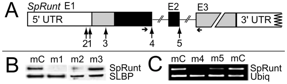
Morpholino antisense oligonucleotides can be used to sequence-specifically block translation [10] or pre-mRNA splicing [11]. We employed both strategies to study
SpRunt function, using morpholinos that target sequences within exon 1 (m1, m2, and m3) and the first two exon-intron junctions (m4 and m5; Figure 1A). To test the effi-cacy of the translation-blocking morpholinos, zygotes were injected with m1, m2, or m3 or a non-specific con-trol morpholino (mC), together with synthetic SpRunt mRNA (>100-fold excess over endogenous levels), and the resulting embryos analyzed by immunoblot using an antibody generated against the N-terminal peptide of SpRunt. Zygotic translation of full-length SpRunt protein from exogenous mRNA is completely blocked by m1 and partially blocked by m2, but not at all by m3, which tar-gets a potential secondary translational start site (Figure 1B). To test the efficacy of the splice-blocking mor-pholinos, we performed RT-PCR on total RNA isolated from embryos injected with m4, m5, or mC. While both m4 and m5 interfere with production of full-length
(Figure 1C). In addition, m5 induces production of a tran-script that retains intron 2 (which encodes a truncated protein with a complete Runt domain, and hence a poten-tially functional protein), and also skipping of exon 2, producing an aberrant transcript that fuses exon 3 directly to exon 1 (data not shown).
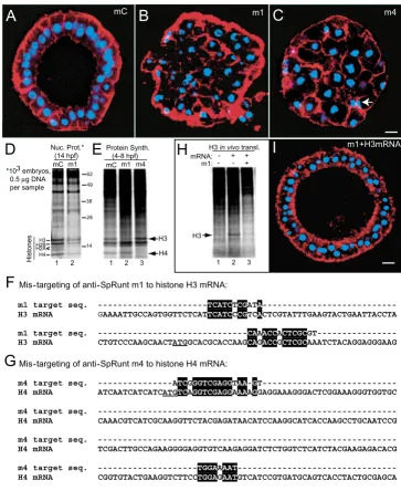
Morpholinos 1 and 4 produce a cleavage stage artifact by mis-targeting to histone mRNA
Embryos injected with anti-SpRunt morpholinos develop two distinctive phenotypes: an early blastula stage arrest (Figure 2) and a later gastrula-stage arrest (Figure 3; see below). We initially interpreted this as reflecting complete versus partial loss of SpRunt function, since the more severe early phenotype is obtained with the two mor-pholinos that are most effective at knocking down SpRunt (that is, m1 and m4; see Figure 1). By late cleavage-stage, all of the nuclei in m1- or m4-injected embryos are abnor-mal (Figure 2B,2C; compare to control in Figure 2A), with a 'cut'-like phenotype indicative of a failure to segregate mitotic chromosomes (Figure 2C, arrow). Counts of nuclei and time-lapse imaging show that these embryos undergo a global mitotic catastrophe during cleavage cycle 7 or 8 (data not shown). For both m1 and m4, this phenotype is obtained in >95% of embryos injected with morpholino doses of ~1–2 µM, comparable to effective dosage in other studies [10,12].
When m1 is co-injected with synthetic H3 mRNA (0.1 mg/ml), translation of the exogenous histone mRNA is indeed blocked (Figure 2H; compare lanes 2 and 3). How-ever, co-injection of sufficiently high levels of H3 mRNA (≥1 mg/ml) rescues m1-injected embryos from mitotic catastrophe (Figure 2I), whereas similar levels of H3 mRNA do not rescue m4-injected embryos (not shown). Co-injection of SpRunt mRNA lacking the m1 target sequence does not rescue either m1- or m4- injected embryos (not shown). We conclude that m1-injected embryos undergo mitotic catastrophe because of anti-sense mis-targeting to histone H3 mRNA, whereas the mitotic catastrophe in m4-injected embryos is probably caused by mis-targeting to histone H4 mRNA. These results show that production of similar defects by two dif-ferent morpholinos that target the same gene does not prove that the defects are caused by knockdown of the intended target.
Morpholinos 2 and 5 cause gastrula stage developmental defects
Embryos injected with m2 or m5 appear relatively normal through mesenchyme blastula stage (not shown), but thereafter develop numerous obvious defects. By late gas-trula stage, the embryos are smaller than controls, and dis-play differentiation and/or patterning defects in all of the major tissue territories: the ectoderm is radialized, the pri-mary mesenchyme cells fail to undergo skeletogenesis,
and the gut rudiment is short, disorganized, and typically evaginates as an exogastrula (Figure 3C,3D; compare to controls in Figure 3A,3B). Pigment cells do eventually develop however, indicating that some differentiation occurs.
Although m2- and m5-injected embryos develop indistin-guishable defects, the coincidental mis-targeting artifact obtained with m1 and m4 compelled us to use a rescue approach to prove that these defects are caused by loss of SpRunt function. Since m5 is a splice-blocking mor-pholino that targets the second exon-intron junction, it would not be expected to block SpRunt translation, and we reasoned that it should therefore be possible to sup-press the defects observed in m5-injected embryos by co-injection of wild-type SpRunt mRNA. This is indeed the case (Figure 3E,3F): in three separate experiments with injection solutions containing 1 mg/ml SpRunt mRNA (~6 kb), 30–50% of embryos had the rescue phenotype depicted in Figure 3, while the remainder of the embryos were somewhat more normal looking than those injected with morpholino alone (for example, with longer exogas-trulated guts). The incomplete penetrance of the rescue is probably due to turnover of the injected mRNA by gas-trula stage. In contrast, only 10–15% of embryos injected with m5 alone display a normal phenotype (probably as a result of under-injection).
Morpholino antisense targeting of SpRunt
Figure 1
Morpholino antisense targeting of SpRunt. (A) Schematic of SpRunt, showing the relative locations of sequences targeted by each of the five morpholinos used in this study. Exons 1–3 are depicted as boxes, with coding regions shaded. The Runt domain is black. Horizontal arrows indicate the positions of primers used for the RT-PCR shown in (C). (B) Immunoblots of protein translated in vivo from injected SpRunt mRNA co-injected with control morpholino (mC), m1, m2, or m3. As a loading control, the same blots were probed with an antibody to the sea urchin stem-loop binding protein (SLBP). (C) RT-PCR of SpRunt from total RNA extracted from early blastula stage embryos injected with mC, m4 or m5 (two separate experiments). RT-PCR of ubiquitin was used to control for RNA levels.
1
2
E1
E2
E3
3
4
SpRunt
5' UTR
3' UTR
5
A
B
mC
mC
m4
SpRunt
m1
C
SpRunt
SLBP
Ubiq
m5
m3
Embryos injected with anti-SpRunt m1 and m4 undergo a mitotic catastrophe caused by antisense mis-targeting to histone mRNA
Figure 2
Embryos injected with anti-SpRunt m1 and m4 undergo a mitotic catastrophe caused by antisense mis-targeting to histone mRNA. (A-C) Confocal images of fixed 15 hr embryos stained with DAPI (blue) and rhodamine phalloidin (red). (A) Control (mC)-injected embryo, (B) m1-injected embryo, and (C) m4-injected embryo. The arrow indicates a pair of 'cut' cells. Bar = 10 µm. (D) SDS PAGE of whole nuclei containing equivalent amounts of DNA (0.5 µg) from 14-hour control (mC) and m1-injected samples, stained with SYPRO Ruby protein stain. The positions of core histones, obtained from the mobility of calf thymus histone standards run on the same gel, are shown on the left of the gel, while the positions of molecular weight stand-ards are indicated on the right. (E) Total protein from 600 mC-, m1-, or m4-injected embryos labeled metabolically with 35 S-Met/Cys from 4 to 8 hours post-fertilization (hpf). Histones are easily identified by their characteristic size and stoichiometry, and by the fact that at this stage they represent ~5–10% of the total protein synthesized in the embryo [27]. The positions of histones H3 and H4 are indicated. (F, G) Sequence alignment between (F) the target sequence for m1 and α-histone H3 mRNA, and (G) the target sequence for m4 and α-histone H4 mRNA. Sequence identities are highlighted in black. The start codons are underlined in each sequence. (H) In vivo translation of synthetic histone H3 mRNA co-injected with m1. The injected zygotes were labeled metabolically with 35S-Met/Cys for 2 hours, during the first cleavage cycle. (I) 15-hr blastula stage embryo, stained as in A-C, showing rescue of m1-induced cell division defects by co-injection of 1 µg/µl H3 mRNA; bar = 10 µm.
A
mCB
m1C
m4D
14 28
H3 H2B H4 H2A
49
38 62
mC m1
Histones
Protein Synth. (4-8 hpf) mC m1 Nuc. Prot.*
(14 hpf)
1 2 *103 embryos,
0.5µg DNA per sample
m4
H3 H4
E
1 2 3
H
H3in vivotransl.I
Mis-targeting of anti-SpRunt m1 to histone H3 mRNA:
m1 target seq. ---TCATCTCGATA
---H3 mRNA GAAAATTGCCAGTGGTTCTCATTCATCCCGTCACTCGTATTTGAAGTACTGAATTACCTA
m1 target seq. ---CAAACCACTCGC
GT---H3 mRNA CTGTCCCAAGCAACTATGGCACGCACCAAGCAGACCGCTCGCAAATCTACAGGAGGGAAG
Mis-targeting of anti-SpRunt m4 to histone H4 mRNA:
m4 target seq. ---ATCGGGTCGAGGTAA-G
T---H4 mRNA ATCAATCATCATCATGTCAGGTCGAGGAAAAGGAGGAAAGGGACTCGGAAAGGGTGGTGC
m4 target seq.
---H4 mRNA CAAACGTCATCGCAAGGTTCTACGAGATAACATCCAAGGCATCACCAAGCCTGCAATCCG
m4 target seq.
---H4 mRNA TCGACTTGCCAGAAGGGGAGGTGTCAAGAGGATCTCTGGTCTCATCTACGAAGAGACACG
m4 target seq. ---TGGAAAAT
---H4 mRNA CGGTGTACTGAAGGTCTTCCTGGAGAATGTCATCCGTGATGCAGTCACCTACTGCGAGCA
F
G
+ +
m1+H3mRNA
+ m1: mRNA: -
-H3
Phenotype of late gastrula stage embryos injected with anti-SpRunt m2 and m5
Figure 3
Phenotype of late gastrula stage embryos injected with anti-SpRunt m2 and m5. DIC images (A-F) and cytometric analysis (G-I) of 48 hour embryos. (A) Control morpholino (mC)-injected embryo, cross section, with the oral side to the left and the gut indicated by an arrowhead. Bar = 20 µm. (B) mC-injected embryo, oral view, with the skeletal spicules indicated by an arrow (sp). (C) m2-injected embryo. (D) m5-injected embryo. (E-F) Embryos co-injected with m5 and SpRunt FL mRNA, showing the rescued development of gut (E) and spicule rudiments (F). (G) DNA histogram of mC-injected embryos. (H) DNA histo-gram of m5-injected embryos; note the accumulation of cells in G2/M. (I) DNA histohisto-gram of embryos co-injected with m5 and SpRunt mRNA. Note the reduced accumulation of cells in G2/M compared to the embryos injected with m5 alone.
A
G
C
D
mC
m2
m5
F
sp gut
B
mC
62% G1
35% S
3% G2/M
E
50% G1
38% S
12% G2/M
m5+SpRunt mRNA
m5+SpRunt mRNA
sp gut
H
I
m5
mC
60% G1
34% S
The small size of SpRunt-depleted embryos suggests that they might be defective in growth and/or cell proliferation. Consistent with such a possibility, embryos injected with m5 were found to contain about half the DNA content of control embryos by 48 hours post-fertili-zation (late gastrula stage; data not shown). We used cytometry to further examine the DNA content in these embryos. Compared to controls of the same age (Figure 3G), m5-injected embryos at 48 hours post-fertilization display an abnormally high proportion of cells in G2/M phase (Figure 3H), a defect that is rescued by co-injection of SpRunt mRNA (Figure 3I), indicating that SpRunt is required for the normal developmental program of cell division. This is consistent with the expression pattern of SpRunt, which is isomorphic with the pattern of cell pro-liferation in the embryo [5].
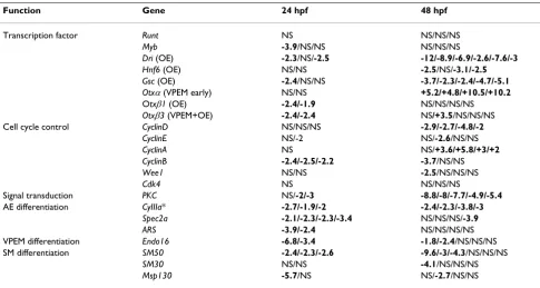
Quantitative RT-PCR (Q-PCR) was used to examine the effect of SpRunt knockdown on gene expression (Table 1). The data in Table 1 indicate that SpRunt is required for the expression of differentiation genes in each of the four major territories of the embryo (that is, oral ectoderm, aboral ectoderm, vegetal plate endomesoderm, and skele-togenic mesoderm). One of the genes whose activity is most dramatically affected by depletion of SpRunt is
SpDri, which encodes a transcription factor that is impor-tant for gastrula stage differentiation of oral ectoderm [16]. Expression of SpGsc, another transcription factor important for oral ectoderm differentiation [17], is also diminished (Table 1). This may be a secondary effect, as
SpGsc is likely to be a target of SpDri [16]. SpRunt is also required for normal expression levels of terminal differen-tiation markers such as CyIIIa in aboral ectoderm, Endo16
in endoderm, and SM50 in skeletogenic mesoderm (Table 1). It is possible that the skeletogenic defects are a non-autonomous consequence of the failure to differentiate ectoderm [18], although the fact that SpRunt is expressed at relatively high levels in skeletogenic mesenchyme [5] is consistent with an autonomous requirement. Moreover, a sequence that contains the Runx binding consensus (TGT/
CGGTT/C) is found upstream of the promoter of the SM50 gene (A.J.R., unpublished observation).
In support of a role in cell proliferation, SpRunt is required for normal levels of cyclins B and D, which are expressed in a proliferation-specific pattern similar to that displayed by SpRunt [19,20]. In addition, SpRunt posi-tively regulates the expression of protein kinase C (PKC; Table 1). Interestingly, human PKCβhas been shown to be a direct target of RUNX1 [21]. Given the wide variety of
Function Gene 24 hpf 48 hpf
Transcription factor Runt NS NS/NS/NS
Myb -3.9/NS/NS NS/NS/NS
Dri (OE) -2.3/NS/-2.5 -12/-8.9/-6.9/-2.6/-7.6/-3
Hnf6 (OE) NS/NS -2.5/NS/-3.1/-2.5
Gsc (OE) -2.4/NS/NS -3.7/-2.3/-2.4/-4.7/-5.1
Otxα(VPEM early) NS/NS +5.2/+4.8/+10.5/+10.2 Otxβ1 (OE) -2.4/-1.9 NS/NS/NS/NS
Otxβ3 (VPEM+OE) -2.4/-2.4 NS/+3.5/NS/NS/NS Cell cycle control CyclinD NS/NS/NS -2.9/-2.7/-4.8/-2
CyclinE NS/-2 NS/-2.6/NS/NS
CyclinA NS NS/+3.6/+5.8/+3/+2
CyclinB -2.4/-2.5/-2.2 -3.7/NS/NS
Wee1 NS/NS -2.5/NS/NS/NS
Cdk4 NS NS/NS/NS
Signal transduction PKC NS/-2/-3 -8.8/-8/-7.7/-4.9/-5.4 AE differentiation CyIIIa* -2.7/-1.9/-2 -2.4/-2.3/-3.8/-3
Spec2a -2.1/-2.3/-2.3/-3.4 NS/NS/NS/-3.9
ARS -3.9/-2.4 NS/NS/NS/NS
VPEM differentiation Endo16 -6.8/-3.4 -1.8/-2.4/NS/NS/NS SM differentiation SM50 -2.4/-2.3/-2.6 -9.6/-3/-4.3/NS/NS/NS
SM30 NS/NS -4.1/NS/NS/NS
Msp130 -5.7/NS NS/-2.7/NS/NS
developmental signaling processes that utilize protein kinase C, it is possible that a significant number of the later defects in SpRunt morphant embryos develop as a consequence of loss of PKC expression.
Although loss of SpRunt function appears to have global effects on development, the expression of a number of genes is not affected, which argues against a non-specific effect on gene expression caused by a general arrest of development. Moreover, loss of SpRunt function has stage-specific effects on the expression of each of genes listed in Table 1, in some cases early, and in others late. The expression of some genes (for example, Spec2a, ARS,
Otxβ3, and Endo16) is affected more strongly at mesen-chyme blastula stage than at late gastrula stage, and the opposite is true for several other genes (for example,
SpDri, cyclinD, and Otxα). A few genes are affected at both stages assayed (for example, CyIIIa, PKC, and SM50). In most cases, knockdown of SpRunt causes a diminishment in gene expression levels, consistent with its previously identified function as an activator of CyIIIa [8]. However, in the cases of Otxα and cyclinA there is an elevation in expression at 48 hours. Normally Otxα levels are greatly diminished at this stage of development [22], and it is possible that this involves SpRunt mediated repression.
The stage-specific effects of SpRunt knockdown on gene expression are consistent with the proposition that SpRunt functions within the context of one or more cis -regulatory modules in each target gene, thereby regulating a temporal and/or spatial sub-element of the target gene's overall expression pattern, as is the case with CyIIIa [8,23]. While further work using chromatin immunoprecipita-tion and cis-regulatory analysis will be necessary to deter-mine which of the affected genes in Table 1 are direct targets of SpRunt, these data demonstrate that SpRunt is required globally for both cell proliferation and differen-tiation, and is likely to be a key node in the gene regula-tory network that coordinates these processes during embryogenesis. Understanding of how this is accom-plished will require a comprehensive analysis of the rele-vant gene regulatory network, and this is in progress.
Conclusions
In both Drosophila and in mice, Runx proteins are expressed in localized domains and are important for the development of specific structures, and are thus often referred to as cell lineage or tissue-specific transcription factors. Unlike sea urchins, both insects and vertebrates have multiple Runx genes [2], which may reflect the greater regulatory network complexity in these organisms (that is, the Runx paralogs may reflect a requirement for expression variants as opposed to functionally distinct proteins). Our results suggest that SpRunt is required for normal development in all of the embryonic tissues. This
would not have been predicted based on its spatial pattern of expression, which in the post-gastrula stage embryo is confined to actively dividing cells [5]. Nonetheless, SpRunt clearly functions autonomously to promote the gastrula stage differentiation of aboral ectoderm [8], wherein its mRNA (but not protein; J.A.C., unpublished) is virtually undetectable [5]. Rather than specifying cell fate per se, SpRunt is probably required as a 'promoter organizer' [24] for the normal functioning of a variety of different territory-specific transcription factors. It is possi-ble that Runx genes are generally required for coordinat-ing cell proliferation and differentiation durcoordinat-ing animal development, a requirement that would be masked by the idiosyncrasies of their mRNA expression patterns as well as by functional redundancies between paralogs in model systems that contain multiple Runx genes.
Methods
Animals, embryo culture, and microinjection
Strongylocentrotus purpuratus were obtained from Marinus, Inc. (Long Beach, CA, USA), Charles Hallohan (Santa Bar-bara, CA, USA), or Pat Leahy (Corona del Mar, CA, USA). Gamete collection, fertilization, embryo culture, and microinjection were carried out as previously described [8]. For metabolic labeling, embryos were incubated in 14 µCi 35S-Met/Cys (>1000 Ci/mmole; APBiotech, Piscata-way, NJ, USA) in a total of 1 ml artificial seawater (ASW) at 15°C.
Morpholino antisense oligonucleotides
The following morpholinos were purchased from Gene-Tools, LLC (Philomath, OR, USA): ACGCGAGTGGTTTG-TATCGAGATGA (m1); GATGAAAGGGCGGGAAAAAATGATT (m2); TAACTGT-TATGTAGGTTGTTCCTCC (m3); ATTTTCCAACTTAC-CTCGACCCGAT (m4); and GGTATGACTTACGTCTGGGTTCTC (m5).
Target site selection and design of each morpholino was performed by GeneTools. The standard control mor-pholino (mC) from GeneTools was used as a non-specific control. Prior to injection, morpholinos were diluted to 150–400 µM in 120 mM KCl.
Synthetic mRNA
sis and linearized with Sac1 for mRNA synthesis. For rescue experiments, the 3' UTR of SpRunt (including the poly A signal and tail) was amplified from cDNA using the primers CAGATCGCTACCTACAACCGTGCAA (forward) and TTTTTTTTTTCAAAGTGAACAGAGTGTTTTA (reverse) and cloned into pGEM-T Easy (Promega, Madison, WI, USA). This plasmid was digested with AatII to release the insert, which was then cloned into the AatII sites of the original SpRunt clone [8] to generate a plasmid contain-ing the full-length sequence of the SpRunt mRNA. Follow-ing verification by sequencFollow-ing, this plasmid was used as a PCR template to amplify full-length SpRunt, using the T7 primer and the reverse primer representing the SpRunt poly A tail (see above), and the resulting amplicon was then used for mRNA synthesis. Capped synthetic mRNA was made with the T7 mMessage Machine from Ambion (Austin, TX, USA).
Microscopy and cytometry
Staged embryos were fixed in 4% formaldehyde in filtered ASW for 20 min to 6 hrs on ice, then washed 3× with PBS and into PBS plus 0.2% Tween-20 (PBST). To detect nuclei and cell boundaries, embryos were stained for 5– 10 mins with rhodamine phalloidin (Molecular Probes, Eugene, OR, USA) at 1:40 and DAPI (Molecular Probes) at 300 nM in PBST, and washed 3× in PBS. Digital images were collected using either a Zeiss Axioplan or a Leica TCS SP2 confocal microscope.
For DNA analysis, embryos grown at 15°C were collected by centrifugation, washed once 1 M Glycine, 2 mM EDTA, pH 8, and then resuspended in calcium-free seawater. The embryos were dissociated into single cells by mild tritura-tion, and stained with propidium iodide (PI) using the PI Stain kit from Sigma (St. Louis, MO, USA). PI fluorescent signal (580/30 nm) was measured using a Cyan flow cytometer (Dako-Cytomation, Fort Collins, CO, USA). Approximately 10,000 events were collected per sample. DNA ploidy analysis was performed using ModFit LT soft-ware (Verity Softsoft-ware House, Topsham, ME, USA).
Isolation of nuclei and DNA fluorometry
Embryos were lysed by incubation for 15 minutes on ice in ~5 volumes of hypotonic lysis buffer (10 mM HEPES, pH 7.9, 1.5 mM MgCl2, 10 mM KCl, 1 mM DTT) supple-mented with a general protease inhibitor cocktail (Sigma), followed by addition of Igepal CA-630 (Sigma) to a final concentration of 0.6% and vortexing for 10 sec-onds. Nuclei were then isolated by centrifugation for 30 seconds at 10,000 g. The nuclear pellet was resuspended in ~5 volumes Buffer D (25 mM Hepes pH 7.5, 5 mM MgCl2, 0.1 mM EDTA, 1 mM DTT, 1 mM Spermidine, 0.1 mM PMSF), and the DNA content was quantitated by
DyNA Quant 200 system (APBiotech).
SDS polyacrylamide electrophoresis (PAGE) and immunoblots
Protein samples (representing equivalent total protein, as determined by the method of Bradford [26]), whole nuclei (representing equivalent total DNA, as determined by fluorometry), or whole 35S-labeled embryos (equiva-lent numbers, as determined by manual counts) were mixed with an equal volume of 2× SDS sample buffer, heated to 95°C for 5 min, and run on a 10% NuPAGE Bis-Tris gel (Invitrogen, Carlsbad, CA, USA). Gels were dried and imaged, stained with SYPRO Ruby (Bio-Rad, Her-cules, CA, USA) and imaged directly, or transferred to a nitrocellulose membrane. Radioactive and fluorescent gels were imaged on a Typhoon 8600 phosphorimager (APBiotech). For immunoblots, the membrane was incu-bated for 1 hr in TBS-T (20 mM Tris-HCl, pH 7.5, 150 mM NaCl, 0.05% Tween-20) with 5% nonfat milk (TBS-TM), and then incubated in TBS-TM containing an appropriate dilution of primary antibody overnight at 4°C. Following four washes for 5 min each with TBS-T, the membrane was incubated for 1 hr at room temperature in TBS-TM con-taining 2 ng/ml of goat anti-rabbit IgG HRP conjugate (Biosource International, Camarillo, CA, USA). The mem-brane was then washed 5× for 5 min each in TBS-T, incu-bated for 4 min in Super Signal West Femto substrate (Pierce Biotech, Rockford, IL), and exposed to Kodak X-OMAT film.
The SpRunt antibody was raised against the N-terminal peptide of SpRunt, and affinity purified on a column bear-ing the peptide antigen.
Reverse transcription and quantitative polymerase chain reaction
Total RNA was isolated from morpholino injected embryos using the RNeasy mini kit (Qiagen, Valencia, CA, USA), and quantified with the RiboGreen Quantification Kit (Molecular Probes). Quantitative, real-time, one-step RT-PCR was performed using the Qiagen QuantiTect SYBR Green RT-PCR kit and the fluorescence measured in a BioRad iCycler. Starting with 50 ng of total RNA and 0.4 µM of each primer the reaction was set up and run follow-ing the Qiagen recommended protocol. For quantitative real-time PCR (Q-PCR) measurements, determination of the ratio of target in the control versus the sample was cal-culated as 1.9∆Ct, where ∆C
ensure that there was only a single product formed under the conditions used for each primer pair. Primer sequences used for the RT-PCR and Q-PCR reactions are listed in the Supplemental Table [see Additional file 1].
Authors' contributions
JAC directed the research, performed microinjections, light microscopy, and embryo dissociations for cytome-try, and drafted the manuscript. CDS performed microin-jections, confocal microscopy, and Q-PCR. JSH performed the cytometry and cell cycle analysis. JJM per-formed metabolic labeling, electrophoresis, and immuno-bloting. AJR made the SpRunt expression constructs and synthetic mRNA and assisted with the RT-PCR and Q-PCR analyses.
Additional material
Acknowledgements
The SLBP antibody used for a loading control was kindly provided by Dr. William Marzluff (University of North Carolina). We thank Dr. Paul Trainor for providing a critique of this manuscript prior to submission. This work was supported by the Stowers Institute for Medical Research.
References
1. Coffman JA: Runx transcription factors and the developmen-tal balance between cell proliferation and differentiation.Cell Biol Int 2003, 27:315-324.
2. Rennert J, Coffman JA, Mushegian AR, Robertson AJ: The evolution of runx genes. I. A comparative study of sequences from phy-logenetically diverse model organisms.BMC Evol Biol 2003, 3:4. 3. Canon J, Banerjee U: Runt and Lozenge function in Drosophila
development.Semin Cell Dev Biol 2000, 11:327-336.
4. Lund AH, van Lohuizen M: RUNX: a trilogy of cancer genes. Can-cer Cell 2002, 1:213-215.
5. Robertson AJ, Dickey CE, McCarthy JM, Coffman JA: The expres-sion of SpRunt during sea urchin embryogenesis.Mech Dev
2002, 117:327-330.
6. Stricker S, Poustka AJ, Wiecha U, Stiege A, Hecht J, Panopoulou G, Vilcinskas A, Mundlos S, Seitz V: A single amphioxus and sea urchin runt-gene suggests that runt-gene duplications occurred in early chordate evolution.Dev Comp Immunol 2003,
27:673-684.
7. Coffman JA, Moore JG, Calzone FJ, Britten RJ, Hood LE, Davidson EH:
Automated sequential affinity chromatography of sea urchin embryo DNA binding proteins.Mol Mar Biol Biotechnol 1992,
1:136-146.
8. Coffman JA, Kirchhamer CV, Harrington MG, Davidson EH: SpRunt-1, a new member of the runt domain family of transcription factors, is a positive regulator of the aboral ectoderm-spe-cific CyIIIA gene in sea urchin embryos. Dev Biol 1996,
174:43-54.
9. Pancer Z, Rast JP, Davidson EH: Origins of immunity: transcrip-tion factors and homologues of effector genes of the verte-brate immune system expressed in sea urchin coelomocytes.Immunogenetics 1999, 49:773-786.
10. Howard EW, Newman LA, Oleksyn DW, Angerer RC, Angerer LM:
SpKrl: a direct target of beta-catenin regulation required for endoderm differentiation in sea urchin embryos.Development
2001, 128:365-375.
11. Draper BW, Morcos PA, Kimmel CB: Inhibition of zebrafish fgf8 pre-mRNA splicing with morpholino oligos: a quantifiable method for gene knockdown.Genesis 2001, 30:154-156. 12. Davidson EH, Rast JP, Oliveri P, Ransick A, Calestani C, Yuh CH,
Minokawa T, Amore G, Hinman V, Arenas-Mena C, Otim O, Brown CT, Livi CB, Lee PY, Revilla R, Schilstra MJ, Clarke PJ, Rust AG, Pan Z, Arnone MI, Rowen L, Cameron RA, McClay DR, Hood L, Bolouri H: A provisional regulatory gene network for specification of endomesoderm in the sea urchin embryo. Dev Biol 2002,
246:162-190.
13. Sullivan E, Santiago C, Parker ED, Dominski Z, Yang X, Lanzotti DJ, Ingledue TC, Marzluff WF, Duronio RJ: Drosophila stem loop binding protein coordinates accumulation of mature histone mRNA with cell cycle progression.Genes Dev 2001, 15:173-187. 14. Pettitt J, Crombie C, Schumperli D, Muller B: The Caenorhabditis elegans histone hairpin-binding protein is required for core histone gene expression and is essential for embryonic and postembryonic cell division.J Cell Sci 2002, 115:857-866. 15. Kodama Y, Rothman JH, Sugimoto A, Yamamoto M: The stem-loop
binding protein CDL-1 is required for chromosome conden-sation, progression of cell death and morphogenesis in Caenorhabditis elegans.Development 2002, 129:187-196. 16. Amore G, Yavrouian RG, Peterson KJ, Ransick A, McClay DR,
David-son EH: Spdeadringer, a sea urchin embryo gene required separately in skeletogenic and oral ectoderm gene regula-tory networks.Dev Biol 2003, 261:55-81.
17. Angerer LM, Oleksyn DW, Levine AM, Li X, Klein WH, Angerer RC:
Sea urchin goosecoid function links fate specification along the animal- vegetal and oral-aboral embryonic axes. Develop-ment 2001, 128:4393-4404.
18. Cavalieri V, Spinelli G, Di Bernardo M: Impairing Otp homeodo-main function in oral ectoderm cells affects skeletogenesis in sea urchin embryos.Dev Biol 2003, 262:107-118.
19. Moore JC, Sumerel JL, Schnackenberg BJ, Nichols JA, Wikramanayake A, Wessel GM, Marzluff WF: Cyclin D and cdk4 are required for normal development beyond the blastula stage in sea urchin embryos.Mol Cell Biol 2002, 22:4863-4875.
20. Thatcher JD, McBride B, Katula KS: Promoter binding factors regulating cyclin B transcription in the sea urchin embryo.
DNA Cell Biol 1995, 14:869-881.
21. Hug BA, Ahmed N, Robbins JA, Lazar MA: A chromatin Immuno-precipitation screen reveals protein kinase C beta as a direct RUNX1 target gene.J Biol Chem 2003, 279:825-830.
22. Yuh CH, Brown CT, Livi CB, Rowen L, Clarke PJ, Davidson EH:
Patchy interspecific sequence similarities efficiently identify positive cis-regulatory elements in the sea urchin.Dev Biol
2002, 246:148-161.
23. Coffman JA, Kirchhamer CV, Harrington MG, Davidson EH: SpMyb functions as an intramodular repressor to regulate spatial expression of CyIIIa in sea urchin embryos.Development 1997,
124:4717-4727.
24. Westendorf JJ, Hiebert SW: Mammalian runt-domain proteins and their roles in hematopoiesis, osteogenesis, and leukemia.J Cell Biochem 1999, Suppl:51-58.
25. Wang ZF, Ingledue TC, Dominski Z, Sanchez R, Marzluff WF: Two Xenopus proteins that bind the 3' end of histone mRNA: implications for translational control of histone synthesis during oogenesis.Mol Cell Biol 1999, 19:835-845.
26. Bradford MM: A rapid and sensitive method for the quantita-tion of microgram quantities of protein utilizing the princi-ple of protein-dye binding.Anal Biochem 1976, 72:248-254. 27. Goustin AS: Two temporal phases for the control of histone
gene activity in cleaving sea urchin embryos (S. purpuratus).
Dev Biol 1981, 87:163-175.
Additional File 1
Sequences of PCR primers used in this study. Click here for file