ELEMENTS WITHIN HOST GENOMES
by
Edward C. Davis, Jr.
A thesis
submitted in partial fulfillment of the requirements for the degree of Master of Science in Computer Science
Boise State University
DEFENSE COMMITTEE AND FINAL READING APPROVALS
of the thesis submitted by
Edward C. Davis, Jr.
Thesis Title: Identification of Small Endogenous Viral Elements within Host Genomes
Date of Final Oral Examination: 04 March 2016
The following individuals read and discussed the thesis submitted by student Edward C. Davis, Jr., and they evaluated his presentation and response to questions during the final oral examination. They found that the student passed the final oral examination.
Timothy Andersen, Ph.D. Chair, Supervisory Committee
Amit Jain, Ph.D. Member, Supervisory Committee
Gregory Hampikian, Ph.D. Member, Supervisory Committee
The author wishes to express gratitude to the members of the supervisory com-mittee for providing guidance and patience.
A parallel string matching software architecture has been developed (incorpo-rating several algorithms) to identify small genetic sequences in large genomes. En-dogenous viral elements (EVEs) are sequences originating in the genomes of viruses that have become integrated into the chromosomes of sperm or egg cells of infected hosts, and passed to subsequent generations. EVEs have been identified in all seven classes of viruses and in the species of all kingdoms of life. Viruses from groups V and VI are considered in this thesis, including HIV and Ebola, within host genomes ranging from bacteria to humans. This database of small endogenous viral elements (SEVEs) contains homology between the viruses and every chromosome of the ten multicellular organisms in this study, including human, chimpanzee, gorilla, mouse, fruitbat, nematode, and thale cress.
ABSTRACT . . . vi
LIST OF TABLES . . . x
LIST OF FIGURES . . . xi
LIST OF ABBREVIATIONS . . . xiii
LIST OF SYMBOLS . . . xv
1 Introduction . . . 1
1.1 Endogenous Viral Elements . . . 1
1.1.1 Conservation of Endogenous Elements . . . 2
1.1.2 Motivating the Search for EVEs . . . 3
2 Literature Review . . . 8
2.1 Biological Computation . . . 8
2.1.1 Substitution Matrices . . . 8
2.1.2 Bioinformatics Toolkits . . . 9
2.2 String Matching . . . 11
2.2.1 Match Table Algorithms . . . 11
2.2.2 Hashing Algorithms . . . 12
2.2.3 Automata Algorithms . . . 13
2.3 Endogenous Viruses . . . 14
2.3.1 Virology . . . 14
2.3.2 Endogenous Retroviruses and Nonretroviruses . . . 15
2.3.3 Potential Functions of Endogenous Viruses . . . 17
3 Methods . . . 20
3.1 Species Selections . . . 20
3.2 Computational Approach . . . 22
3.2.1 FTPScanner . . . 23
3.2.2 GenomeScanner . . . 24
3.2.3 MatchDatabase . . . 31
4 Results . . . 34
4.1 Overview . . . 34
4.2 Ebolavirus . . . 37
4.2.1 Overview . . . 37
4.2.2 Ebola SEVEs by Species . . . 39
4.2.3 Ebola SEVEs by Viral Gene . . . 42
4.3 Human Immunodeficiency Virus 1 . . . 42
4.3.1 Overview . . . 42
4.3.2 HIV-1 SEVEs by Species . . . 46
4.3.3 HIV-1 SEVEs by Viral Gene . . . 46
4.4 Simian Immunodeficiency Virus . . . 48
4.4.1 Overview . . . 48
4.4.2 SIV SEVEs by Species . . . 50
4.5 Measles Morbillivirus . . . 53
4.5.1 Overview . . . 53
4.5.2 Measles SEVEs by Species . . . 54
4.5.3 Measles SEVEs by Viral Gene . . . 56
4.6 Influenzavirus A . . . 57
4.6.1 Overview . . . 57
4.6.2 Influenza A SEVEs by Species . . . 59
4.6.3 Influenza A SEVEs by Viral Gene . . . 59
4.7 SEVEs in miRBase . . . 61
4.8 Randomly Generated Genome . . . 62
4.9 Chromosome Bands . . . 63
4.10 Most Frequent SEVE Sequences . . . 65
4.11 Scalability and Efficiency . . . 67
5 Conclusions . . . 69
5.1 Future Work . . . 69
5.2 Summary . . . 71
REFERENCES . . . 73
3.1 Viral Genome Sizes . . . 24
3.2 String Algorithm Benchmark Results . . . 31
4.1 Ebolavirus Gene Products . . . 37
4.2 HIV-1 Gene Products . . . 43
4.3 SIV Gene Products . . . 49
4.4 Measles Morbillivirus Gene Products . . . 53
4.5 Influenzavirus A H7N9 Gene Products . . . 57
3.1 Example Viral Subsequences . . . 25
3.2 GenomeScanner Parallel Architecture. . . 27
3.3 UML diagram of StringSearch class hierarchy. . . 28
3.4 UML diagram of Threading hierarchy. . . 29
3.5 Example of Complementary Viral Subsequences. . . 29
3.6 JSON Output . . . 30
4.1 Ratio of SEVE sequences to host genome sizes by host and virus species. 35 4.2 Ratio of SEVE sequences to host genome sizes by host and virus species with Mouse / HIV-1 outlier excluded. . . 36
4.3 Zaire ebolavirus SEVE match count by host name and chromosome. . . 40
4.4 Zaire ebolavirus SEVE match count per viral gene and normalized by gene size. . . 43
4.5 Human immunodeficiency virus 1 SEVE match count by host name and chromosome. . . 47
4.6 Human immunodeficency virus 1 SEVE match count per viral gene and normalized by gene size. . . 48
4.7 Simian immunodeficiency virus SEVE match count by host name and chromosome. . . 51
4.8 Simian immunodeficency virus SEVE match count per viral gene and normalized by gene size. . . 52
4.10 Measles morbillivirusSEVE match count per viral gene and normalized
by gene size. . . 56
4.11 Influenzavirus A SEVE match count by host name and chromosome. . . 60
4.12 Influenzavirus A SEVE match count per viral gene and normalized by gene size. . . 61
4.13 Ratio of SEVE sequences to host genome sizes by host and virus species with random organism included. . . 63
4.14 HIV-1 SEVE sequence matches by human chromosome bands. . . 64
4.15 HIV-1 SEVE sequence matches in human chromosome 2 bands. . . 65
4.16 Most frequent SEVE sequences in the MatchDatabase. . . 66
4.17 GenomeScanner scalability graph including the file sizes from three input genomes (Human, Mouse, and Orangutan) versus running time, indicating a clear linear relationship. . . 67
4.18 GenomeScanner efficiency graph of subsequence size k fit against run-ning time using human chromosome 22 and the HIV-1 virus. . . 68
EVE – Endogenous Viral Element
SEVE – Small Endogenous Viral Element
ERV – Endogenous Retroviral Element
ENRV – Endogenous Non-Retroviral Element
HERV – Human Endogenous Retrovirus
EDI – EVE-derived Immunity
HIV – Human Immunodeficiency Virus
SIV – Simian Immunodeficiency Virus
HTLV – Human T-cell Leukemia Virus
LINE – Long Interspersed Nuclear Element
SINE – Short Interspersed Nuclear Element
UTR– Untranslated Region
ORF – Open Reading Frame
ncDNA– Noncoding DNA
ncRNA – Noncoding RNA
lncRNA – Long noncoding RNA
mRNA – Messenger RNA
ceRNA – Competing Endogenous RNA
RISC – RNA-induced silencing complex
LTR– Long Terminal Repeat
ENCODE – Encyclopedia of DNA Elements
GPCR – G-protein Coupled Receptor
CD – Cluster of Differentiation
≤ Less than or equal to
O Big-O
α Alpha
β Beta
µ Mu
CHAPTER 1
INTRODUCTION
1.1
Endogenous Viral Elements
The sequencing of the complete human genome by the Human Genome Project ranks among the most momentous achievements of modern science. One of the surprising results was the relatively small percentage of actual protein encoding genes, a mere 1.5% according to Nature [1]. The remainder consists of various types of noncoding DNA (ncDNA), including introns (approximately 6%), regulatory sequences (8-20%), mobile elements such as long interspersed nuclear elements (LINEs) and short inter-spersed nuclear elements (SINEs), and noncoding RNA (ncRNA) such as the 5’ and 3’ untranslated regions (UTRs) of mRNAs. Other sequences encode for small RNAs, such as small interfering RNAs (siRNA) and micro RNAs (miRNA).
viruses. Much research has been focused on the study of ERVs because retroviruses, Group VI on the Baltimore classification, are the only class of viruses that must insert their genome into the host chromosome as a requisite part of their life cycle. However, endogenous viral elements (EVEs) have been identified from all seven groups of viruses in the Baltimore classification [4]. The mechanisms for the incorporation of nonretroviral sequences into host genomes are not well understood, but it has been suggested that rogue nucleic acids from viral infections are inserted with the help of the reverse transcriptase and integrase enzymes of the retrotransposons that function similarly to those in retroviruses [5].
1.1.1 Conservation of Endogenous Elements
particularly significant given rapid viral mutation rates.
Conserved viral sequences could act as agents of infection, aiding and abetting future viruses in the infection of the host by providing homologous targets for viral integration. Alternatively, the sequences might confer some type of antiviral defense or immunity to the host. One potential mechanism could be the production of small RNA molecules derived from the viral sequences that interfere with some stage of viral reproduction when expressed. Such RNA interference has been observed in plants, invertebrates, and mammals [7]. It is also possible that the small RNAs interfere with immune system function of the host. As an example, a recent study by Chuong et al. indicates that ERV transcripts affect the transcriptional regulation of the interferon network (IFN) [8].
There are many constraints imposed on viral sequences, so the location of the sequences within the viral genomes is also important. Viruses must maintain compact genomes to be contained within a tiny icosahedral capsid with a mean diameter of 5 nm [9]. Of particular interest from an immunological perspective are the viral glycoprotein (GP) genes. The glycoproteins form on the viral envelope and in many cases are what allow viruses to evade the immune system and gain entry into the host cell via endocytosis. The glycoproteins enable the immune system to determine the critical distinction between self and non-self. Further increasing the difficulty of integration is that to gain access to the genomic DNA viral sequences must also pass the double nuclear membrane of the eukaryotic cell.
1.1.2 Motivating the Search for EVEs
Ebola filoviruses that cause human hemorrhagic fever. Other recent epidemics of note include the Rift Valley bunyavirus outbreak in Kenya in 2006 and the H1N1 influenza pandemic of 2009. Measles, caused by the morbillivirus, is another high profile infection enjoying a recent resurgence [10].
Retroviruses are of particular interest due to their innate reverse transcriptase activity and high levels of virulence. A relative of the well known HIV virus is the similarly structured Simian immunodeficiency virus (SIV). Together such immune targeting viruses are members of the lentivirus family. Other retroviruses can even lead to cancer by converting proto-oncogenes into oncogenes. Examples of these include the human T-cell leukemia viruses (HTLV) [11], and the mouse mammary tumor virus (MMTV) [12].
Another class of viruses capable of host genome endogenization are the negative sense single-stranded RNA (-ssRNA) viruses, Group V on the Baltimore classification. They are referred to as negative sense because their genomes are encoded in the 3’ to 5’ direction, opposite of the 5’ to 3’ direction of the mRNA transcripts to be translated on ribosomes. Therefore, an RNA replicase enzyme is required to generate the mRNA transcript that will be translated into viral proteins. Examples of -ssRNA viruses include theZaire ebolavirus of the family Filoviridae, the bornavirus of family Bornaviridae, measles of family Paramyxoviridae, and rabies of the family Rhabdoviridae.
by Francis Crick (DNA → RNA → protein). No such mechanism is known to exist in prokaryotes [13] and such viruses target vertebrates specifically. Lentiviruses and filoviruses infect many mammalian species, and bornaviruses are able to infect mammals or birds [14].
Endogenous viral elements can be divided into two broad categories, endogenous retroviruses (ERVs) that rely on retroviral reverse transcriptase encoded by the virus itself for insertion, and endogenous non-retroviruses (ENRVs) that require other means of insertion. The process by which ERVs are endogenized is well understood. The reverse transcriptase enzyme generates the double-stranded DNA copy of the viral RNA genome, complete with identical flanking long terminal repeats (LTRs). The dsDNA copy of the genome is transported to the centrosome along microtubules, enters the nucleus through a nuclear pore, and covalently binds to the genomic DNA. Once integrated, it begins the latent proviral stage of its lifecycle where it is indistinguishable from the host DNA. In this way, the mutation rate slows to the host’s neutral rate [15].
Most likely, it is the mRNA molecules that become endogenized in the germ line rather than the viral genomes themselves. The reverse transcriptase activity of the retrotransposons within the host genome, such as LINEs, are the most likely candidates for endogenization.
Whether EVEs confer immunity or encourage susceptibility to viral infection remains uncertain. One hypothesis suggests they become incorporated into EVE-derived immunity genes (EDI). The Fv1 and Fv4 genes in mice have been shown to act as inhibitors of murine leukemia [17]. Another hypothesis is that the sequences encode for small RNA elements that either work to block viral RNA translation or as miRNAs acting as competing endogenous RNA (ceRNA). Such elements encoded by expressed pseudogenes have been studied with respect to human cancers [18]. Given the established link between viruses and cancer, such a mechanism could also exist in viruses.
CHAPTER 2
LITERATURE REVIEW
2.1
Biological Computation
The relationship between biology and computer science dates back at least fifty years to the 1960s. From Sanger’s successful sequencing of the insulin protein, to Watson and Crick’s discovery that the DNA molecule is the coding language for life, it quickly became apparent that biomolecules are information carriers, much like a silicon transistor. This revelation created a conceptual link between molecular biology and Shannon’s information theory [21]. Zuckerkandl and Pauling compared nucleic and amino acid sequences to semantemes, the fundamental unit of information in linguistics. They coined the term semantides, a fundamental chemical unit [22]. This gave rise to the field ofpaleogenetics, now better known as molecular evolution. The once controversial idea that phylogenetic relationships could be inferred simply from sequence analysis, combined with the advent of the molecular clock, helped form the foundation of the field of bioinformatics [23].
2.1.1 Substitution Matrices
sub-stitution matrix for sequence alignment [24]. PAM matrices remain in use today, with the PAM250 being the most common. The BLOcks SUbstitution Matrix or BLOSUM is another important substitution matrix that applies observed rather than extrapolated local alignment scores as PAM does [25]. The BLOSUM62 matrix is a notable example.
Whereas substitution matrices employ heuristic techniques for pairwise compar-isons, the Smith and Waterman algorithm for local sequence alignment always pro-duces the same results. It is a dynamic programming algorithm that makes use of matrices to reward matches and penalize gaps [26].
The FASTA set of programs for pairwise sequence similarity scoring was created by Lipman and Pearson [27]. FASTA programs allow direct comparison of nucleotide and amino acids sequences by performing translation on the fly. The RDF2 program evaluates similarity scores with a shuffling method that permits the preservation of the original sequence. The LFASTA program generates dot matrix plots of similarity greater than a given threshold and supports a number of different scoring matrices. The best sequences are evaluated by collecting the top ten sequences and rescoring them. One of the most enduring contributions of this work is the FASTA file format, which is now ubiquitous within the field of bioinformatics.
2.1.2 Bioinformatics Toolkits
segment pair (MSP) score. BLAST sacrifices the accuracy of Smith-Waterman for speed, but with greater sensitivity than FASTA. The BLAST algorithm filters out low complexity regions (meaning highly repetitive), then converts the query sequence into ak-word list (k = 11 for nucleotides, 3 for amino acids). Each word is compared with a substitution matrix such as BLOSUM62 and those obtaining scores greater than a threshold T are kept. The remainder are added to a tree used to search the database for exact matches that are then extended with gaps considered to yield high score segment pairs (HSPs). These high scoring pairs are evaluated for significance and a Smith-Waterman alignment is performed on the highest scoring of all. An e-value is calculated from these alignments based on gap penalties and those achieving a value greater than the threshold E (expected value) are reported to the user.
Other noteworthy bioinformatics tools include Thompson’s ClustalW method for multiple sequence alignments (MSA) [30], Hidden Markov Models using Bayesian networks [31], and genetic algorithms [32]. For the automated construction of phyloge-netic trees, there is Hall’s Molecular Evolutionary Gephyloge-netics Analysis using maximum likelihood (MEGA) [33].
2.2
String Matching
The online exact string matching problem has broad applications in computer science, not merely in computational biology or chemistry but also in text and speech analysis, digital signal processing, databases, and compression. Generally stated, it is the task of finding all occurrences of a pattern string p of length m within a given text t of length n over an alphabet Σ of size σ. The worst case lower bound of the string matching problem is O(n).
2.2.1 Match Table Algorithms
O(n) time for an overall complexity ofO(m+n). Given the obvious assumption that
m ≤n, the overall time is simplified to O(n).
The distinction of creating the first string matching algorithm to achieve sublinear average time complexity belongs to Boyer and Moore [35]. The success of the Boyer-Moore algorithm is the innovative revelation that the end of a string (the suffix) should be used to scan for matches rather than the first because it allows more of the text to be skipped. Matches continue back to front until the first character of the pattern is matched. Similarly to the KMP algorithm, a preprocessing table based on the pattern is constructed in linear time O(m), and is accessible in constant time O(1). The overall worst case performance of the algorithm is O(m+n) when the
pattern does not occur in the text (p 6⊂ t), and O(mn) when the pattern does occur in the text (p ⊂ t).
2.2.2 Hashing Algorithms
2.2.3 Automata Algorithms
Another class of algorithms capable of achieving sublinear average time complexity are those that make use of factor automata. The automata are data structures that can identify all factors of a given pattern p. The Backward-Oracle-Matching (BOM) algorithm from Allauzen, Rochemore, and Affinot is one of the more efficient exam-ples, particularly for long patterns (largem) [37]. In an attempt to combine the best of both worlds, Faro and Lecroq introduced the Extended-Backword-Oracle-Matching (EBOM) fast string matching algorithm [38]. It is a variant of the Boyer-Moore algorithm with the suffix lookup table replaced with an automata based oracle like BOM. The oracle is a deterministic finite automaton that accepts all of the suffixes of a word. The automaton is built with the reverse of the pattern p in O(m) time and searches with a sliding window moving from right to left, hence it is a backward oracle match. The worst case time complexity is quadraticO(mn) like Boyer-Moore, but the average time complexity is O(nlogm/m).
2.2.4 Suffix Trees
2.3
Endogenous Viruses
Viral genomes are among the most rapidly evolving in nature. This allows them the flexibility to keep one step ahead of host immune systems, quickly adapting and crossing interspecies boundaries. Such rapid mutation gives researches the opportu-nity to observe evolution in nearly real-time by sequencing viral isolates. However, it becomes much more difficult to track viral evolution across great expanses of time. Fortunately, viruses tend to leave behind markers of their passage in the genomes of the hosts they infect. These molecular “fossils” can be analyzed by viral archaeologists to gain a greater understanding of both viral and host evolution.
2.3.1 Virology
association with cancer. A Class VII for pararetroviruses (e.g., hepatitis B) was later added, but otherwise Baltimore’s system of classification has required very little modification.
2.3.2 Endogenous Retroviruses and Nonretroviruses
In one of the early treatments of this topic, Katzourakis and Gifford provide a rather exhaustive analysis of endogenous viral element integration in animal genomes, both retroviral and nonretroviral [14]. They performed in silico analysis (i.e., BLAST searches) in a wide array of animal and viral genomes. Homologous sequences were observed between DNA, RNA, and RT viruses, within animal hosts ranging from insects to vertebrates, including mammals and birds. Both nuclear and cytoplasmic replicating viruses were covered. Phylogenetic analyses were also performed with wide ranging results. The function of EVEs and whether they are advantageous or deleterious to the host remained unanswered.
In a related work by Horie, et al., the authors perform a comprehensive search for endogenous bornavirus-like elements (EBLs) [16]. Despite being nonsegmented -ssRNA viruses that replicate their genomes in the cytoplasm, bornaviruses can cause persistent infections in the nuclei of host cells. This means their mRNA transcripts also find their way into host genomes via endogneous germline integrations just like their retroviral counterparts. They provide a review of the presence of EBLs in eukaryotic genomes, including invertebrates. In terms of host function, they note the existence of endogenous nucleoprotein sequences in mice impacting the murine leukemia virus, the remnants of open reading frames in primates, and the fragments ofenv genes in endogenous retroviruses that resulted in the development of placental mammals. Experiments were even conducted to insert modern bornavirus DNA into cultured mouse cells, albeit with limited success.
Lee et al. also considered the ERV-L mutation that gave rise to the mammalian placenta [15]. The authors conducted a study tracking ERV lineage back to 104-110 Mya. Other sequences, selfish genetic elements (SGEs), are found inserted within the ERV sequences. For example, the ERV-L endogenous retroviral gene has homologs in the chromosomes of four mammalian species, including boar, horse, chimpanzee, and human (on chromosome 17). The study included multiple bioinformatics methods, such as BLAST, MUSCLE, Needle, and RepeatMasker.
on the tell-tale signs of poly-G caps and poly-A tails surrounding the elements, they were identified as former viral mRNA transcripts that had been endogenzed, likely with the help of the reverse transcriptase enzyme from retrotransposons such as long interspersed elements (LINEs). The estimates of the number of integrations are admittedly low due to the limitations of the bioinformatics techniques applied in the analysis.
In a recent review paper, Aiewsakun and Katzourakis explain that endogenous viral elememts from all seven viral groups from the Baltimore classification have been identified within the genomes of eukaryotic organisms [4]. They provide several different dating techniques to trace viral-host interaction routes throughout evolution. This can be accomplished by comparing orthologs or paralogs, assuming the host neutral mutation rate, and augmentation with geographic data from known host migration patterns.
2.3.3 Potential Functions of Endogenous Viruses
into the cell (glycoprotein) or disrupting viral replication (gag genes) and immune system anticipation (super antigen orsag genes). The functions of many other EVEs remain unknown.
In addition to EDIs, another potential function of EVEs is the encoding of small interfering RNAs (siRNAs) or micro RNA (miRNAs), one of the emerging areas of study in genetics. They appear to be derived from pseudogenes (genes that have lost their regulatory sequences) or other noncoding regions. According to Kalyana-Sundaram et al., the traditional model of post-transcriptional modification may be incomplete [18]. The model holds that introns are excised from RNA tran-scripts, leaving only the exons to be spliced together in various combinations by the spliceosome to form mature mRNA transcripts. However, endogenous siRNA or miRNA binding sites may provide another level of control. Analysis of miRNA recognizing elements (MREs) in pseudogones has been limited by their similarity to analogous sequences encoding genes. The authors provide an analysis of pseudogene transcription from 280 normal tissue samples and thirteen cancerous ones. They found pseudogene expression to be prevalent, even ubiquitous, and in some cases possibly cancer-specific. They propose a connection to the recently discovered competitive endogenous RNA (ceRNA) networks in the transcriptome. Although this work did not cover viruses specifically, EVEs could be potential ceRNAs given the relationship between retroviruses and cancer (e.g., HTLV).
CHAPTER 3
METHODS
3.1
Species Selections
Exhaustive identification of all potential endogenous viral elements in a particular genome is challenging due to the considerable size of vertebrate genomes and the rapid mutation rate of viruses. Here a sample size is generated by subdividing several viral genomes into small fragments of only 18 base pairs, or about the size of a siRNA sequence, with a step size of three base pairs. All of the chromosomes of a given host genome are then scanned for all occurrences of each viral fragment sequence. The primary objective of this research has been to assemble an initial database containing a representative sample of all small endogenous viral elements across multiple viruses and multiple host genomes.
The focus has been on viruses from groups V and VI of the Baltimore classification. The lentiviruses HIV-1 and SIV were selected to represent the retroviruses. These viruses were chosen because their genomes have been well studied and endogenous retroviruses are already known to encompass 8% of the human genome. In terms of potential clinical importance, there are also primates known to possess immunity to SIV, such as the sooty mangabey [44].
and its presence as a blood-borne pathogen. For a nonretrovirus to be endogenized, its genome or the mRNA produced from it must be present within the nucleus of a sperm or egg cell while a retrotransposon is active (assuming that the aforementioned ENRV insertion hypothesis is correct). Such an event seems much more likely to occur in the presence of a virus that can be sexually transmitted and is therefore already in the vicinity of the gonads where germ cells reside. The morbillivirus responsible for measles infections was selected as another representative from group V (-ssRNA viruses) as something of a control against the ebolavirus, as it is known to be highly infectious to humans but not known to be sexually transmitted.
The Influenza A (H7N9) virus from the Orthomyxoviridae family was the final se-lection from group V due to the long, complex history between influenza and humans. The influenza genome is segmented, whereas the ebolavirus and morbillivirus genomes are not. The H7N9 genome is from a 2013 outbreak of H7N9 in China. The virus is known to infect birds as well as mammals (the A is for avian). Particularly virulent strains emerge when genetic recombination occurs between avian and mammalian versions of influenza [45].
The set of host genomes selected for inclusion in this study reveals a strong primate bias. The first on the list was the GRCh38 version of the human genome, consisting of 22 autosomal chromosomes along with the sex chromosomes X and Y. The genomes of the nearest living genetic relatives of Homo sapiens have also been analyzed, including the chimpanzee (Pan troglodytes), the gorilla (Gorilla gorilla), the orangutan (Pongo pygmaeus), and the gibbon (Nomascus leucogenys). The most recent common ancestor of humans and chimpanzees dates to at least 13 million years ago or as early as 4 Mya.
organisms from more distant branches of the current phylogeny are also included. Arguably the best studied member of class Mammalia is the house mouse, also known asMus musculus. The GRCm38.3 version of the mouse genome was the most recent at the time of this writing. Venturing away from class Mammalia and even phylum Chordata, within the Ecdysozoa are the phyla Nematoda and Arthropoda. Drawn from them are the genomes of the nematode worm C. elegans and the pioneering fruitfly of T.H. Morgan’s lab, Drosophila melanogaster.
Representing the other two eukaryotic kingdoms Viridiplantae and Fungi are the model genomes of the thale cress plant, Arabidopsis thaliana, and the haploid yeast Saccharomyces cerevisiae. The E. coli bacteria is the representative model organism for all prokaryotes.
The work of Pourrut et al. shows that fruit bats may act as reservoirs for theZaire ebolavirus [46]. The black flying fox or Pteropus alecto is known to be a host for the Ebola and Nipah viruses, both from group V of the Baltimore classification, and the SARS virus from group IV (+ssRNA) [47]. The black flying fox genome has been sequenced but not annotated, meaning that the raw sequencing data are available as scaffold files, but have not been compiled into chromosomes [48]. Nevertheless, the number of viral matches for the flying fox is of interest from an emerging infection point of view, and so have been included in the study.
3.2
Computational Approach
string search tool called the GenomeScanner, and the last a set of tools for storying and analyzing the output data called theMatchDatabase.
3.2.1 FTPScanner
One of the common places where genomes are stored for bioinformatics research is on the NCBI FTP site.1 The files are freely available for any industrious coder
to download. The two primary file formats are FASTA (.mfa or fa extension) and Genbank (.gbk extension). The FASTA format is the simplest, consisting of a single comment line beginning with a greater than character followed by a description. All other lines are sequences of nucleotides or amino acids. Genbank files contain more detailed information, including annotations, but are more complex and therefore more difficult to parse.
The contents of the FTP server are arrayed in a sprawling filesystem with many directories and subdirectories. In order to simplify the navigation of this hierarchy, the first software component constructed for this project was the aptly named FTPScan-ner and is implemented in the Java language. Its purpose is to scan the contents of the FTP server for genome files in FASTA and Genbank format and download or update the files on the local filesystem if desired. Upon encountering a new species, the code performs an automated Wikipedia search to fetch pertinent information about the organism such as kingdom, phylum, class, etc., and then stores the information in a JSON file database, along with the paths to the genomic files.
Once the required genomic data have been acquired from the NCBI FTP server, they can be passed to the next component, the genome scanner.
1
3.2.2 GenomeScanner
The primary component of the software framework implemented for this project is the GenomeScanner, a massively parallel string parser and searching engine. Built to be as fast as possible, it is implemented in the C++ programming language. Linux is the target operating environment but the source code could certainly be compiled for another platform.
The user interface is command line driven with an input argument pointing to the data file path where the genome files are stored on the filesystem (-fp). The second input argument is a text file containing a line delimited list of viral genome files in FASTA format (-vf). The third argument is a similar list containing all of the host genome files to be searched (-hf).
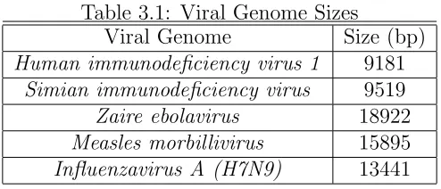
Table 3.1: Viral Genome Sizes
Viral Genome Size (bp) Human immunodeficiency virus 1 9181
Simian immunodeficiency virus 9519
Zaire ebolavirus 18922
Measles morbillivirus 15895
Influenzavirus A (H7N9) 13441
1: GGTCTCTCTGGTTAGACC 2: CTCTCTGGTTAGACCAGA 3: TCTGGTTAGACCAGATCT ...
n-2: AATAAAGCTTGCCTTGAG n-1: AAAGCTTGCCTTGAGTGC n: GCTTGCCTTGAGTGCTTC
Figure 3.1: Example of the first three and last three viral subsequences of the HIV-1 virus given parameters of k=18 and s=3.
is also a step size parameters that configures the scanner to step ahead byscharacters (nucleotides) when generating the next subsequence. The default value of s is three, but other values can be specified from the command line with the -step argument provided that s is less than k. Figure 3.1 illustrates an example of this technique by indicating the first three and the last three subsequences generated from the HIV-1 genome. The default step size of three was selected due to the large number of host genomes and viruses selected for the study.
The host genomes are vast compared to the tiny viral ones. The primate genomes (human, chimpanzee, gorilla, and orangutan) all contain approximately 3×10⁹ base
pairs. This is not an unreasonable amount of data to be read into memory on any machine with sufficient memory, but not without sacrificing some parallel processing capabilities. For that reason, the GenomeScanner reads the host genome files in discrete blocks. Host files are generally stored as one file per chromosome because a chromosome is simply one continuous DNA molecule. The blocks are measured in number of lines of a FASTA file. The block size defaults to one hundred thousand (1×10⁵) but can be specified from the command line via the -bs argument. Since a
byte of space, the average size of a host genome block is about seven megabytes.
With the input data properly subdivided into viral subsequences (the proverbial needles) and host genome blocks (the corresponding haystacks), the actual substring matching can be performed. This problem is essentially an instance of the online exact string matching problem. An excellent review of the problem space has been provided by Faro and Lecroq [49].
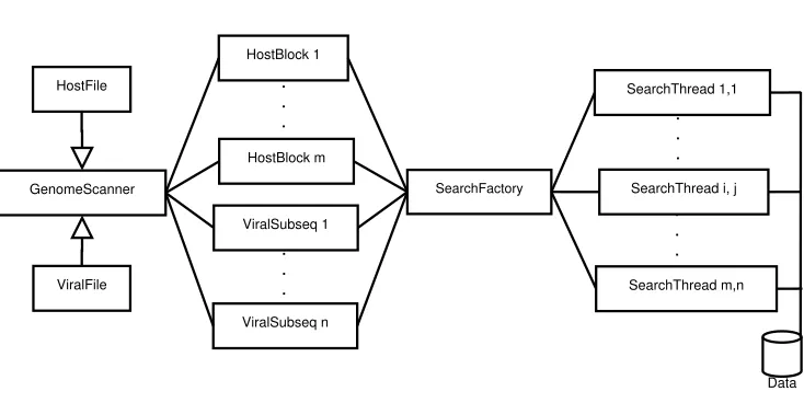
The GenomeScanner contains implementations of several algorithms designed to solve this problem. These include preprocessing algorithms such as the Knuth-Morris-Pratt (KMP) algorithm [34] and the Boyer-Moore (BM) algorithm [35]. Also included is the Rabin-Karp (RK) randomized algorithm [36] and an implementation of Ukkonen’s online suffix tree construction algorithm [39]. Preliminary benchmarks indicated that the implementation of Faro and Lecroq’s extended backward oracle match algorithm (EBOM) [38] yielded the fastest search results within the current architecture. The string matching algorithm to be applied can also be specified from the command line using the -sa argument. The EBOM algorithm is the default option due to the performance.
GenomeScanner SearchFactory SearchThread 1,1 HostFile ViralFile ViralSubseq 1 ViralSubseq n . . . HostBlock 1 HostBlock m . . .
SearchThread i, j
. . . . . . SearchThread m,n Data
Figure 3.2: GenomeScanner Parallel Architecture.
The parallel architecture of the GenomeScanner engine is greatly inspired by the Java programming language. First, there is a Runnable interface with a virtual Run
method to be implemented by all implementing classes.
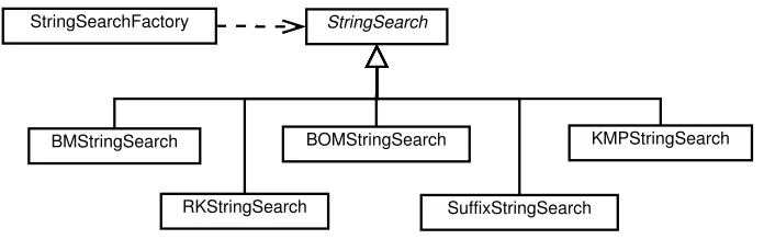
An abstract classStringSearchencapsulates the data and methods for each string search, including the block to be searched, the subsequence to be matched, the block size, beginning and end, and a string identifier for each of the source files, e.g., the host genome and viral genome file names. It also implements theRunnableinterface, which simply invokes the Search method. The infrastructure is intentionally generic so the code can easily be extended to any alphabet or search space (e.g., amino acid sequences or written text). Each subclass of the StringSearch class implements a different string matching algorithm when it overrides the Search method. Each implementation is responsible for updating the vector containing the indices of all matches.
StringSearch StringSearchFactory
BMStringSearch BOMStringSearch KMPStringSearch
RKStringSearch SuffixStringSearch
Figure 3.3: UML diagram of StringSearch class hierarchy.
for example (Figure 3.3).
Parallelism in the code is implemented (or not) via the thread interface class
ThreadInt. Each thread consists of a named identifier, a Runnable target object and a boolean running status. There are three implementing subclasses of the thread interface, a SimpleThread, which simply executes the code sequentially and returns (sequential), a PThread implemented with Linux pthreads, and MPIThread
implemented with the Message Passing Interface (MPI). The pthread and the MPI versions of the application are compiled separately as different dependencies are required (MPI programs are executed withmpiexec). The pthread version is intended for single machines with multiple cores, with each thread running on one processor core. In the MPI implementation, the zero rank (root) process acts as a delegator, passing data to the other worker nodes with nonzero rank to search each block for subsequence matches.
The GenomeScannerprogram generates an abundant number of threads. In order to avoid the overhead of frequent creation and destruction of threads, the thread pool design pattern has been implemented in the ThreadPool class to allow the reuse of thread objects (Figure 3.4).
ThreadInt ThreadPool
SimpleThread PThread MPIThread
Runnable
Figure 3.4: UML diagram of Threading hierarchy.
of genomic data from host genomes, it associates each subsequence i and block j with a thread from the thread pool and invokes a search with the appropriate search algorithm. However, it is not sufficient to search for the viral subsequence alone but also the complement. Sequences are saved in FASTA or Genbank files just as they would be read in the 5’ to 3’ direction. In other words, the way they would be read from mRNA molecules during translation. This allows researchers to search for start and stop codons, to find expressed sequences or pseudogenes within the genome. In the case of endogenous viral elements, it cannot be known whether the mRNA or the viral genome itself was inserted into the host genome. Thus, each viral subsequence actually results in two search threads, one for the 5’ to 3’ direction, and another for 3’ to 5’ (complementary). Figure 3.5 provides an example sequence.
To ensure that there are no dependencies between running search threads, the threads should not need to report their results back to the master node. To that end, each thread is responsible for writing its own data to disk. TheStringSearchclass is serializable to the JSON file format by implementing theJsonizable interface class. When a thread finishes its search and if it has found any matches, it will generate
5’-GGTCTCTCTGGTTAGACC-3’ 3’-CCAGAGAGACCAATCTGG-5’
its own file name with the JsonFile method and write its contents to it with the
Jsonizemethod. An example of the output is given in Figure 3.6.
The GenomeScanneralso maintains a log file inspired by the log4jlogger so that the process can be monitored while it is running. The logs and data are written to the file path specified by the-fpargument provided to theGenomeScannerwhen the executable is launched.
{
"matches": [6407392], "block": 1,
"begin": 1, "end": 100311, "dir": "5",
"textID": "NC_004354_chrX", "pattID": "NC_001498",
"pattern": "CCGAAGTTGGCCTTGTCG" }
Figure 3.6: JSON output from a match within the X chromosome of Drosophila melanogaster and the Measles morbillivirus.
All of the match data for this project were collected by running theGenomeScanner
across all five viral species and twelve host genomes on a Beowulf cluster with four nodes. Command lines for each host, virus pair (60 pairs) were generated and executed. The data were logged to the JSON flat file database.
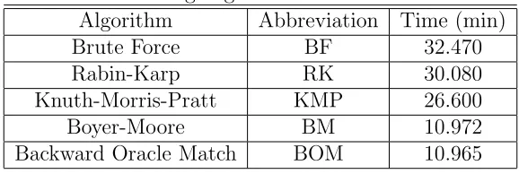
in the benchmark were brute force, Knuth-Morris-Pratt, Boyer-Moore, Rabin-Karp, and Backward Oracle Match. The brute force algorithm was implemented using the
find method of the string class in the C++ standard template library.
Table 3.2: String Algorithm Benchmark Results Algorithm Abbreviation Time (min)
Brute Force BF 32.470
Rabin-Karp RK 30.080
Knuth-Morris-Pratt KMP 26.600
Boyer-Moore BM 10.972
Backward Oracle Match BOM 10.965
The results are summarized in Table 3.2. The Boyer-Moore and Backward Oracle Match algorithms have approximately the same running time and are both well ahead of the other string matching algorithms.
3.2.3 MatchDatabase
The third and final component of this software framework is the MatchDatabase. Not quite as structured as the other two packages, it is a collection of data process-ing scripts implemented in the Python programmprocess-ing language. This decision was motivated by the desire to take advantage of the excellentBiopython bioinformatics package developed by Cock et al. [50], and also the scipy and pandas scientific computing packages.
The first priority was to cross reference the output from the GenomeScanner
the Biopython package. This proved prohibitively slow due to the large number of matches.
NCBI databases can be downloaded using the update blast db command that comes bundled with the BLAST+ toolkit [51]. Using this function, the nt (nu-cleotide), nr (non-redundant), andrefseq databases were downloaded to the filesys-tem on the Beowulf cluster. The executables of the BLAST+ package were built locally on the system as well. Python code was developed to wrap the calls to the blastn command and capture the output. The results are returned in XML format, and additional code was developed to convert the XML data into JSON format and update the files in the MatchDatabase. The mpi4py package is an MPI implementation for the Python language [52]. The resulting seveBlasterMPI
program was capable of running multiple BLAST searches in parallel for match, filter the data to those entries that pertained to the specific host and virus, and update the JSON files in the MatchDatabase.
BLAST search results for viral sequences contained the viral isolate from which the sequence was derived but lacked the actual viral genes. TheviralGenomeReader
program was developed to read the viral entries from the MatchDatabase and the corresponding Genbank file for each virus. Combining the annotations from the Genbank file with the match data allowed the actual location of the match within the viral genome to be determined, such as the nucleoprotein (NP) or glycoprotein (GP). Once the MatchDatabase entries were completed, the data could be analyzed. ThematchDataWriterprogram is the last major component of theMatchDatabase
CHAPTER 4
RESULTS
4.1
Overview
The final version of the MatchDatabase contains 47,480 total records. These consist of the verified exact matches of 18 base pair in length, derived from the five viral genomes across the twelve eukaryotic genomes. Since the step size parameter was set to 3 base pairs in the step parameter passed to the GenomeScanner, these data represent one third of all possible SEVE sequences from each virus.
The reduced sampling was deemed necessary, as even the smallest virus (HIV-1) contains over 9,000 base pairs requiring a total of 18,000 subsequence searches through every host genome to consider all possible sequences. Reducing the sample size enabled the collection of data across several viruses and numerous hosts. The total run time required to scan the entire human genome for every possible 18 bp SEVE match from the HIV-1 virus (step size of one) with the existing algorithm is 45 hours, so a step size of three reduces that time to 15 hours. The step size of one results in 2,745 unique SEVE sequences while the step size of three produces 1,450. This means that one third of the run time yields nearly one half of all sequences.
with six billion nucleotides to share homology with a random viral sequence than the genome of a haploid yeast with only 120 million.
The haploid yeast S. cerevisiae genome contained no viral matches, and the E. coli bacteria only two, one from the Ebolavirus and another from SIV. Therefore, these two organisms will be omitted from the following summary figures and tables.
The number of SEVE matches in each host species are normalized by length (18 in this case). The host genome sizes are normalized by million of base pair (Mbp). In this way, the number of matches can be compared between genomes.
Figure 4.1: Ratio of SEVE sequences to host genome sizes by host and virus species.
between the mouse and HIV-1 genomes. BLAST search data confirm that this sequence is very prevalent in the mouse genome. The complementary sequence
3’-TCTCTCTCTGTCTCTGTC-5’ represents another 2,873 of the 9,672 total matches between the mouse and HIV-1 genomes. The two sequences together comprise 88% of the matches. Further analysis of the sequences will be included later, but for now they will be omitted so that the mouse and HIV-1 data are more comparable to the other sets.
Figure 4.2: Ratio of SEVE sequences to host genome sizes by host and virus species with Mouse / HIV-1 outlier excluded.
4.2
Ebolavirus
4.2.1 Overview
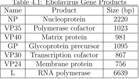
TheZaire ebolavirus is a filovirus (filo meaning filamentous) named for a tributary of the Congo river known as the Ebola. The virus is 970 nm long and 80 nm in diameter. The genome encodes seven viral proteins in the order described in Table 4.1.
Table 4.1: Ebolavirus Gene Products
Name Product Size (bp)
NP Nucleoprotein 2220
VP35 Polymerase cofactor 1023
VP40 Matrix protein 981
GP Glycoprotein precursor 1095 VP30 Transcription cofactor 867 VP24 Membrane protein 756
L RNA polymerase 6639
The lifecycle of the Ebolavirus, like many viruses, consists of seven stages. These are attachment to the host cell membrane, gaining entry to the cell cytoplasm, transcription of the viral genome into messenger RNAs, translation of mRNAs into viral proteins, replication of the viral genome, the assembly of genomes into proteins into new virions, and finally the exit of mature virions from the cell.
The glycoprotein (GP) facilitates attachment to the host receptors DC-SIGN and DC-SIGNR [54]. Phosphatidyl serine on the viral membrane surface then binds to the HAVCR1 cell receptor, inducing the cell to initiate apoptic mimicry by signal transduction, and permitting the virion to enter the cell via macropinocytosis.
The RNA polymerase from the L gene binds to the 3’-OH leader of the viral RNA transcribes it into an mRNA complete with 5’ cap and 3’ poly-A tail. The glycoprotein is cleaved by the furin enzyme in the host cell into GP1 and GP2 proteins. The furin enzyme, also known as the paired basic amino acid cleaving enzyme (PACE), is a calcium-dependent serine endoprotease expressed in the cells of many tissue types, including neuroendocrine, liver, gut, and brain. The human FURIN gene is found on chromosome 15 [55].
GP1 promotes fusion of the viral membrane with the vesicle membrane by in-teracting with host NPC1, allowing the ribonucleocapsid to enter the cytoplasm. NPC1 (Niemann-Pick disease, type C1) is a transmembrane protein responsible for mediating cholesterol transfers in mammalian cells. The human version is located on chromosome 18.
Viral genome replication can begin once sufficient nucleoproteins have been trans-lated to encapsulate the newly produced genomes. Viral genes are typically organized by the quantity of a protein product required. In Ebola, for example, many more copies of the NP protein are required than of the L polymerase. Therefore, the NP gene is the first after the 3’ leader and the L gene is the last before the 5’ trailer.
The VP30 zinc-binding protein is necessary for activation of viral transcription and is associated closely with the nucleocapsid complex [58]. VP24 promotes viral survival by suppressing the the production of alpha/beta interferon (IFN-α/β) [59]. The role of the 5’ trailer sequence of the Ebola genome is not completely understood. However, transcripts with the 5’ trailer deleted have been shown to be deficient in replication, indicating that the trailer is important for viral genome replication [60].
4.2.2 Ebola SEVEs by Species
Grouping the number of ebolavirus matches by host chromosome and normalized by the host chromosome size (Mbp) as a measure of relative homology (Figure 4.3) reveals that the chimpanzee and even more so the mouse contain a relatively high degree of homology compared to the other species.
According to a study of ebolavirus infections in 47 different mouse lineages by Rasmussen et al., the mice displayed a range of symptoms from full hemorrhagic fever to none at all [61]. Those that developed the lethal fever exhibited low levels of activity for the Tie1 and Tek genes that increased the permeability of their membranes and resulted in significant inflammatory responses. Importantly for this study, the mouse adapted version of the ebolavirus (MA-EBOV) does not lead to the fatal syndrome in lab mice. That includes GRCm38.3 genome from which these data are produced. Similarly, not all humans are susceptible to the ebolavirus hemorrhagic fever.
Congo [62]. The mortality rates could be as high as 95% for gorillas and 77% for chimpanzees.1
Figure 4.3: Zaire ebolavirus SEVE match count by host name and chromosome.
The most frequently occurring sequences in the chimpanzee are on chromosome 1 and the Y chromosome. The top two sequences in chromosome 1 are 5’-AAAAATTTAAA-AATAAAT-3’ and3’-TTTTTAAATTTTTATTTA-5’, which occur 206 times and 466 times, respectively. The two sequences are also complementary. The 5’ to 3’ sequence is located within the 5’ header of the Ebola genome. BLAST searches returned no significant results for the 5’ to 3’ sequence, however the 3’ to 5’ sequence was found in the CAMK2N1 gene for the calcium/calmodulin-dependent protein kinase II inhibitor 1 protein on chromosome 1. The sequence can be found on every chromosome in the Pan troglodytes genome.
The same sequences are also the most frequent Ebola matches in the human genome, with 92 matches for the 5’ and 252 matches for the 3’ to 5’ sequence.
3’-TTTTTAAATTTTTATTTA-5’ can be found in the CECR2 gene in humans, a cat eye syndrome candidate. The gene encodes a protein containing a bromodomain involved in chromatin remodeling that may also play a role in DNA damage response [63].
Another often occurring sequence is5’-TATTGAGCAGTATTGAAA-3’with 57 matches on human chromosome 15. The sequence comes from the VP24 gene for the Ebola membrane protein. BLAST searches indicate that the sequence occurs only in non-coding regions.
Other notable Ebola SEVE matches in the human genome include 5’-ATTATTTAA-AATTCTTTC-3’ in the TRIM37 motif, 5’-AAAACAAAACTGATCTTT-3’ in the GRIN2B glutamate receptor on chromosome 12, and3’-TTACAAAGATGGCCTTAG-5’in the PKC-potentiated PP1 inhibitory protein (PPP1R14A gene) on chromosome 19.
The most frequent match for the chimpanzee Y chromosome is 5’-TCAACCACCACCT-GGACC-3’ with 14 matches. The sequence is located within the VP35 polymerase cofactor gene in the Ebola genome. The sequence is also found on the human Y chromosome, within the inverted repeat IR2 of the Y palindromes P1, P2, and P3.
The now familiar 5’-AAAAATTTAAAAATAAAT-3’ and 3’-TTTTTAAATTTTTATTTA-5’
sequences also make numerous appearances within the mouse genome, occurring 40 and 157 times each. The 3’ sequence is found in the RRNAD1 gene for ribosomal RNA adenine dimethylase located on chromosome 1.
Ebola nucleoprotein gene, also located within the CXCL14 gene that encodes the chemokine (C-X-C motif) ligand 14. This protein is involved in immunuregulatory and inflammatory responses, and is related to the Akt signalling pathway [64]. The human version is located on chromsome 5.
An additional sequence from the nucleoprotein is 5’-GAAAAAGAGGCCATGAAT-3’, found 68 times in the mouse genome. BLAST results located it within the MAP3K13 gene that codes for a member of the serine/threonine protein kinase family that can activate MAPK8 or MAP2K7 MAP kinase cascades, which indicates a likely role in the JNK signalling pathway [65].
4.2.3 Ebola SEVEs by Viral Gene
Having considered the SEVE matches from the host perspective by chromosome, it is also useful to group by viral gene. The number of matches in D. melanogaster, A. thaliana, and C. elegans are negligible. The majority of the Ebolavirus SEVE matches occur within the 5’ trailer at the end of the genome. In the M. musculus genome, the sequence5’-TGAGTTCCAGGCCAGCCT-3’discussed in the preceding section accounts for the large number of matches from the VP35 polymerase cofactor protein. The role of the 5’ genome header in viral infectivity is not well understood.
4.3
Human Immunodeficiency Virus 1
4.3.1 Overview
Figure 4.4: Zaire ebolavirus SEVE match count per viral gene and normalized by gene size.
proteins encoded by the genome are summarized in Table 4.2. The protein coding section is flanked on either side by long terminal repeat (LTR) regions 600 bp in length. The 5’ end also contains a primer binding site (PBS) and the 3’ end includes a polypurine tract (PPT).
Table 4.2: HIV-1 Gene Products
Name Product Size (bp)
gag-pol Protease, RT, RNaseH, integrase 4308 gag Group-specific antigen 1503 vif Viral infectivity factor 579
vpr Viral protein R 291
tat Transcriptional activator 2595 rev Regulator of expression 2685
vpu Viral protein U 249
env Envelope surface glycoprotein 2571 asp Antisense protein (unknown) 570
nef Negative factor 621
incubation periods. The infection process begins with attachment by the gp120 glyco-protein to the host cell surface receptors DC-SIGN [19], Heparan Sulfate Proteoglycan [66], and the CD4 receptors of the helper T cells [19]. Host cell entry is mediated via clathrin-dependent endocytosis with the transmembrane glycoprotein gp4 facilitating dynamin-dependent fusion with the endosome. The envelope spike encoded by the env gene consists of three copies of gp120 and gp41 to form a trimer of heterodimers.
Once the nucleocapsid enters the cytoplasm, the +ssRNA viral genome is tran-scribed into linear dsDNA by the viral reverse transcriptase (RT) enzyme. The dsDNA must be transported to the host nucleus along with the viral integrase encoded by the pol gene. The integrase enzyme randomly integrates the viral DNA into the nuclear host DNA to form a provirus, accomplished by hijacking the DNA repair mechanisms of the host cell [67]. The provirus may become latent, awaiting later activation, or be transcribed immediately into new viral genomes.
The 5’-LTR of the provirus contains promoter elements that are bound by the RNA polymerase II enzyme of the host to begin transcription. Some of the transcripts will be unspliced and others will be spliced by post-transcriptional modification in the spliceosome. The unspliced transcripts will either become future RNA genomes or be translated after the transcripts are exported from the nucleus. The spliced transcripts will be immediately translated to produce Tat, Rev, and Nef proteins.
the nucleus for another cycle.
The unspliced transcripts are then translated into Env, Gag, and Gag-pol polypro-teins. Cleavage of the Env proteins by the viral protease yields the envelope proteins TM and SU, as well as the accessory proteins Vif, Vpu, and Vpr. New virions are assembled and the genomes packaged at the host plasma membrane. The virions are released via exocytosis by budding. The precusor polyproteins translated from the unspliced transcripts are cleaved by the viral protease to form mature virions.
The Tat protein (Trans-Activator of Transcription) is a kinase that greatly in-creases transcription rate of viral dsDNA by phosphorylating cell factors [68]. Tat can also be absorbed by nearby uninfected T cells, inducing apoptosis, and accelerating the demise of the host immune system [69].
The Nef protein is a negative regulatory factor that helps active T cells to increase the likelihood of infection. It acts as an enzyme to lower the activation energy of CD4+ lymphocytes. The T cell receptor response (TCR) renders the cells susceptible to infection by other virions [70]. Nef expression is not strictly required for HIV infection to occur.
The Vif protein, or viral infectivity factor, inhibits the antiviral activity of the APOBEC3G protein by marking it for degradation via ubiquitination. APOBEC3G is a cytidine deaminase that mutates viral mRNAs by deaminating the cytosine nucleotides into uracil. Vif is necessary for viral replication because otherwise the deaminase will enter the budding virions and scramble their genomes before they reach the next target cell [71].
and can also suspend dividing in the G2 phase leading to apoptosis [72].
The Vpu protein (Viral Protein Unique) induces the degradation of the CD4 viral receptor in the endoplasmic reticulum, resulting in a downregulation of CD4 expression. This results in the prevention of unintentional CD4-Env binding in the ER to faciliate the proper formation of virions within the cell. The Vpu protein itself is not packaged into new virions [73].
The role of the asp (antisense protein) remains unclear, though recent evidence suggests it may be involved in the process of autophagy [74].
4.3.2 HIV-1 SEVEs by Species
In Figure 4.5, the data indicate that the least significant degree of homology exists be-tween the Mus musculus genome and the HIV-1 virus. According to Zheng et al., lab mice do not exhibit symptoms when infected with HIV due to a post-transcriptional block [75]. The mouse version of the protein (mp32) is a nuclease that actively cleaves HIV mRNA transcripts. The human version (p32) does not cleave the transcripts. When the human p32 protein is introduced to their genomes, the mice become susceptible to infection.
The primate species have similar levels of relative homology, with exceptions in the gibbon chromosome 24, orangutan chromosome 19, chimpanzee Y chromosome, and human chromsomes 20 and 21. A pair of complementary sequences from therev gene are the cause of these high values and will be discussed in the next section.
4.3.3 HIV-1 SEVEs by Viral Gene
Figure 4.5: Human immunodeficiency virus 1 SEVE match count by host name and chromosome.
the rev gene, with an exceptionally high ratio in the P. alecto genome. The large number of matches from the HIV-1 rev gene in the MatchDatabase are due to two complementary sequences, 5’-AGAGAGAGACAGAGACAG-3’ having 8,715 matches and
human version is located on chromosome 5.
Since the fruitbat genome has not been annotated, the chromosome data is un-available. However, these same two sequences, 5’-AGAGAGAGACAGAGACAG-3’ and
3’-TCTCTCTCTGTCTCTGTC-5’ occur 739 and 515 times respectively. Clearly these are the most significant sequences from the HIV-1 virus in host genomes.
Figure 4.6: Human immunodeficency virus 1 SEVE match count per viral gene and normalized by gene size.
4.4
Simian Immunodeficiency Virus
4.4.1 Overview
Multiple lineages of SIV exist, including SIVcpz (chimpanzee), from which HIV-1 evolved, SIVsm (sooty mangabey) from which HIV-2 evolved, SIVagm (African green monkey), and several others. The data presented here are from the SIVcpz genome, which is only known to naturally infect Pan troglodytes [77]. Unlike HIV in humans, SIV in primates is not always pathogenic, but can lead to the fatal Simian AIDS (SAIDS).
Table 4.3: SIV Gene Products
Name Product Size (bp)
gag SIV2 Glycoprotein 1 1554
pol Protease, RT, RNaseH, integrase 3033 vif Viral infectivity factor 639
vpx Viral protein X 300
vpr Viral protein R 306
tat Transcriptional activator 2574 env Envelope surface glycoprotein 2601
nef Negative factor 558
The SIV life cycle is similar to the HIV-1, as the two viruses are closely related. Like HIV, attachment is mediated via the gp160 glycoproteins in the viral envelope binding to the CD4 molecules in the T cell membranes [78]. The glycoproteins are encoded by thegag gene.
4.4.2 SIV SEVEs by Species
The chimpanzee genome contains the greatest number of SEVE matches of any of the primates in the study. This could be significant as only chimpanzees are susceptible to SIV (the SIVcpz strain specifically). Indeed it is suspected by Worobey et al. that HIV-1 is a derivative of SIVcpz that crossed the species barrier due to bushmen hunting chipanizees for food [80]. The SEVE matches in the chimpanzee genome are well distributed with each only occurring only a few times. The ex-ceptions to this are 3’-TGGTGTTTGGTTTTTCGT-5’, 5’-AAAGAAAGGAAAATAGAA-3’, and
3’-GTGTTAAAATTTTCTTTT-5’, each occurring 26, 22, and 16 times, respectively. These results indicate that a high degree of viral homology does not confer immunity to the host.
Mice are unlikely to be infected by SIV if they are not naturally infected by HIV [81]. Figure 4.7 contains the SEVE matches by host name and chromosome. The unexpectedly high degree of homology in the mouse Y chromosome is due to two sequences,3’-GTTTATTGTGTATAAGAA-5’ from thetat gene with 87 matches, and
Figure 4.7: Simian immunodeficiency virus SEVE match count by host name and chromosome.
4.4.3 SIV SEVEs by Viral Gene
Most of the SEVE matches from the SIV virus occur within the chimpanzee genome, particularly from the pol, tat, and vif viral genes (Figure 4.8). Of the matches within the pol gene, there are three protein coding genes. The SEVE sequence
5’-AAAGAAGGGAAAGCAGGA-3’ is contained within the KLHL33 gene for kelch-like family member 33 from chromosome 14. SEVE 5’-TTGTGGTATAACCTGTTG-3’ resides in the GTF2A1 gene also on chromosome 14 that encodes the general transcrip-tion factor TFIIA. Lastly, the sequence 5’-AGAGACCAAGCAGAGAAA-3’ is found in the THSD7A gene on chromosome 7, encoding the thrombospondin type I glycoprotein necessary to create blood platelets (thrombocytes).
From the SEVEs in the HIV-1tatgene, the sequence5’-CAAGACTATCCATGTGGG-3’
glycoprotein 1, a sperm cell surface receptor. Another is3’-ATTGACTGTTATACTGTC-5’, contained within the ADAM18 gene on chromosome 8, encoding the disintegrin and metalloproteinase domain 18. Only one sequence from the SIV vif SEVEs is significant, 3’-TATATTCAAGTGTTTGAT-5’, contained within the HOOK3 chimpanzee gene on chromosome 8. HOOK3 codes for the hook microtubule-tethering protein 3.
There are also a few significant matches from the tat gene within the mouse genome. One is SEVE seqence 3’-GTCCAGGGTACCTTCTTT-5’from chromosome 16 for the LOC105246101 long noncoding RNA. Another is 3’-TGATACTTCTCGTTGGTT-5’
located within the Gnpda2 gene on chromosome 5 for the glucosamine-6-phosphate deaminase 2 protein. Finally, there is sequence 3’-TACATTGTCTTTACAAGA-5’ from the Gpr126 gene for G protein coupled receptor (GPCR) 126 on chromosome 10.
4.5
Measles Morbillivirus
4.5.1 Overview
TheMorbillivirus, commonly known as the measles, has a spherical capsid of 150-300 nm with a -ssRNA genome that is 15-16 kb in size and encodes eight proteins as described in Table 4.4.
Table 4.4: Measles Morbillivirus Gene Products
Name Product Size (bp)
N Nucleocapsid 1581
P/V/C Phosphoprotein 1524 M Matrix protein 1008 F Fusion protein 1653 H Hemagglutinin 1854 L RNA Polymerase 6552
The Morbillivirus life cycle is similar to other group V viruses like theEbolavirus. Attachment occurs through the hemaglutinin (H) protein on the viral surface to the cell surface receptors. Three such receptors for the measles H protein have been identified in humans. One is the CD46 complement regulatory protein (cluster of differentiation 46), an inhibitory receptor encoded by theCD46 gene. Another is the signalling lymphocyte activation molecule (SLAM) encoded by the SLAMF1 gene. The third is the Nectin-4 cellular adhesion molecule encoded by the PVRL4 gene [82]. All three genes are located on chromosome 1.
Following the binding of the receptor by the H protein, the F protein trimer conformation changes, allowing fusion with the plasma membrane to occur [83]. The ribonucleocapsid is then released into the cytoplasm via endocytosis.
polyadenyla-tion to form mature mRNA transcripts. The gene that encodes the phosphoprotein P also contains two overlapping genes for the V and C proteins. The mRNA for the V protein is an edited version of the P mRNA and the C protein is a result of leaky scanning. The process of leaky scanning involves a weak start codon (e.g., ACG) and a small upstream open reading frame (uORF), allowing the ribosome to occasionally skip the weak codon and translate multiple proteins from one mRNA [84].
Replication begins when sufficient nucleoproteins have been translated. The nu-cleocapdsid (N) binds to the matrix protein (M) near the plasma membrane. The P protein is a polymerase cofactor that binds the N proteins and helps position them for assembly. The V and C proteins are viral infectivity factors that are not strictly required for propagation [85]. Exocytosis is facilitated by host ESCRT proteins (endosomal sorting complex for transport), and the virion is released through budding [86].
4.5.2 Measles SEVEs by Species
There are no known animal hosts for the measles virus (MeV), though it is believed to have evolved from the rinderpest virus of cattle [87]. Other viruses belonging to theMorbillivirus, such as distemper, can infect dogs, cats, and cetaceans (Figure 4.9). Recent research indicates that although the instances of infection are rare in the wild, viruses such as measles and influenza from humans are capable of crossing species and infecting apes and monkeys, including chimpanzees [103].
CD150 interferon (IFN) pathways disrupted have been engineered to study measles infections in mouse models [104].
There are four SEVE sequences that account for most of the matches between measles and mice. The 3’-GGGGTGATTGGGAGGAGT-5’ sequence from the matrix pro-tein (M) gene occurs 68 times, frequently in chromosomes 1, 3, 8, 10, and X. Sequence
5’-CAGCAACTGCATGGTGGC-3’ from the hemagglutinin protein (H) accounts for 73 matches, mostly within chromosomes 1, 2, and 7. SEVEs5’-AAGAAAAGGAGATCAAGG-3’
and5-TAGCAACAGTGTACTCAT-3from the polymerase (L) contribute 63 and 35 matches, respectively, the former from chromosomes 1, 6, X and the latter from 7, 10, and 17. None of these appeared within protein coding regions.
The Y chromosome of the chimpanzee appears as an outlier in Figure 4.9 because it contains 14 SEVE matches in a very small chromosome, three of which occur three times each.
4.5.3 Measles SEVEs by Viral Gene
Most of the Measles SEVE matches occur within the 3’-OH Leader of the viral genome by a significant margin (Figure 4.10). That could be significant since the 3’-OH leader is where the L polymerase binds at the beginning of transcription. Only one of the 3’-OH matches occurs a significant number of times, with 3’-TTGTCCCAGCCCCTCTTC-5’
having twelve appearances.
Two of the sequences do appear in protein coding genes inHomo sapiens and other primates. The3’-TTGGATCCTAACGACTTT-5’sequence occurs within theNUCB2 gene on chromosome 11, encoding the nucleobindin 2 regulator for glucose transporter 4 (GLUT4) [105]. Another interesting sequence is 3’-TTGTCCCAGCCCCTCTTC-5’, residing within the ARRB2 gene on chromosome 17, responsible for coding arrestin
β 2 protein believed to play a role in the agonist-mediated desensitization of GPCRs [106].
4.6
Influenzavirus A
4.6.1 Overview
The Influenzavirus is an enveloped virus with a spherical or filamentous capsid of 80-120 nm in diameter with a -ssRNA genome that is 13-14 kb in size and encodes twelve proteins in eight segments, as described in Table 4.5. Unlike the other group V viruses, the influenza genome is segmented, with segments ranging in length from 890 to 2,340 nucleotides. Influenza viruses are classified by the hemagglutinin and neuraminidase receptor proteins expressed in the envelopes, H7N9 in this case. The viruses are also categorized by the natural host, such as avian, bovine, or swine (e.g., H1N1). According to the Centers for Disease Control (CDC), researchers have identified 11 neuraminidasae subtypes and 18, for a total of 198 possible influenza combinations.
Table 4.5: Influenzavirus A H7N9 Gene Products
Name Product Segment Size (bp)
PB2 PB2 Polymerase 1 2280
PB1 PB1 Polymerase 2 2274
PB1-F2 Apoptotic factor 2 273
PA PA Polymerase 3 2151
PA-X PA-X protein 3 760
HA Hemagglutinin 4 1683
NP Nucleocapsid protein 5 1497
NA Neuraminidase 6 1398
M2 Matrix protein 2 7 982
M1 Matrix protein 1 7 760
NEP Nuclear export protein 8 838 NS1 Nonstructural protein 1 8 654