Islamic RepubUc of Irnn Bahar &. Tabl:slan 1370 Spring &. Summer 1991
CORRELATION BETWEEN EXPRESSION OF
CLASS I ANTIGEN AND DEVELOPMENT OF
TUMOUR
INFILTRATING
LYMPHOCYTES
(TILS) FROM TUMOUR BIOPSIES OF PATIENTS
WITH BLADDER CANCER
A.M.E. N OURI,· A.V.L. DOS SANTOS,·· AND
A. SHAMSA
•••Depts.of· Medical Oncology and ··Immunology of The Royal London Hospital,UK, and ••• Marhad U'I;versity of Medical Sciences, Mashad, Islamic Republic of Iran.
ABSTRACT
This report presents the findings of TIL development from tumour
biopsies of patients with bladder cancer. Analysis of tissue sections showed
the presence ofT cells in intra-epithelial layer in 8 of 12 cases investigated. In
a larger group of patients, TILs were established from
7of 20 cases and
expansion of these cells in Interleukin-2 (IL-2) in the presence of conditioned
medium (CM) resulted in increase cell number to as many as
100
fold. Cell
surface antigen profile of the cells showed characteristics of activated T cells
and showed cytotoxic activity against tumour cell lines
K256
and Daudi. In
one case where TILs and tumour cell line from the same individual was
established, it was found that TILs showed low levels of cytotoxicity against
the autologous tumour cells. Investigation of correlation between class I
antigen expression and TIL development revealed that all the
7cases of
positive TILs were established from tissue expressing normal levels of free
chain of class I antigens. These results are indicative of the presence of
IL-2-expandable T cells in tumour biopsy of patients with bladder cancer and
demonstrate that the successful expansion of these cells correlates with the
normal expression of class I antigens.
MJ/RI, Vol.5, No. 1,2, 9-14, 1991
INTRODUCTION
Recent reports demonstrating that treament of patients with neoplastic diseases with IL-2 result in durable complete remission has restimulated interest in the immunological mechanism(s) involved in tumour rejection.' This has led to the suggestion that the therapeutic efficacy of IL-2 might be due to the clonal expansion ofT cells activated by tumour specific antigen (s). The most significant report supportin� such a hypothesis has been the fIndIngs of Itoh, et al. who
Address for correspondence: AM E Nouri. Medical Oncology, The Royal Loncion Hospital, WlJiteclwpel, Londoll EI lAD.
9
showed that IL-2-expandable TILs can be developed from tumoursof patientswithmelanoma.Furthermore, in more than 50% of these cases, TILs showed MHC restricted killing against the autologous tumour target with little or no killing of allogeneic tumours. In addition, Topalian, et aldemonstrated that compared with autologous IL-2-activated peripheral lymphocy ets, TILs were 50 to 100 times more active in tumour killing.)
These observations militate against the escape of tumour cells from immunological controls. One possi ble explanation for the inability of TILs to inhibit tumour progression may in part be due to the abnormal expression of MHC class I antigens by tumour cells.
Class I Antigen and Bladder Cancer
a
u.s
1.0
0.5
1.0
1.5
o 2 , 6
Days of culture 1
:
)
1 I
1 1
1 1
Fag.l.lncrease in TIL numberinrcsponse to IL-2 and eM for FB (a) and FS (b) with increase in the time of culture.
The first clear indication for the loss of these antigens in malignancy was reported by Hui et al. 4 Using animal model, these authors demonstrated that the transfec tion of missing class I gene into tumour cells lacking these antigens led to a reversal of their tumourgenicity. H uman aberrant expression of these antigens have been reported in a number of neoplastic diseases. 5-1 In our own studiesS investigating bladder tumours, 14 of 18 cases showed some degree of abnormality and this seemed to be correlated with the invasiveness of the tumor.
These observations prompted us to investigate whether TILs can be established from bladder tumour and furthermore, whether there is any correlation between TIL growth and the expression of class I antigens.
MATERIALS AND METHODS
Operative specimens from the Urology Depart ment of the Royal London Hospital were used im mediately after operation. The tissues were divided into two portions the smaller of which was snap frozen and kept in liquid nitrogen for tissue sectioning. The second portion was washed, minced and the resulting cell suspension and tissue fragments were used for developing TILs and tumour celliines.
The cell preparation was incubated overnight in :i Il.
015,000
z o
�
o
�10.000
o�
a: " l;-i!;
5.000
o
10,000
100,000
500,000
CEll NOIWEll
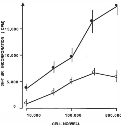
Fig.l. Proliferative response omLs from FS to IL-2 (lOOulml) alone ( D--<l ) and IL-2 plus eM (5%, ______ ) for different cell numbers per well.
RPMI containing 10% fetal calf serum in the presence or absence of recombinant IL-2 (100 u/ml, Biogen). After the incubation the non-adherent cells were re moved, spun down and resuspended in fresh IL-2 and cultured in a separate flask. In some cases the cell suspension which at this stage contained a large amount of cell debris, was layered onto density gradient solu
tion and after centrifugation, interface cells, ie; mainly lymphocytes were removed and cultured in medium with IL-2. In the cases where successful TILs were established, cells were kept in IL-2 medium and feed ing continued every two or three days by adjusting the cell number to 0.5 x 10⁶/ml. After two weeks of culture and the initial expansion of IL-2-receptor bearing T cells, conditioned-medium (eM, 5% v/v, see below) was added in order to increase the rate of cell prolifera tion. The adherent cells were fed until confluence and were expanded by trypsinising the cells and sub culturing into new flasks.
Table 1. Ceil surface pheDotype oCTILs from different individuals
w6/J2 HB55 CD4 CD8 CDJ NT
FS 98 95 45 50 87 -\!e
WIL 98 98 1·2 33 80 -ve
IF 95 83 23 40 89 ·ve
FB 100 70 4 30 87 -ve
AW 89 B6 27 36 83 -vo
LR 100 93 24 29 69 -vo
Phenotypic characteristics of TILs from six different individuals. Cells were analysed after being in culture for more than 30 days.
�
J
� u �
'H �
"
� "
0-35
]0
25
211
"�----7---�----�----�--�
, ! , !:: 3 4 :; h
Tim!! uf incubation (hr)
Fig.3. Time course of target (Oaudi) killing by effector cells (FS) at Err ratios of 25/1 ( ..-), 12.5/1 ( ____ ), 6.5/1 ( 0--0 )
and 3.211 ( ><---K).
Conditioned Medium
This was' prepared by activating normal peripheral blood mononuclear cells (2X 10⁶/ml) with I?HA (2 ug/m!) for two hours at 37"C. The cells were washed three times and culturing continued for a further 36 hrs at above cell number. After the incubation the cell-free supernatant was removed, aliquoted and frozen until us�.
Cytotoxicity
Established human tumour cell lines Daudi (EBV transformed B cell line), Molt 4 (T cell line), U937 (monocytic like cells) and K562 (myelocytic cell) or our own established bladder line were labelled with 51Cr
Table U. TIL (WIL) phenotype after different times of culture
Date 12111188 1/12188 22112188
cm 76% 98% 80%
CD4 35% 75% 1-2%
CDB 38% 30% 33%
W6I32 93% ND 98%
HB55 70% 100% 98%
control ·ve ·ve ·ve
percent positive cells, -ve denotes negative staining.
,,u
50
40
20
IO
II
3211 6.511 12.511 2511
Err Ratios
Fig.4. Results are expressed in percent specific killing of Daudi ( )t---l(), Molt 4 (A-£), U937 (--...) and K562 ( ..--.... ) by TILs (WIL) at different effector/target ratios.
(250 uCi/target) for 1 hr at 37"C followed by extensive washes. These cells i.e., target cells (T), were mixed (three replicates/treatment) with TILs i.e. effector cells (E) to give EIT ratios of 3.211,6.511 and 25/1 in round-bottomed microtitre plates. The cell mixtures were incubated for different length of times after which cell-free supernatants were removed and counted us ing a gamma counter. The specific killing activity for each treatment was calculated using standard formu lae.
Fluorescent Staining
Cell suspension was prepared in round-bottomed tube to give 0.5 x 10⁶/tube. After centrifugation, super natant was discarded and cells were resuspended in 50 ul of appropriate antibody and incubated for 45 mins at room temperature. Cells were washed in PBS and FITC-conjugated rabbit anti-mouse i.e. 2nd antibody (1/50 dilution, Dakopatts) was added and incubation continued for a further45 mins. Afterthreewashes, the cell pellets were used for FACS analysis.
Tissue Staining
. Frozen sections were cut using a cryostat at a th�ckness of 5 urn, placed on microscope slides, air dned and at _40" until use. A peroxidase anti peroxidase staining method was employed as pre VIOusly reported by Nouri, eLat.8
Monoclonal antibodies
The monoclonal antibodies (Mabs) used as primary
Class I Antigen and Bladder Cancer
reagents in the form of tissue culture supernatants, are listed as follows together with their specificities: W6/32 detects all B 2 m-associated HLA-A,B,C antigens· HC10 detects non-B 2 associated HLA-A,B,C antigens,1O L243 detects HLA-DR antigen, l1 anti CD3, -CD4 and /CD8 (Ortho- pharmaceutical) detect total T, T helper and T suppressor/cytotoxic lympho cytes subsets respectively.
Cell Proliferation
Proliferation of cells was measured by incorpora tion of tritiated thymidine (3H-Tdr, 0.1 uCilwell, Amersham) into cells. TILs were dispensed into round-bottomed microtitre plates at 0.5 x 10⁶/well in three replicates and incubated in the presence or the absence of stimulus for 48 hr, the last 4 hr of which was in the presence of 3H-Tdr. The degree of 3H-Tdr uptake by the cells was measured by harvesting the cells onto filter paper and counting radioactivity in a scin tillation counter.
RESULTS
Primary Cell Culture
In total, 20 cases with predetermined stage of tumour invasiveness were chosen for establishing TILs of which 7 showed positive growth. The pattern of cell proliferation of two such TILs
(FB
and FS) over aperiod of 10 days are presented in Fig. 1. As can be seen, the cell numbers increased at least twofoldsevery48 hrs and in the case of one individual (FS), this reached 37.2, folds over a period of 10 days. Analysis of TIL prolif eration of a representative TILs (FS) to IL-2 alone and IL-2 plus CM is shown in Fig.2. As can be seen, the cell number of IL-2-activated cells increased further in the presenceof CM. ThusatO.5x 106celliwell, the3H-Tdr uptake of IL-2-activated cells increased from 5,200 cpm to 19,800 cpm indicating the additive effect ofCM. This increase was not due to the carried over PHA which might have been present in the CM, since a 0.1 ug/ml equivalent to 5%, PHA had no stimulatory activity on fresh lymphocytes (data not shown).
Cytotoxicity
Cytotoxic activity of TILs from an individuals (FS) against Daudi cells after different times of incubation was investigated, the results of which are presented in Fig.3. As can be seen, at all the Err ratios the longer the incubation period the greater the degree of tumour killing. Furthermore, as the ratios of EIT increased the degree of cell killing also increased. Thus at 4 hrs the specific killing at 3.211 and 25/1 ratios were 10.2, 19.625 and 44% respectively and the 4 hr incubation was chosen for subsequent experiments. The standard deviation between the replicates of these and
subse-quent experiments were all below 10%.
The ability of cultured TILs (expanded in vitro for more than two weekS) to kill different well established allogeneic human cell lines was investigated, the results of one individual (WIL) TIL are presented in FigA. As expected there was a direct correlation between Err
ratios and tumour killing. Daudi cells were found to be the most sensitive targets followed by Molt 4 and U937 and K562 the least sensitive. Thus, the percent killing for Daudi cells at 25/1, 12.5/1 and 3.211 were 62,59, 44 and 35% respectively. The degree of cytotoxicity against Daudi cells byTILs from the other 6 individuals at 25/1 Err ratio varied from 26% to 62%. In addition, TILs from WIL (same individual from which perma nent cell line was established) were found to be capable of killing autologous tumour cells by 15% at 25/1 EIT ratio compared with 7% against epithelial cell line SKV14 (tumour cell of similar origin) indicating the low level of cytotoxicity of the TILs against autologous tumour cells.
TIL pheuotypes
The results of cell surface phenotyping for different TILs after beiug in culture for more than 30 days were assessed, the results of which are presented in Table I. As can be seen, the percentage of CD3, class I and II positive cells was greater than 69% for all the six cases. The percent CD8 positive cells was between 29% to 50%, whereas CD4 positive cells showed greater variability ranging from 1-2% (WIL) to 45% (FS). Furthermore, in all the cases, the percent CD4 positive cells was lower than CD8 positive cells. TILs from one individual (WIL) frozen at different times of culture was a'nalysed for cell surface markers, results of which are presented in Table II. As can be seen, the percent CD3 and CD8 positive cells remained relatively con stant throughout the culture period, whereas CD4 positive cells showed an initial increase followed by decrease. Thus the percentage of CD4 positive cells at
"
' . .
> " " ... .
.... -
:
'. . .... ... • J ... . • •
Fig.S CD3 positive cells nre demonstrated here in bladder tissue section of FS. Positive cells are present in both tumour stroma (st) and in the intra-epithelial (ie) areas,
12111/88, 1112188 and 22112188 were 35, 37 and 1-2% respectively. The percentage-of class I and II positive cells in the same culture period remained 'above 70%, indicating either the preferential expansion of CD8 cells or depletion of CD4 positive cells. The analysis of CD3 positive cells in 12 tumour biopsies showed the presence of both CD4 and CD8 cells in tumour stroma (11 cases) and within the tumour epithelial (8 cases) and example of which is shown in Fig.S (FS).
Tn.. Development and Class I Antigen Expression Analysis of class I antigen on tumour biopsies using HClD Mab showed comparable intensity of antigen expression between tumour and tumour stroma in 14 of 20 and in the remaining 6 cases the tumour showed weak or negative staining. The number of positive TIL growth among the above two groups were 7 of 14 and 0 of 6 respectively, indicating that TILs can be more successfully developed from tumours expressing nor mal levels of class I free heavy chain.
DISCUSSION
The results of this investigation have revealed that (a) there are activated T lymphocytes in tumour biop sies of some bladder cancer patients and they can be expanded in vitro in response to IL-2 to as many as hundred folds; (b) they express phenotypes of normal activated T cells; (c) they are capable of lysing estab lished allogeneic tumour cell lines; (d) TILs from one individual from whom tumour cell line was established, showed low levels of cytotoxicity against the auto logous tumour cells; (e) there seem to be a correlation between the expression of free chain of class I antigens on tumour and the success of Tn.. growth.
It is well established that IL-2 acts on its receptor expressed on activated T cells, resulting in their clonal expansion.12 The findings of this study implies that there are activated T cells in bladder tumours of some of these patients capable of proliferating in response to IL-2. This conclusion was reinforced by the observa tion that there are T cells present at the tumour site in 8 of12 cases investigated. The most convincing evidence demonstrating the capacity of TILs to kill autologous tumour cells in a MHC-restricted fashion, has been reported in melanoma. 2Taken this and the demonstra tion that lymphocyte infiltration into tumour is a good
. 13 14 ' th prognostic factor for tumour regressIOn, . nses e question why bladder tumours escape cytolytic effect of the cytotoxic T cells. One possible explanation for this might be the frequent abnormal expression of class I antigens in these tumours. This is based on our earlier study demonstrating that 14 of 18 cases IDveslIgated showed some degree of abnormality in expression of
13
class I antigens." It is tempting to argue that the failure of clinical efficacy of IL-2 in these patients may in part be due to this abnormality.
The low level cytotoxicity of WIL TILs against autologous tumour cell line is in agreement with the above conclusion, since analysis of MHC class I anti gens on these tumour cells demonstrated the absence of HLA-B locus antigens (B7 and B44).IS
Hence the loss-of these antigens may have rendered tumour cells resistant to cytolytic activity of cytotoxic T cells. The finding that TILs were more successfully developed from tumours expressing normal levels of class I antigens seems to be a novel observation. Although, the mechanism(s) for this is not clear, it is possible to envisage that T cells infiltrate and possibly proliferate at the tumour site where there is normal expression of class I antigens acting as an associative molecules for the presentation of putative neo antigen( s). This possibility is currently under investiga tion.
In conclusion, the findings of these studies have demonstrated that TILs can be developed from about 30% of bladder tumours in sufficient quantities for immunological analysis. It is envisaged that such a approach may provide opportunities for re introducing the missing class I antigens by transfecting appropriate genes into autologous tumour cells and investigating the nature of putative neo-antigen(s) which may be present on the tumour cell and these studies are currently in progress.
REFERENCES
1- Rosenberg SA, Lotze MT, Muul LM, ct. al: A progress report on the treatment of 157 patients with advanced cancer using lymphokine-nctivated killer cells and interleukin-2 or high-dose interlcukin·2 alone. N Eng! J Med 316:889, 1987.
2- Itoh K, Platsoucas CD. Balch eM: Autologous tumour specific cytotoxic T lymphocytes in the infiltrate of human metastatic melanomas: activation by interleukin-2 and autologous tumour cells, and involvement of the T cell receptor. J Exp Med 168:1419,1988.
3-Topalian SL. Muul LM. Solomon D, Rosenberg SA: Expansion of human tumour infiltrating Iympbocytes for use in immunother apy trials. J Immunol Methods 102:127,1987.
4- Hui K. Grosveld F, Festenstein H: Rejection of transplantable AKR leukacia cells following MHC DNA-mediated transforma tion. Nature 311:750,1984.
5- Fleming KA, ,McMichael A, MortonJA, WoodsJ, McGecJOD: Distribution ofHlA class I antigens in normal human tissue and in mammary cancer. J Clio PathoI34:179.1981.
6-Masucci MG. Torsteiddottir S, Colombani1, Baruthar C, Klein E, Klein G: Down-regulation of class I antigens and of the Epstein Barrvirus-encoded latent membrane protein in Burkitt lympho ma lines. Proc Nat Acad Sci 84:4567, 1987.
7- Momburg F, Degener T, Bacchus E, Moldenhauer G, Hammerl ing 01. MoUer P. Loss of HLA-A.B,C and de novo expression of I-U.A-D in oolorectal cancer. Int J Cancer 37:179, 1986
8- Nouri AME, Smith MEF, Crosby D, Oliver RID: Selective and
Class I Antigen and Bladder Cancer
non_selectiveloss ofimmunoregulatorymolecules(HLA-A,B,C antigens and LFA-3) in transitional cell carcinoma. Br J Cancer 62:603,1990.
9- Brodsky FM, Paraham P ,Barnstable CJ, Crumpton MJ, Bodmer WF: Monoclonal antibodies for analysis of the HLA system. Immunol Rev 47:3, 1979.
10- Starn NJ. Spits H. Ploegh HL: Monoclonal antibodies raised against denatured HLA-B locus heavy chain pennit biochemical characterization of certain HLA-C locus products. J Immunol 137:2299,1986.
11-Lampson L. Levy R: Two population ofIa molecules on a human
B cell line. J ImmunoI125:293, 1980.
12-Gillis S, Smith KS: Long term culture of tumour-specific cytolytic "Fcells. Nature 268:154, 1977.
13-'Dayan AD, Marshall AME: Immunological reaction in mail against ccrtain tumours. Lancct 11.1102,1964.
14w Tsujihashi H, UejimaS, Akiyama T, KuritaT: Immunohistochcw mical detection of tissue infiitratinglymphocytcsin bladder. Ural In,44:5,1989.
15- Noun AME. Bcrgbaum A. Lederer E, Crosby D, Shamsa A Oliver RID. Characteristics of newly developed bladder tumour cell line. (In press, Eur J Cancer).