Sex differences in the morphological failure
patterns following hip resurfacing arthroplasty
Hinsch
et al.
R E S E A R C H A R T I C L E
Open Access
Sex differences in the morphological failure
patterns following hip resurfacing arthroplasty
Andrea Hinsch
1†, Eik Vettorazzi
2†, Michael M Morlock
3, Wolfgang Rüther
4, Michael Amling
5and Jozef Zustin
1*Abstract
Background:Metal-on-metal hybrid hip resurfacing arthroplasty (with a cementless acetabular component and a cemented femoral component) is offered as an alternative to traditional total hip arthroplasty for the young and active adult with advanced osteoarthritis. Although it has been suggested that women are less appropriate candidates for metal-on-metal arthroplasty, the mechanisms of prosthesis failure has not been fully explained. While specific failure patterns, particularly osteonecrosis and delayed type hypersensitivity reactions have been suggested to be specifically linked to the sex of the patient, we wished to examine the potential influence of sex, clinical diagnosis, age of the patient and the size of the femoral component on morphological failure patterns in a large cohort of retrieved specimens following aseptic failure of hip resurfacing arthroplasty.
Methods:Femoral remnants retrieved from 173 hips with known patient’s sex were morphologically analyzed for the cause of failure. The results were compared with the control group of the remaining 31 failures from patients of unknown sex. The odds ratios (OR) and 95% confidence intervals (CI) of the following morphologically defined variables were calculated using logistic regression analysis: periprosthetic fractures (n = 133), osteonecrosis (n = 151), the presence of excessive intraosseous lymphocyte infiltration (n = 11), and interface hyperosteoidosis (n = 30). Logistic regression analysis was performed both unadjusted and after adjustment for sex, age, the size of the femoral component, and preoperative clinical diagnosis.
Results:Femoral remnants from female patients had a smaller OR for fracture (adjusted OR: 0.29, 95% CI 0.11, 0.80,
Pfor difference = 0.02) and for the presence of osteonecrosis (adjusted OR: 0.16, 95% CI 0.04, 0.63,Pfor difference = 0.01). However, women had a higher OR for both the presence of excessive intraosseous lymphocyte infiltration (adjusted OR: 10.22, 95% CI 0.79, 132.57,Pfor difference = 0.08) and interface hyperosteoidosis (adjusted OR: 4.19, 95% CI 1.14, 15.38,Pfor difference = 0.03).
Conclusions:Within the limitations of this study, we demonstrated substantial sex differences in distinct failure patterns of metal-on-metal hip resurfacing. Recognition of pathogenically distinct failure modes will enable further stratification of risk factors for certain failure mechanisms and thus affect future therapeutic options for selected patient groups.
Background
Gender medicine is a novel and rapidly evolving research discipline. Indeed, there has been an almost linear increase in the literature incorporating sex/gender differences [1]. Within the last few years, lively discus-sion regarding possible sex differences has also been initiated in the orthopedic surgeon community. Serious
concerns have arisen regarding the potential adverse biological reactions to metal-bearing surfaces and parti-cular prosthesis designs such as hip resurfacing arthro-plasty. In fact, metal-on-metal technology is now used in over one-third of all hip arthroplasties performed in the United States [2]. In recent years, hip resurfacing arthroplasty has become an accepted alternative to tradi-tional stemmed total hip arthroplasty in young adults worldwide [3], although patient selection is important in order to avoid failure [4-11]. Most authors [2-6,8-12] consider men under the age of 65 with osteoarthritis to be the best candidates for hip resurfacing. However, * Correspondence: [email protected]
†Contributed equally 1
Institute of Pathology, University Medical Center Hamburg-Eppendorf, Germany
Full list of author information is available at the end of the article
recent reports from centers that design hip resurfacing arthroplasty [7,13] suggest that the smaller size of the femoral component rather than female sex is linked with worse outcomes for this procedure.
In our earlier studies on failed hip resurfacing arthro-plasty, we observed some sex differences in a large col-lection of retrieved prostheses: men were more frequently revised for postnecrotic fractures [14], and the extent of osteonecrosis was larger than in specimens obtained from women [14]. However, women were more frequently revised for unexplained persistent groin pain, which was attributed to a suggested hypersensitiv-ity reaction after the index surgery [15]. In the present study, we calculated the ORs for morphologic failure modes in the entire cohort after adjustment for sex, age, the size of the femoral component, and preoperative clinical diagnosis. We asked: is the previously reported sex dimorphism really linked with the sex of the patient?
Methods Data collection
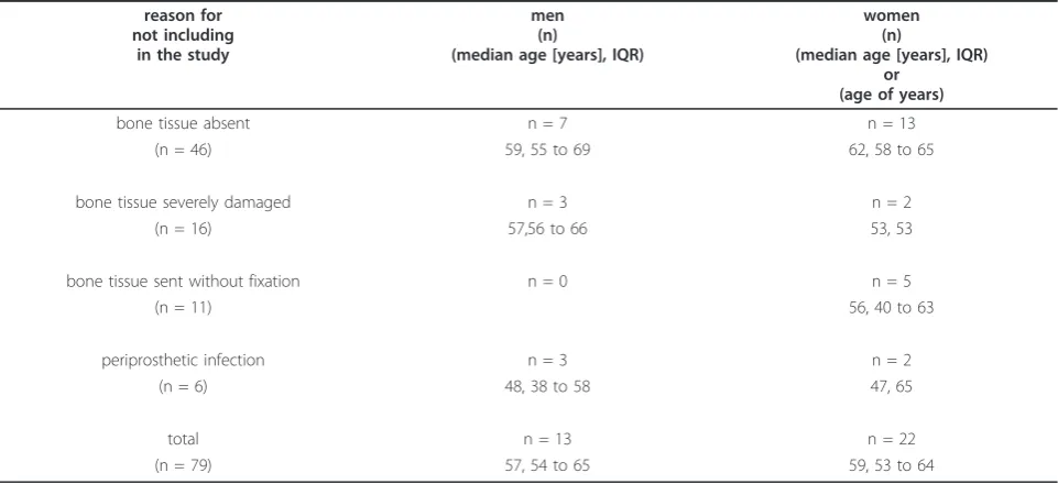
In an international multi-surgeon retrieval study on total hip resurfacing arthroplasty (THRA), we obtained 283 specimens between January 2004 and February 2010. During the planning of the design of this study in 2003, the suggested primary objective was a tribological inves-tigation of the prosthesis surface in order to demon-strate the potential wear-induced failures as they were frequently reported in the second generation of (metal-on-polyethylene) THRA. Therefore, several specimens, preferentially from the early phase of the Hamburg retrieval study on THRA were obtained without bone tissues or without using any standard fixation method
for bone tissue (Table 1). Later on, when we presented preliminary results of morphological analyses of the first dozen standard analyzed retrieved hips and specifically focused on the issue of histopathological changes within the periprosthetic tissues and the potential adverse reac-tions to metal material, the discipline of the cooperating surgeons in the submission of basic clinical data sub-stantially improved. Altogether, 46 specimens did not contain bone remnant tissues under the cup at all; in 16 cases focal rests (mostly less than 2 cm2) of the bone tissue were severely mechanically damaged and 11 speci-mens contained osseous tissue but were sent without fixation and the histopathology was non-informative. We also obtained 31 cases with minimal clinical data; particularly the data on sex were completely missing. Finally, six cases were revised for periprosthetic infec-tions and were not included in further analyses. After excluding all 79 cases with septic complications, insuffi-cient quality of fixation of the femoral remnant bone tis-sue and hips with invalid demographic data, the present study cohort contained 85 women (median age 56 years old, interquartile range (IQR) 49 to 60) and 88 men (median age 56 years old, IQR 51 to 60; P = 0.584; Table 2). Valid clinical data were obtained for the majority of the specimens in the study cohort: 97.1% (168) for age, 93.6% (162) for the duration of implanta-tion, and 82.1% (142) for the preoperative clinical diag-nosis. Most hips were treated for advanced stages of primary osteoarthritis (71.8%). Other conditions were developmental hip dysplasia (11.3%), femoral head osteonecrosis (7.0%), posttraumatic arthritis (4.9%), and rheumatoid arthritis (4.9%). The remaining 31 cases with unknown patient sex but informative results on the
Table 1 Cases not included in the present study
reason for not including
in the study
men (n)
(median age [years], IQR)
women (n)
(median age [years], IQR) or
(age of years)
bone tissue absent n = 7 n = 13
(n = 46) 59, 55 to 69 62, 58 to 65
bone tissue severely damaged n = 3 n = 2
(n = 16) 57,56 to 66 53, 53
bone tissue sent without fixation n = 0 n = 5
(n = 11) 56, 40 to 63
periprosthetic infection n = 3 n = 2
(n = 6) 48, 38 to 58 47, 65
total n = 13 n = 22
(n = 79) 57, 54 to 65 59, 53 to 64
Hinschet al.BMC Medicine2011,9:113 http://www.biomedcentral.com/1741-7015/9/113
morphological analyses made up the control group (Table 2).
The specimens came from patients with five hip resur-facing femoral systems (Table 2): 125 Articular Surface Replacements (ASR™;DePuy Orthopaedics Inc, Warsaw, IN), 15 DUROM® (Zimmer Inc, Warsaw, IN), 14 Cor-met™(Corin Group PLC, Cirencester, UK), 10 Birming-ham Hip Resurfacing (BHR™; Smith & Nephew, London, UK), and 9 ReCAP® (Biomet Inc, Warsaw, IN).
All revisions were unilateral. One hundred and four-teen revisions (66%) out of a total of 173 cases with valid data on patient sex were performed for peripros-thetic fractures, 45 (26%) for non-fractural causes, and 14 (8%) for acetabular loosening (Table 3). Several cases had more than one reason for revision surgery, for example several hips with pseudoarthrosis hidden under the femoral component caused by chronic fracture were clinically or radiographically classified as loosening of the femoral component.
Morphological classification of failure patterns
Each specimen was cut using a water-cooled band saw and analyzed macroscopically, contact radiographically and microscopically according to a high standard sampling protocol as described previously [14-18]. Briefly, the
femoral heads within situfemoral components were cut in the coronal plane and X-rayed and documented photo-graphically. A second section was oriented perpendicular to the first. The coronal plane and the anterior section were embedded without decalcification in their full length and microscopically analyzed. Each case was examined macroscopically, microscopically and by contact radiogra-phy. In our previous work, we proposed classifications for both periprosthetic fractures [18] and the loosening of the femoral component [17] based mostly on the macroscopic and contact radiographic findings which were subse-quently confirmed microscopically (for example osteone-crosis, pseudoarthrosis). Histopathological analyses also revealed findings that could not be recognized by macro-scopic assessment (for example intraosseous lymphocyte infiltration, hyperosteoidosis of the interface bone trabecu-lae). We summarized all the results of the histopathologi-cal analyses, both macroscopic and microscopic, and proposed classification schemas under the term“ morpho-logical patterns”of THRA failure.
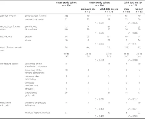
Briefly, the periprosthetic fractures were morphologi-cally classified [18] as postnecrotic, when advanced osteonecrosis was found in the complete femoral rem-nant proximal to the fracture line [14,15], or as biome-chanical, when the bone tissue from both sides of the Table 2 Demographic and clinical characteristics of the study cohort
entire study cohort entire study cohort n = 204
valid data on sex n = 173 unknown sex
n = 31
valid data on sex n = 173
men n = 88
women n = 85
age
(median age [years], IQR)
56, 50 to 60
51, 48 to 60
56, 50 to 60
56, 51 to 60
56, 49 to 60 P= 0.531 P= 0.584
clinical diagnosis osteoarthritis 107 5 102 54 48
avascular
necrosis of femoral head
10 0 10 8 2
rheumatoid arthritis 7 0 7 3 4
arthritis secondary to trauma
7 0 7 3 4
developmental hip dysplasia
17 1 16 6 10
P= 0.867 P= 0.288
THRA design ASR™ 143 18 125 66 59
DUROM® 16 1 15 8 7
Cormet™ 20 6 14 6 8
BHR™ 14 4 10 4 6
ReCAP® 11 2 9 4 5
P= 0.127 P= 0.877 duration of implantation
(median in situ time [days], IQR)
147, 51 to 399
127, 25 to 570
147, 55 to 384
124, 54 to 327
182, 55 to 445 P= 0.724 P= 0.373 size of the femoral component
(median diameter [mm], IQR)
46, 44 to 50
46, 44 to 50
46, 42 to 48
50, 48 to 52
fracture line was proven viable by histopathology. In cases of acute fracture, no reparative reaction was pre-sent. In hips with chronic fracture, either the fracture callus (union) or pseudoarthrosis (non-union) was detected microscopically (Figure 1A) [18].
Advanced osteonecrosis was defined macroscopically by yellowish colored areas of the bone and confirmed microscopically by the presence of trabeculae without stainable osteocytes, disorganized bone marrow, and bordering fibrosis (Figure 1B). Because all osteonecrotic lesions showed contact with the surface of the femoral remnant under the prosthesis, we also measured the vertical distance between the bone remnant surface and bordering fibrosis [14].
Excessive intraosseous lymphocyte infiltration was characterized microscopically by the finding of more than 300 lymphocytes within one high power field of the microscope in areas with maximum intraosseous lymphocyte infiltration (Figure 1C) [15].
Interface hyperosteoidosis was defined microscopically by the presence of widened osteoid seams on the trabe-cular surface at the bone-cement interface. These areas represented compact but somewhat irregular non-mineralized bone tissue within lamellar structured viable superficial bone trabeculae (Figure 1D). These were oriented mostly parallel to the surface of the cement in the vicinity of the cement mantle and also next to inter-trabecular cement interdigitations, irrespective of the direction of the intratrabecular lamellae [17].
Failures were defined as clinical complications leading to the revision surgery with loss of the THRA device. One hundred and thirty-three (65%) out of a total of 204 cases provided reproducible results of the morpho-logical analyses showing failure due to periprosthetic fracture. Seventy-one hips were revised for reasons other than the fracture: loosening of the acetabular component (n = 15), loosening of the femoral compo-nent (n = 10), cement-socket debonding (n = 3), Table 3 The prevalence of distinct failure patterns in the study cohort
entire study cohort n = 204
entire study cohort n = 204
valid data on sex n = 173 unknown sex
n = 31
valid data on sex n = 173
men n = 88
women n = 85
cause for revision periprosthetic fracture 133 19 114 65 49
non-fractural cause 71 12 59 23 36
P= 0.683 P= 0.026 periprosthetic fracture
pattern
postnecrotic biomechanic
73 60
9 10
64 50
41 24
23 26 P= 0.619 P= 0.086
osteonecrosis present 174 23 151 80 71
absent 30 8 22 8 14
P= 0.093 P= 0.151 extent of osteonecrosis
[mm]
7.4, 6.4, 7.6, 15.3, 6.2,
2.9 to 19.5
2.1 to 6.8
3.1 to 21.3
3.6 to 24.2
2.6 to 14.6 P= 0.171 P= 0.008 non-fractural causes Loosening of the
acetabular component
15 1 14 4 10
Loosening of the femoral component
10 3 7 2 5
cement-socket debonding
3 0 3 2 1
Collapsed osteonecrosis
5 2 3 0 3
Metallosis 2 1 1 0 1
Unexplained groin pain
36 5 31 14 17
P= 0.249 P= 0.417 unexplained
groin pain
excessive lymphocyte infiltration
14 3 11 1 10
P= 0.451 P= 0.021
interface hyperosteoidosis 37 7 30 8 22
P= 0.457 P= 0.005
Hinschet al.BMC Medicine2011,9:113 http://www.biomedcentral.com/1741-7015/9/113
collapsed osteonecrosis (n = 5), macroscopic visible metallosis (n = 2) and unexplained groin pain (n = 36). Even though several potential causes of the groin pain have been discussed in the literature (for example femoro-acetabular impingement or hypersensitivity reac-tion), we did not obtain any further specific information and included such cases in the group of ‘unexplained groin pain’.
Statistical methods
Descriptive statistics were performed to describe the median and interquartile range (IQR). As time to revi-sion surgery, the vertical extent of osteonecrosis, and age deviated from a normal distribution, a non-para-metric analytical method was used (Mann-Whitney-U test). Logistic regression analysis was used to estimate odds ratios (OR) and 95%- confidence intervals (95% CI). In order to evaluate the possible influence of other variables on the failure pattern of THRA, the size of the femoral component (women commonly need smaller sized prostheses) and clinical diagnoses, logistic regres-sion analysis was also performed after adjustment for sex, age, size of the femoral component, and clinical diagnoses. In the adjusted models, age (in years) and the femoral component size (in millimeters) were used as continuous covariates; for the categorical variables sex and clinical diagnosis all categories were compared to a reference category. We used a global F-test for clinical diagnosis to overcome the problem of sparse subgroups. Although the main focus of our study did not lie in reporting distinct morphological failure patterns for dif-ferent clinical diagnoses, but instead in investigating the potential cofounders for the examined sex effect, we included these factors in our adjusted models.
Results
Periprosthetic fracture was the reason for the revision surgery for 65 (73.9%) men and 49 (57.6%; OR: 0.482, 95% CI: 0.254, 0.915;P= 0.026) women. The time to fail-ure of male patients (medianin situtime 85 days, IQR 45 to 185) did not differ significantly from thein situtime in women (medianin situtime 65 days, IQR 33 to 206;P = 0.250). Logistic regression analysis with adjustment confirmed the trend for lower ORs for female patients (adjusted OR: 0.290, 95% CI: 0.105, 0.798;P= 0.017), but increased ORs for older persons (adjusted OR: 1.049, 95% CI: 1.001, 1.098;P= 0.048) with THRA that failed due to periprosthetic fracture (Table 4).
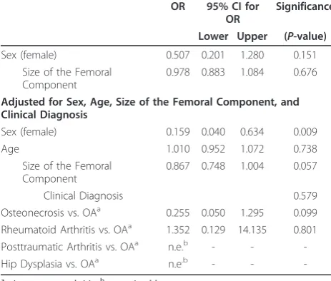
Osteonecrosis was detected in the femoral remnants of 80 (90.9%) male and 71 (83.5%; OR: 0.507, 95% CI: 0.201, 1.280; P = 0.151) female patients. The vertical extent of osteonecrosis was, however, significantly larger in the femoral remnants of male patients (median verti-cal extent of osteonecrosis 15.3 mm, IQR: 3.6 to 24.2) compared with female patients (median vertical extent of osteonecrosis 6.2 mm, IQR: 2.6 to 14.6;P = 0.008). Moreover, 41 (63.1%) out of 65 hip fractures in men were defined as postnecrotic, with a slightly lower fre-quency in female patients (23 (46.9%) out of 49 peri-prosthetic fractures were postnecrotic, P = 0.086). Interestingly, after adjusting for sex, age, and size of the femoral component, the logistic regression analysis revealed lower ORs for the occurrence of osteonecrosis within the femoral remnants for female patients (adjusted OR: 0.159, 95% CI: 0.040, 0.634; P = 0.009) compared with men (Table 5).
Excessive intraosseous lymphocyte infiltration of femoral remnant bone tissue was observed in 11 (6.4%) hips. Ten patients with unexplained groin pain and excessive
lymphocyte infiltration of the femoral remnant were women (OR: 11.600, 95% CI: 1.451, 92,731;P= 0.021). In addition, larger femoral components had a lower OR (OR: 0.810, 95% CI: 0.689, 0.953;P= 0.011). After adjusting the analysis, a similar strong correlation was detected for excessive lymphocyte infiltration in women (adjusted OR: 10.216, 95%CI: 0.787, 132.574;P= 0.076), but not for the size of the femoral component (adjusted OR: 0.971, 95% CI: 0.779, 1.210;P= 0.792; Table 6).
Hyperosteoidosis at the bone-cement interface was observed in 30 (17.3%) out of all 173 cases. Of these, 22
(73.3%) were women (OR: 3.492, 95% CI: 1.457, 8.368;P = 0.005). The relationship between interface hyperos-teoidosis and the size of the femoral component was not significant (OR: 0.918, 95% CI: 0.836, 1.009; P = 0.076). After adjusting the analysis for sex, age, the size of the femoral component and clinical diagnosis, the interface hyperosteoidosis showed a strong association with female sex (adjusted OR: 4.190, 95% CI: 1.142, 15.376;P= 0.031; Table 7).
Discussion
Summary of main findings
We investigated the possible sex differences in failure patterns of the current generation of metal-on-metal hip resurfacing arthroplasty. We analyzed morphologically distinct failure modes in a large collection of retrieved hips and performed statistical analyses. We observed substantial sex differences in the failure patterns of hip resurfacing arthroplasty: male hips showed more fre-quent osteonecrosis with larger lesions than those of women and osteonecrosis led to fracture more fre-quently in men. On the other hand, the bone remnants of women were more likely to contain excessive lym-phocyte infiltrations and to show interface hyperosteoi-dosis, both of which were linked to unexplained persistent groin pain associated with suggested hyper-sensitivity reaction.
Explaining the results and comparing them with those of other studies
Following improvements in metallurgy and surgical technique, patient selection remains an important tool with which to positively influence the outcome of Table 4 Periprosthetic fractures in the cohort of retrieved
hip resurfacing arthroplasty
OR 95% CI for OR
Significance
Lower Upper (P-value)
Sex (female) 0.482 0.254 0.915 0.026 Size of the Femoral
Component
1.031 0.959 1.109 0.408
Adjusted for Sex, Age, Size of the Femoral Component, and Clinical Diagnosis
Sex (female) 0.290 0.105 0.798 0.017
Age 1.049 1.001 1.098 0.048
Size of the Femoral Component
0.948 0.848 1.060 0.349
Clinical Diagnosis 0.060
Osteonecrosis vs. OAa 0.154 0.034 0.698 0.015 Rheumatoid Arthritis vs. OAa 0.656 0.117 3.684 0.632 Posttraumatic Arthritis vs. OAa 4.798 0.481 47.889 0.182 Hip Dysplasia vs. OAa 0.568 0.180 1.794 0.335
a
primary osteoarthritis
Logistic regression analysis and a global F test (for clinical diagnosis).
Table 5 Osteonecrosis in retrieved hip resurfacing arthroplasty
OR 95% CI for OR
Significance
Lower Upper (P-value)
Sex (female) 0.507 0.201 1.280 0.151 Size of the Femoral
Component
0.978 0.883 1.084 0.676
Adjusted for Sex, Age, Size of the Femoral Component, and Clinical Diagnosis
Sex (female) 0.159 0.040 0.634 0.009
Age 1.010 0.952 1.072 0.738
Size of the Femoral Component
0.867 0.748 1.004 0.057
Clinical Diagnosis 0.579
Osteonecrosis vs. OAa 0.255 0.050 1.295 0.099 Rheumatoid Arthritis vs. OAa 1.352 0.129 14.135 0.801 Posttraumatic Arthritis vs. OAa n.e.b - - -Hip Dysplasia vs. OAa n.e.b - -
-a
primary osteoarthritis;b
not estimable
Logistic regression analysis and a global F test (for clinical diagnosis)
Table 6 Excessive lymphocyte infiltration of bone remnant tissue in retrieved hip resurfacing arthroplasty
OR 95% CI for OR Significance Lower Upper (P-value)
Sex (female) 11.600 1.451 92.731 0.021 Size of the Femoral
Component
0.810 0.689 0.953 0.011
Adjusted for Sex, Age, Size of the Femoral Component, and Clinical Diagnosis
Sex (female) 10.216 0.787 132.574 0.076
Age 0.969 0.891 1.055 0.471
Size of the Femoral Component
0.971 0.779 1.210 0.792
Clinical Diagnosis 0.886
Osteonecrosis vs. OAa 3.546 0.288 43.597 0.323 Rheumatoid Arthritis vs. OAa 1.982 0.161 24.437 0.593 Posttraumatic Arthritis vs. OAa n.e.b - - -Hip Dysplasia vs. OAa n.e.b - -
-a
primary osteoarthritis;b
not estimable
Logistic regression analysis and a global F test (for clinical diagnosis).
Hinschet al.BMC Medicine2011,9:113 http://www.biomedcentral.com/1741-7015/9/113
metal-on-metal hip arthroplasty. Although men under the age of 65 with osteoarthritis are considered to be the best candidates for hip resurfacing based on data from registries and larger centers [2-6,8-12], such data are mostly relatively unstructured and do not provide an adequate answer to the question, how do other fac-tors such as the diameter of the prosthesis, age of the patient or clinical diagnosis influence the prosthesis fail-ure? Recently, McBryde and associates computed a mul-tivariate Cox proportional hazard survival model, and found that increased risk was related to differences in the size of the femoral component in their cohort of 48 failures (out of a total of 2,123 implanted hips) [13]. Similarly, in their study cohort of 1,107 resurfaced hips, Amstutz and collaborators reported a higher revision rate in women, although the effect of sex disappeared after adjustment for component size and surgical tech-nique [7]. In contrast to clinical studies, we analyzed a large cohort of standardly analyzed retrieved hip resur-facing arthroplasties and focused on several morphologi-cally well-defined lesions within the remnant tissue that had previously been suggested to show some degree of sexual dimorphism. It seems likely that further classifi-cation of characteristic failure modes into subgroups will enable further insight into the different biological reactions to prostheses in men and women. Similarly to our results [14], Little and colleagues [19] also found osteonecrosis in the majority of fractures in male patients. Moreover, the suggestion that female patients suffer from hypersensitivity reactions to prostheses more frequently than males is generally accepted [15,17,20,21].
Limitations of the study
We recognize several important limitations to the pre-sent study. First, we were unable to estimate the total population of patients with implanted THRA operated on by the cooperating surgeons, and therefore the pre-valence, preoperative and postoperative functional scor-ings and other possible risk factors remain unknown. Moreover, we cannot exclude that some surgeons did not send all their retrieved hips to our laboratory or that some revision surgeries were possibly performed by other than our cooperating surgeons (selection bias). However, to reduce further selection bias, all cases with informative morphological findings were included in the current study and we also present our complete data on all specimens submitted to the Hamburg retrieval study on hip resurfacing arthroplasty. Furthermore, as only 8 to 15 failures were obtained for four of the five studied designs, we did not further differentiate between the dif-ferent designs of prostheses. However, it must be noted that in our study cohort, association of THRA design with distinct failure modes was not observed. To mini-mize classification bias, all specimens were processed according to highly standard schema and we did histolo-gical analysis from three quadrants from each retrieved hip. In our previous work, we also investigated inter-and intra-observer agreement for qualitative diagnoses such as the presence of osteonecrosis [14,18] and the final diagnoses were assigned by consensus between two investigators (MA, JZ). In terms of the morphological changes associated with the potential delayed-type hypersensitivity reaction, it should be kept in mind that there is no consensus about the specific histopathologi-cal features of this complication. While some investiga-tors suggested that anterior solid granulomatous pseudotumors [20,21] are specific for hypersensitivity, newer data observed malpositioning leading to the accu-mulation of metal wear particles directly within such lesions [22-24]. In the few cases in our study cohort that seemed to be associated with metal hypersensitivity, we observed proliferative desquamative synovitis linked with joint effusion under pressure and excessive intraosseous lymphocyte infiltration [15]. Because confi-dence intervals for some categories were quite wide, a reclassification of a single case (for example in a group of 11 cases showing excessive lymphocyte infiltration) may possibly change these substantially. To overcome the problem of inter-observer variability in semiquanti-tative diagnoses (for example moderate versus severe lymphocyte infiltration), we therefore defined intraoss-eous excessive lymphocyte infiltration quantitatively as more than 300 cells in one high power field [15], which represented a very conservative cutoff value. We also reported interface hyperosteoidosis [17] occurring pre-ferentially in failures in female patients, but its possible Table 7 Hyperosteoidosis of the bone trabeculae at the
bone-cement interface in retrieved hip resurfacing arthroplasty.
OR 95% CI for OR
Significance
Lower Upper (P-value)
Sex (female) 3.492 1.457 8.368 0.005 Size of the Femoral
Component
0.918 0.836 1.009 0.076
Adjusted for Sex, Age, Size of the Femoral Component, and Clinical Diagnosis
Sex (female) 4.190 1.142 15.376 0.031
Age 0.496 0.928 1.037 0.471
Size of the Femoral Component
1.003 0.876 1.037 0.961
Clinical Diagnosis 0.733
Osteonecrosis vs. OAa 2.909 0.586 14.430 0.191 Rheumatoid Arthritis vs. OAa n.e.b - - -Posttraumatic Arthritis vs. OAa n.e.b - - -Hip Dysplasia vs. OAa 0.771 0.180 3.312 0.727
a
primary osteoarthritis;b
not estimable
association with the hypersensitivity reaction remains unclear until specific tests for metal allergy are available.
Implications for research and clinical practice
In the current study, we demonstrated that detailed clas-sification of distinct failure patterns of prostheses might help to explain the differing pathogenesis of such com-plications and enable future stratification of risk factors as well as different therapeutic strategies for certain patient populations (gender medicine and/or persona-lized medicine). Specific diagnostics of (as minimally invasive as possible) and therapy for (immunomodula-tory instead of operative) the hypersensitivity reaction to prostheses remains an important issue for future inter-disciplinary research in orthopedics.
Conclusions
Within the limitations of this study, we can conclude that, we demonstrated a substantial sex difference in distinct failure patterns of metal-on-metal hip resurfa-cing. The recognition of pathogenically distinct failure modes will enable further stratification of risk factors for certain failure mechanisms and will influence future therapeutic options for selected patient groups.
Note
The study was supported by DePuy Orthopaedics, Inc, Warsaw IN (MA, MM); Smith&Nephew, London, UK (MA, MM); Corin Group PLC, Cirencester, UK (MA, MM); Zimmer Inc, Warsaw, IN (MA, MM); and Biomet Inc, Warsaw, IN (MA, MM).
Abbreviations
CI: confidence interval; IQR: interquartile range; OR: odds ratio; THRA: total hip resurfacing arthroplasty.
Acknowledgements
The authors wish to thank to all cooperating orthopedic surgeons.
Author details
1
Institute of Pathology, University Medical Center Hamburg-Eppendorf, Germany.2Department of Medical Biometry and Epidemiology, University Medical Center Hamburg-Eppendorf, Germany.3Biomechanics Section, TUHH University of Technology Hamburg-Harburg, Germany.4Department of Orthopaedics, University Medical Center Hamburg-Eppendorf, Germany. 5Institute of Osteology and Biomechanics, University Medical Center Hamburg-Eppendorf, Germany.
Authors’contributions
JZ is the lead investigator of the study and developed its design, carried out data acquisition, and supervised data analysis and interpretation. AH and EV carried out data analysis and interpretation and helped to prepare the manuscript. MM, WR and MA provided important intellectual contributions to the study and manuscript preparation. All authors read, edited and approved the final manuscript.
Competing interests
The authors declare that they have no competing interests.
Received: 29 March 2011 Accepted: 13 October 2011 Published: 13 October 2011
References
1. Oertelt-Prigione S, Parol R, Krohn S, Preissner R, Regitz-Zagrosek V:Analysis of sex and gender-specific research reveals a common increase in publications and marked differences between disciplines.BMC Med2010,
8:70.
2. Bozic KJ, Kurtz S, Lau E, Ong K, Chiu V, Vail TP, Rubash HE, Berry DJ:The epidemiology of bearing surface usage in total hip arthroplasty in the United States.J Bone Joint Surg Am2009,91:1614-1620.
3. Shimmin A, Beaule PE, Campbell P:Metal-on-metal hip resurfacing arthroplasty.J Bone Joint Surg Am2008,90(3):637-654.
4. Nunley RM, Della Valle CJ, Barrack RL:Is patient selection important for hip resurfacing?Clin Orthop Relat Res2009,467:56-65.
5. Bozic KJ, Pui CM, Ludeman MJ, Vail TP, Silverstein MD:Do the potential benefits of metal-on-metal hip resurfacing justify the increased cost and risk of complications?Clin Orthop Relat Res2010,468:2301-2312. 6. Beaule PE, Dorey FJ, LeDuff M, Gruen T, Amstutz HC:Risk factors affecting
outcome of metal-on-metal surface arthroplasty of the hip.Clin Orthop Relat Res2004, ,418:87-93.
7. Amstutz HC, Wisk LE, Le Duff MJ:Sex as a Patient Selection Criterion for Metal-on-Metal Hip Resurfacing Arthroplasty.J Arthroplasty2011,
26:198-208.
8. Prosser GH, Yates PJ, Wood DJ, Graves SE, de Steiger RN, Miller LN:
Outcome of primary resurfacing hip replacement: evaluation of risk factors for early revision.Acta Orthop2010,81(1):66-71.
9. Seyler TM, Marker DR, Boyd HS, Zywiel MG, McGrath MS, Mont MA:
Preoperative evaluation to determine candidates for metal-on-metal hip resurfacing.J Bone Joint Surg Am2009,91(Suppl 6):32-41.
10. Maguire CM, Seyler TM, Boyd HS, Lai LP, Delanois RE, Jinnah RH:Hip resurfacing-keys to success.Bull NYU Hosp Jt Dis2009,67:142-145. 11. Della Valle CJ, Nunley RM, Raterman SJ, Barrack RL:Initial American
experience with hip resurfacing following FDA approval.Clin Orthop Relat Res2009,467:72-78.
12. Corten K, MacDonald SJ:Hip resurfacing data from national joint registries: what do they tell us? What do they not tell us?Clin Orthop Relat Res2010,468:351-357.
13. McBryde CW, Theivendran K, Thomas AM, Treacy RB, Pynsent PB:The influence of head size and sex on the outcome of Birmingham hip resurfacing.J Bone Joint Surg Am2010,92:105-112.
14. Zustin J, Sauter G, Morlock MM, Ruther W, Amling M:Association of osteonecrosis and failure of hip resurfacing arthroplasty.Clin Orthop Relat Res2010,468:756-761.
15. Zustin J, Amling M, Krause M, Breer S, Hahn M, Morlock MM, Ruther W, Sauter G:Intraosseous lymphocytic infiltrates after hip resurfacing arthroplasty: a histopathological study on 181 retrieved femoral remnants.Virchows Arch2009,454:581-588.
16. Morlock MM, Bishop N, Ruther W, Delling G, Hahn M:Biomechanical, morphological, and histological analysis of early failures in hip resurfacing arthroplasty.Proc Inst Mech Eng H2006,220:333-344. 17. Zustin J, Hahn M, Morlock MM, Ruther W, Amling M, Sauter G:Femoral
component loosening after hip resurfacing arthroplasty.Skeletal Radiol 2010,39:747-756.
18. Zustin J, Krause M, Breer S, Hahn M, von Domarus C, Ruther W, Sauter G, Morlock MM, Amling M:Morphologic analysis of periprosthetic fractures after hip resurfacing arthroplasty.J Bone Joint Surg Am2010,92:404-410. 19. Little CP, Ruiz AL, Harding IJ, McLardy-Smith P, Gundle R, Murray DW,
Athanasou NA:Osteonecrosis in retrieved femoral heads after failed resurfacing arthroplasty of the hip.J Bone Joint Surg Br2005,87:320-323. 20. Pandit H, Vlychou M, Whitwell D, Crook D, Luqmani R, Ostlere S,
Murray DW, Athanasou NA:Necrotic granulomatous pseudotumours in bilateral resurfacing hip arthoplasties: evidence for a type IV immune response.Virchows Arch2008,453:529-534.
21. Pandit H, Glyn-Jones S, McLardy-Smith P, Gundle R, Whitwell D, Gibbons CL, Ostlere S, Athanasou N, Gill HS, Murray DW:Pseudotumours associated with metal-on-metal hip resurfacings.J Bone Joint Surg Br2008,
90:847-851.
22. Kwon YM, Ostlere SJ, McLardy-Smith P, Athanasou NA, Gill HS, Murray DW:
“Asymptomatic”Pseudotumors After Metal-on-Metal Hip Resurfacing Hinschet al.BMC Medicine2011,9:113
http://www.biomedcentral.com/1741-7015/9/113
Arthroplasty Prevalence and Metal Ion Study.J Arthroplasty2010,
92:356-361.
23. Kwon YM, Glyn-Jones S, Simpson DJ, Kamali A, McLardy-Smith P, Gill HS, Murray DW:Analysis of wear of retrieved metal-on-metal hip resurfacing implants revised due to pseudotumours.J Bone Joint Surg Br2010,
92:356-361.
24. Campbell P, Ebramzadeh E, Nelson S, Takamura K, De Smet K, Amstutz HC:
Histological features of pseudotumor-like tissues from metal-on-metal hips.Clin Orthop Relat Res2010,468:2321-2327.
Pre-publication history
The pre-publication history for this paper can be accessed here: http://www.biomedcentral.com/1741-7015/9/113/prepub
doi:10.1186/1741-7015-9-113
Cite this article as:Hinschet al.:Sex differences in the morphological failure patterns following hip resurfacing arthroplasty.BMC Medicine 20119:113.
Submit your next manuscript to BioMed Central and take full advantage of:
• Convenient online submission
• Thorough peer review
• No space constraints or color figure charges
• Immediate publication on acceptance
• Inclusion in PubMed, CAS, Scopus and Google Scholar
• Research which is freely available for redistribution