R E S E A R C H
Open Access
Mucosal exposure to cockroach extract induces
allergic sensitization and allergic airway
inflammation
Narcy G Arizmendi
1, Melanie Abel
1, Lakshmi Puttagunta
2, Muhammad Asaduzzaman
1, Courtney Davidson
1,
Khalil Karimi
3, Paul Forsythe
3and Harissios Vliagoftis
1*Abstract
Background:Allergic sensitization to aeroallergens develops in response to mucosal exposure to these allergens. Allergic sensitization may lead to the development of asthma, which is characterized by chronic airway
inflammation. The objective of this study is to describe in detail a model of mucosal exposure to cockroach allergens in the absence of an exogenous adjuvant.
Methods:Cockroach extract (CE) was administered to mice intranasally (i.n.) daily for 5 days, and 5 days later mice were challenged with CE for 4 consecutive days. A second group received CE i.n. for 3 weeks. Airway
hyperresponsiveness (AHR) was assessed 24 h after the last allergen exposure. Allergic airway inflammation was assessed by BAL and lung histology 48 h after the last allergen exposure. Antigen-specific antibodies were assessed in serum. Lungs were excised from mice from measurement of cytokines and chemokines in whole lung lysate. Results:Mucosal exposure of Balb/c mice to cockroach extract induced airway eosinophilic inflammation, AHR and cockroach-specific IgG1; however, AHR to methacholine was absent in the long term group. Lung histology showed patchy, multicentric damage with inflammatory infiltrates at the airways in both groups. Lungs from mice from the short term group showed increased IL-4, CCL11, CXCL1 and CCL2 protein levels. IL4 and CXCL1 were also increased in the BAL of cockroach-sensitized mice in the short-term protocol.
Conclusions:Mucosal exposure to cockroach extract in the absence of adjuvant induces allergic airway sensitization characterized by AHR, the presence of Th2 cytokines in the lung and eosinophils in the airways.
Background
Atopy and allergic diseases affect more than 30% of the population worldwide. A study in 10 European countries showed that if we only take into account respiratory allergic conditions they still have a prevalence between 11.7% and 36.6% [1]. The economic burden of these dis-eases is also very high [2]. Despite intense efforts over the last 3 decades, the mechanisms controlling the develop-ment of allergic sensitization are still poorly understood. Animal models have been shown to be invaluable in allowing us to understand the pathogenesis of allergic conditions, especially asthma.
Animal models of asthma have a number of limitations, including the physiological relevance of the allergen used and potential differences between humans and animals in the development of allergic immune responses. There is also controversy regarding the utility of murine models of asthma as predictors of the response of human asthma to therapeutics [3]. Some of these difficulties arise from inadequacies of the murine models we use. However, other differences come from the way we use murine models and the kinds of responses we expect them to predict.
These limitations make it imperative that we use more than one model of asthma to understand specific ques-tions and that we tailor the model to the question asked. The development of new murine models, especially of models that use clinically relevant allergens, may allow us * Correspondence: [email protected]
1
Pulmonary Research Group, Department of Medicine, University of Alberta, Edmonton, AB Canada
Full list of author information is available at the end of the article
to overcome some of these problems. In addition, models using relevant allergens may be proven to be invaluable for our understanding of the development of allergic sen-sitization. To this end researchers have developed a num-ber of different models in mice.
Cockroach allergens are important sensitizing agents that may contain significant allergenic activity and are an important cause of asthma exacerbations in many parts of the World [4]. Several studies indicate that early life exposure to cockroach allergens leads to the develop-ment of specific allergic sensitization to cockroaches. Childhood sensitization has been associated with an increased risk for persistent asthma and bronchial hyper-responsiveness and with a greater loss of lung function [5]. In this regard, a strong relationship between indoor allergic sensitization and exacerbation of asthma symp-toms has been demonstrated for cockroach [6]. Although most of the common cockroach allergens are not serine proteinases, we inhale these allergens along with a variety of cockroach-derived proteins with serine proteinase activity. These cockroach-derived proteins we inhale are from several sources, including cockroach saliva, feces, cast skins, debris and dead bodies [7].
Here we present a model that uses a physiological aller-gen, cockroach allergens present in whole body cockroach extracts, to cause allergic sensitization, and also a physio-logical airway mucosal route of sensitization. The objective of the study was to describe in more detail than what is available in the literature the characteristics of allergic air-way inflammation and airair-way hyperresponsiveness that develop as a response to mucosal sensitization to cock-roach allergens.
Methods
Animals
Male BALB/c mice and C57Bl/6 mice (6-8 weeks old) were purchased from Charles River Laboratories. All mice were housed in virus- and Ab-free conditions and main-tained on a 12-h light-dark cycle. All experiments described in this manuscript were approved by the Univer-sity of Alberta Health Sciences Laboratory Animal Ethics Committee (Edmonton, AB, Canada).
Intranasal administration of whole cockroach extract (CE)
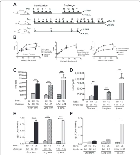
Lyophilized frozen whole body extracts from German cockroach (Blattella germanica) were obtained from Greer Laboratories, (Lenoir, NC, USA) and were resuspended in sterile normal saline and stored at 4°C. Following light anesthesia with ketamine (75 mg/kg) and acepromazine (2.5 mg/kg), mice were given 50μg of whole cockroach extract dissolved in 25μl of sterile normal saline intrana-sally (i.n.) using the short-term and long term protocols
shown in Figure 1A. Control mice were given 25μl of
sterile normal saline at the same time points. Other mice
were sensitized with an i.p. injection of 50μg of CE and 4 mg of Al(OH)3 in 0.5 ml of 0.9% sterile saline and then challenged with CE i.n., as shown in Figure 1A. In this case an i.p. injection of 0.5 ml of 0.9% sterile saline solu-tion without CE was used as a negative control.
Evaluation of AHR and allergic airway inflammation
Twenty-four hours after the final i.n. CE administration, we measured enhanced pause (Penh) to increasing doses of methacholine by non-invasive whole-body plethysmo-graphy (Buxco Electronics, Wilmington, NC, USA) to determine AHR as described [8]. Mice were euthanized 48 h after the last challenge for blood collection by cardiac puncture and lung lavage. The lungs were lavaged five times with 1 ml of isotonic phosphate-buffered saline pH 7.4 and 5 ml of this bronchoalveolar lavage (BAL) fluid was collected. Lungs were then removed and kept frozen until being processed for protein analysis. Protein analysis of lung lysates and BAL fluid was performed by Eve Tech-nologies Corporation (Calgary, Canada) using a multiplex assay based on color-coded polystyrene beads.
The BAL fluid was centrifuged at 300 g for 5 min. Total cells were counted and then cytospins of 5000 cells were prepared and stained with Diff-Quick (Fisher Scientific Co, Kalamazoo, MI, USA). Airway inflammation was assessed by counting the number of inflammatory cells in the BAL fluid as previously described [9]. The cellular composition of bronchoalveolar lavage fluid was also assessed by FACS analysis. BAL cells were stained with anti-CD3, CD11c, CD19, MHCII, and CCR3 antibodies (BD Pharmingen, San Diego, CA). Data were acquired using a FACS Canto (Becton Dickinson, Oakville, Canada) and analyzed using FlowJo software (TreeStar, Ashland, OR). Lymphocytes were identified as Forward Scatter low/ Side Scatter low cells expressing CD3 (T cells) or CD19 (B cells). Granulocytes were recognized as Side Scatter high cells and eosinophils were defined as Side Scatter high cells that express the eotaxin receptor CCR3 and inter-mediate levels of CD11c but very low to undetectable levels of MHCII, CD19 and CD3. Neutrophils were detected in a similar scatter profile as eosinophils but lacked CCR3 expression. Dendritic cells were identified as CD3-CD19-cells that express high levels of MHCII and CD11c.
Detection of cockroach specific IgG1 and IgG2a
A
B
C
D
F
E
0 10 20 30 40
0 1 2 3 4 5 Saline-saline ip sens.-in challenge
***p<0.001 CE-CE *** Methacholine (mg/ml) Pe nh
0 10 20 30 40
0 1 2 3 4 Saline-Saline CE-CE Sens.-ChallengeLong term
Methacholine (mg/ml) P enh Day: 15 AHR 16 BAL Day: 24 AHR 25 BAL 15 AHR 16 BAL Day: Sensitization Challenge
1 6 12 14
11 14
19
8 9 10 11 12 22 23
1 2 3 4 5 12 13
15 18
1 2 3 4 5 16 17
Day: 15 AHR 16 BAL Day: 24 AHR 25 BAL 15 AHR 16 BAL Day: Sensitization Challenge
1 6 12 14
11 14
19
8 9 10 11 12 22 23
1 2 3 4 5 12 13
15 18
1 2 3 4 5 16 17
Sal Sal CE Sal CE Sal CE
0 50000 100000 150000 200000 Sens.
Challenge Sal CE Sal CE
***
Short term Long term CE *** in CE in Sal 600000 900000 1200000 ip sens. *** To ta lce lls
Sal Sal CE Sal CE Sal CE 0 25000 50000 75000 *** *** *** 400000 600000 Sens.
Challenge Sal CE Sal CE
Short term Long term
CE in Sal in CE
ip sens. E os ino phi ls
Sal Sal CE Sal CE Sal CE
0.00 0.25 0.50 0.75
1.00 *** *** ***
Sens.
Challenge Sal CE Sal CE
Short term Long term
CE in Sal in CE
ip sens. Ig G1 (A bs 40 5 nm )
0 10 20 30 40 0 1 2 3 4 Saline-CE Sens.-Challenge *p<0.05,**p<0.01
CE-CE Saline-Saline Short term * ** Methacholine (mg/ml) P enh
Sal Sal CE Sal CE Sal CE
0.00 0.25 0.50
0.75 ***
Sens.
Challenge Sal CE Sal CE
Short term Long term
CE in Sal in CE
ip sen. Ig G 2a (A bs 405 nm )
Canada) or Biotin Rat anti-mouse-IgG2a (BD Pharmin-gen) in PBS/10% FBS was added and plates were incu-bated for 1 h at 24°C, followed by the addition of a 1:1000 dilution (in PBS/10% FBS) of Horseradish peroxi-dase-conjugated streptavidin (Jackson ImmunoResearch Laboratories, Inc., Burlington, ON, Canada). TMB (BD Pharmingen) was added as a substrate, and allowed 20 min at 24°C to develop. The reaction was stopped by the addition of 2 N H2SO4, and the absorbance was mea-sured at 405 nm on a Power Wave XS (BioTek Instru-ments Inc., Winooski, VT, USA) ELISA reader. Results are shown as absorbance units.
Histological analysis
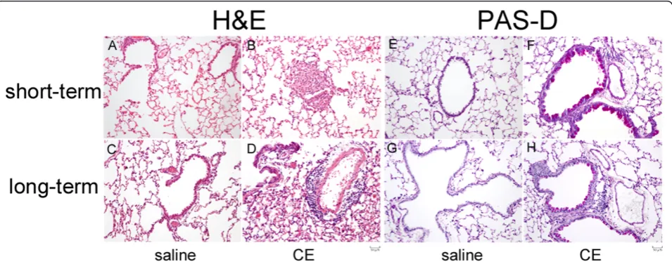
Animals were euthanized and the trachea intubated with a polyethylene catheter. Lungs were inflated with 10% neu-tral buffered formalin (Sigma-Aldrich, St. Louis MO, USA) for 10 min at a constant pressure of 20 cm water and then removed from the animal and placed in fresh 10% neutral buffered formalin for 24 h at 4°C before embedding in par-affin blocks. Sections (4μm) were stained with H&E and PAS-D and analyzed using a Nikon Eclipse E-600 Micro-scope with 2X, 10X, 20X and 40X objectives. Pictures were taken using an 11-megapixel DXM 1200C Nikon Digital Camera.
Statistical analysis
Values are expressed as“mean ± S.E.M”. Statistical differ-ences in the mean values among treatment groups were determined using a paired Studentttest. In all cases, a value for p < 0.05 was considered statistically significant.
Results
We have developed a model for mucosal sensitization of mice using whole body cockroach extract. Groups of mice received 50μg of CE intranasally after anesthesia on 5 consecutive days, rested for 5 days and then received 50μg of CE intranasally daily for 4 more days. Airway hyperresponsiveness (AHR) was assessed 24 h after the last allergen exposure and allergic airway inflammation was assessed 48 h after the last allergen exposure using BAL and lung histology (Figure 1A - short term protocol). To extend the duration of exposure to allergen we sensi-tized another group of mice with 50μg of CE intranasally 5 days a week for 3 weeks, and for 2 consecutive days on the fourth week before evaluation for AHR and allergic airway inflammation as before (Figure 1A - long term pro-tocol). Control groups of mice received saline on the same schedule. To compare our models with better-established models of parenteral sensitization in the presence of an adjuvant, a group of mice were sensitized with CE extract and Al(OH)3i.p. and then challenged with CE i.n. (Figure 1A - i.p.).
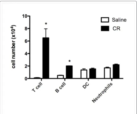
Mice sensitized and challenged with CE using the short term protocol developed AHR (Figure 1B) and increased numbers of inflammatory cells (Figure 1C), primarily eosinophils (Figure 1D), in the airways, compared to mice who received saline only. FACS analysis also revealed an increase in T and B lymphocytes in mice that received CE in the short term protocol compared to mice that received saline, but no differences in the numbers of dendritic cells and neutrophils (Figure 2). Total lympho-cytes in the BAL also increased in the mice that received CE in the long term protocol (from 416 ± 126 cells per mouse in mice receiving saline to 1807 ± 320 cells per mouse in mice receiving CE, n = 9, p < 0.02), but CE again had no effect on neutrophil numbers. Mice exposed to CE in the long term protocol showed less pronounced allergic responses. There was no evidence of AHR in the long term protocol and the numbers of eosinophils were diminished compared to the short term protocol (Figure 1). In both the short and long term protocols allergic air-way inflammation was lower than what was seen with i.p. sensitization to CE in the presence of Al(OH)3(Figure 1C and 1D).
We also evaluated the presence of antigen-specific antibodies. Mice receiving CE developed cockroach-spe-cific IgG1 (Figure 1E), the levels of which were not dif-ferent between the short term, long term and i.p. models. Mice that underwent the short term and long term protocols had no evidence of increased cockroach-specific IgG2a, while mice undergoing the i.p. protocol did develop antigen-specific IgG2a (Figure 1F).
mucin containing epithelial cells (goblet cells) in the airways.
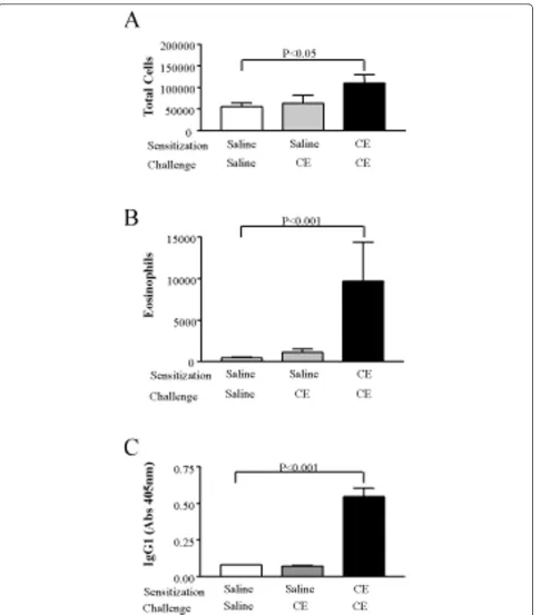
We also sensitized C57Bl/6 mice using the short term protocol. Because we detected a large difference between mice receiving saline in the sensitization phase and CE in the challenge phase vs. mice receiving CE in both phases in Balb/c mice, we used only these two groups for the C57Bl/6 experiments. Mice that received CE in both phases showed increased total cells (Figure 4A) and eosinophils (Figure 4B) in BAL fluid, and increased cockroach-specific IgG1 (Figure 4C) compared to mice that received saline during the sensitization phase and CE during the challenge phase. These mice also showed evidence of AHR (data not shown). This indicates that the cockroach extract has similar effects in Balb/c and
C57Bl/6 mice and that both strains can be sensitized to CE intranasally.
We then studied the presence of cytokines and chemo-kines in the lung tissue and airways of cockroach-sensi-tized and challenged mice. Lungs were removed from mice undergoing the short term protocol and then lyzed to measure cytokines and chemokines using a multiplex assay based on color-coded polystyrene beads. IL-4, CCL11 (eotaxin), CXCL1 (KC) and CCL2 (MCP-1) were increased in the lungs of sensitized and challenged mice (Figure 5). An increase in IL-4 and CXCL1 was also seen in the BAL fluid of these mice, but CCL11 and CCL2 were not increased in the BAL fluid (Figure 5). In all cases there were significantly lower levels of the cytokines in the BAL fluid compared to the lung lysate. In general Figure 2Numbers of T lymphocytes, B lymphocytes, dendritic cells and neutrophils in the BAL fluid of mice sensitized and
challenged with cockroach allergens using the short term protocol. BAL fluid from cockroach sensitized and challenged mice and mice
the protein assay was not sensitive enough to pick up most of the cytokines and chemokines analyzed in BAL fluid and for most of those that could be measured in the BAL fluid there was no difference between naïve and allergen sensitized and challenged mice (data not shown). A number of other cytokines/chemokines were also significantly increased in the lung tissue of mice that were sensitized and challenged with CE. IL-5 and IL-13 were increased, as expected, but IL-12p40, GM-CSF,
CCL3 (MIP-1a), CCL4 (MIP-1b) were also increased
(data not shown). Also G-CSF, IL-17A, CXCL9 (MIG), and CXCL10 (IP10) were elevated in the lung tissue (Figure 6). G-CSF and IL-17A were also increased in the BAL fluid of these mice (although the levels were signif-icantly lower than the levels in the lung tissue), but CXCL9 and CXCL10 were not increased in the BAL
fluid (Figure 6). However, IL-10, IFNg, RANTES and
VEGF were not increased following sensitization and challenge with CE.
Discussion
In this manuscript we present a detailed analysis of the airway inflammation present in a model of mucosal sen-sitization to cockroach allergens. Mice sensitized to cock-roach through the intranasal route in the absence of an adjuvant developed all the expected characteristics of asthma; they developed AHR, eosinophilic airway inflam-mation and allergen-specific IgG1 antibodies after expo-sure to allergens over a period of 2 weeks. These mice also showed increased levels of Th2 cytokines and a number of chemokines in the lung tissue. Mucosal expo-sure to CE led to allergic inflammation in both Balb/c and C57Bl/6 mice, although the latter had lower numbers
of eosinophils accumulating in the airways. Mice exposed to CE for longer periods, 17 intranasal exposures over 31/2 weeks, showed lower AHR and less eosinophils in the BAL compared to mice treated with the short term model. Histological evaluation of the short and long term models showed changes compatible with allergic airway inflammation. The changes seen in the lungs stained with H&E were quite mild compared to other murine models of asthma. CE induced inflammation in both the short and long term models compared to non-sensitized mice. The inflammation seen in the long term model was more diffuse with smaller aggregates of inflammatory cells than what was seen in the short term model. PAS-D staining of the lungs of CE sensitized and challenged mice showed increased goblet cells in the airways in both models com-pared to non-sensitized mice. The exact reason for decreased levels of inflammation in the long term proto-col are not known. Other groups using various models have shown that longer exposure to allergens can lead to tolerance and therefore have decreased levels of inflam-mation compared to the allergic inflaminflam-mation seen fol-lowing shorter exposure periods [10,11]. It is possible that a similar effect is seen in our model.
allergens without any added adjuvant, similar to models using house dust mite allergens. Our model presented here and in our previous work [15] differs from that used by Page and colleagues [16,17] because we used whole cockroach body extract, rather than cockroach frass, to induce allergic sensitization. However, the two models have many similarities. Our aim here was to describe this model in more detail and analyze various aspects of the inflammatory environment induced by mucosal sensitiza-tion to cockroach allergens.
Our data indicate that i.n. sensitization generates a more Th2-like IgG response (increased antigen-specific IgG1, but not IgG2a) compared to murine models that use i.p. sensitization (both IgG subtypes were increased). In accordance with our data, Phipps et al., were unable to detect allergen-specific IgG2a in i.n. sensitized mice [18], while Ng et al., only detected allergen-specific-IgG2a in the serum of mice when an allergen was given i.n. with a strong Th1 polarizing stimulus [19]. These data indicate that mucosal sensitization to cockroach extract leads to a more Th2 skewed model of airway inflammation com-pared to mice sensitized intraperitoneally in the presence
of an adjuvant. Our model therefore, induced a strong allergic phenotype, which supports the validity and value of this model to study mechanisms of allergic sensitization.
We also analyzed the cytokines and chemokines pre-sent in the lungs of mice sensitized and challenged with our short term protocol. We identified increased levels of Th2 cytokines (IL-4, IL-5 and IL-13) as would be expected, as well as increased levels of IL-17A and G-CSF. Increases in IL-17A and G-CSF have been shown previously in a slightly different model of sensitization to cockroach allergens, although in that case the increase was seen in cultured lung cells stimulated with conA [16,17].
present in human induced sputum 24 h after allergen challenge and correlates with endothelial progenitor recruitment in the airways [21]. Furthermore, a molecular phenotype characterized by the presence of CXCL1, RANTES, IFNg, IL-12 and IL-10 separated children with severe asthma from those with moderate asthma [22]. Further work on the presence and role of CXCL1 (KC) in murine models of allergic inflammation may allow us to better understand the pathogenesis of disease and vascular remodeling during asthma.
Cockroach is a complex mix of many proteins includ-ing serine proteinases. A number of common environ-mental allergens, including cockroach allergens, have enzymatic activity, which may skew the immune response toward the Th2 phenotype [23] and mediate, or at least participate in, the development of allergic sensitization. We have published evidence that the serine proteinase activity of cockroach allergens and their ability to activate Proteinase-Activated Receptor-2 (PAR-2) may be an important determinant of the ability to induce allergic sensitization [15]. Developing a model of allergic sensiti-zation using cockroach extract will allow us to dissect the
complexity of the extract and to identify the ability of individual antigens to induce allergic sensitization or to function as effective allergens. For example, one cock-roach allergen, Per a 10, has been cloned and its proteo-lytic activity has been shown to be important for the development of allergic inflammation and AHR [24]. Availability of many different models of mucosal sensiti-zation to be used in comparative studies may also allow us to better understand the basic mechanisms that parti-cipate in allergic sensitization and allergic airway inflam-mation. There is already evidence that different allergens induce different activation pathways in the airways in murine models [25] and comparing more models may expand our knowledge in this field.
These studies also allow us to better understand the regulation of the mucosal immune system in the lung. Mucosal immunology has been, for decades, an area of study in gastrointestinal diseases and has allowed an understanding of many facets of the immune system [26,27]. Translation of these studies into the airways will allow us to obtain a similar understanding of the airway mucosal immune responses.
Conclusions
In conclusion, we have presented a detailed analysis of a model of allergic sensitization using mucosal exposure to cockroach allergens, which is functional in both Balb/ c and C57Bl/6 mice. This model may allow us to better understand the role of cockroach allergens in allergic disease and in the inner city asthma epidemic.
List of Abbreviations Used
AHR: airway hyperresponsiveness; BAL: bronchoalveolar lavage; CE: cockroach extract; H&E: hematoxylin and eosin; i.n.: intranasal; i.p.: intraperitoneal; PAS-D: periodic acid shift with diastase
Acknowledgements
This study was funded by grants from CIHR, Alberta Innovates - Health Solutions and The Lung Association of Alberta and Northwest Territories.
Author details
1
Pulmonary Research Group, Department of Medicine, University of Alberta, Edmonton, AB Canada.2Department of Laboratory Medicine and Pathology,
University of Alberta, Edmonton, AB Canada.3Brain-Body Institute and
Department of Medicine, McMaster University, Hamilton, ON Canada.
Authors’contributions
NGA carried out most of the animal studies, participated in the study design and drafted the manuscript. MA carried out animal studies and participated in the study design. LP performed all histological analysis and prepared the histological pictures. MA performed the cytokine/chemokine analysis. CD participated in the studies analyzing airway inflammation and helped draft the manuscript. KK performed the flow cytometry studies and contributed in study design. PF participated in the study design and helped draft the manuscript. HV participated in the study design and helped draft the manuscript. All authors read and approved the final manuscript.
Competing interests
The authors declare that they have no competing interests.
Received: 26 April 2011 Accepted: 14 December 2011 Published: 14 December 2011
References
1. Dahl R, Andersen PS, Chivato T, Valovirta E, de Monchy J:National prevalence of respiratory allergic disorders.Respir Med2004,98:398-403. 2. Reed SD, Lee TA, McCrory DC:The economic burden of allergic rhinitis: a
critical evaluation of the literature.Pharmacoeconomics2004,22:345-61. 3. Finkelman FD, Wills-Karp M:Usefulness and optimization of mouse
models of allergic airway disease.J Allergy Clin Immunol2008,121:603-6. 4. Rosenstreich DL, Eggleston P, Kattan M, Baker D, Slavin RG, Gergen P,
Mitchell H, McNiff-Mortimer K, Lynn H, Ownby D,et al:The role of cockroach allergy and exposure to cockroach allergen in causing morbidity among inner-city children with asthma.N Engl J Med1997,
336:1356-63.
5. Platts-Mills TA, Rakes G, Heymann PW:The relevance of allergen exposure to the development of asthma in childhood.J Allergy Clin Immunol2000,
105:S503-8.
6. Litonjua AA, Carey VJ, Burge HA, Weiss ST, Gold DR:Exposure to cockroach allergen in the home is associated with incident doctor-diagnosed asthma and recurrent wheezing.J Allergy Clin Immunol2001,107:41-7. 7. Helm R, Pomes A:Cockroach and Other Inhalant Insect Allergens.In
Allergens and Allergen Immunotherapy..3 edition. Edited by: Lockey RF, Bukantz SC. Marcel Dekker, Inc; 2004:271-296.
8. Ebeling C, Lam T, Gordon JR, Hollenberg MD, Vliagoftis H: Proteinase-activated receptor-2 promotes allergic sensitization to an inhaled antigen through a TNF-mediated pathway.J Immunol2007,179:2910-7. 9. Forsythe P, Ebeling C, Gordon JR, Befus AD, Vliagoftis H:Opposing effects
of short- and long-term stress on airway inflammation.Am J Respir Crit Care Med2004,169:220-6.
exposure can induce persistent tolerance.Am J Respir Cell Mol Biol2007,
36:573-84.
11. Goplen N, Karim MZ, Liang Q, Gorska MM, Rozario S, Guo L, Alam R:
Combined sensitization of mice to extracts of dust mite, ragweed, and Aspergillus species breaks through tolerance and establishes chronic features of asthma.J Allergy Clin Immunol2009,123:925-32.
12. Campbell EM, Charo IF, Kunkel SL, Strieter RM, Boring L, Gosling J, Lukacs NW:Monocyte chemoattractant protein-1 mediates cockroach allergen-induced bronchial hyperreactivity in normal but not CCR2-/-mice: the role of mast cells.J Immunol1999,163:2160-7.
13. Thomas MS, Kunkel SL, Lukacs NW:Regulation of cockroach antigen-induced allergic airway hyperreactivity by the CXCR3 ligand CXCL9.J Immunol2004,173:615-23.
14. Berlin AA, Hogaboam CM, Lukacs NW:Inhibition of SCF attenuates peribronchial remodeling in chronic cockroach allergen-induced asthma. Lab Invest2006,86:557-65.
15. Arizmendi NG, Abel M, Mihara K, Davidson C, Polley D, Nadeem A, El Mays T, Gilmore BF, Walker B, Gordon JR,et al:Mucosal allergic sensitization to cockroach allergens is dependent on proteinase activity and proteinase-activated receptor-2 activation.J Immunol2011,
186:3164-72.
16. Page K, Zhou P, Ledford JR, Day SB, Lutfi R, Dienger K, Lewkowich IP:Early immunological response to German cockroach frass exposure induces a Th2/Th17 environment.J Innate Immun2011,3:167-79.
17. Page K, Ledford JR, Zhou P, Dienger K, Wills-Karp M:Mucosal sensitization to German cockroach involves protease-activated receptor-2.Respir Res
2010,11:62.
18. Phipps S, Lam CE, Kaiko GE, Foo SY, Collison A, Mattes J, Barry J, Davidson S, Oreo K, Smith L,et al:Toll/IL-1 signaling is critical for house dust mite-specific Th1 and Th2 responses.Am J Respir Crit Care Med2009,
179:883-93.
19. Ng N, Lam D, Paulus P, Batzer G, Horner AA:House dust extracts have both TH2 adjuvant and tolerogenic activities.J Allergy Clin Immunol2006,
117:1074-81.
20. Jones CP, Pitchford SC, Lloyd CM, Rankin SM:CXCR2 mediates the recruitment of endothelial progenitor cells during allergic airways remodeling.Stem Cells2009,27:3074-81.
21. Imaoka H, Punia N, Irshad A, Ying S, Corrigan CJ, Howie K, O’Byrne PM, Gauvreau GM, Sehmi R:Lung-Homing of Endothelial Progenitor Cells in Human Asthmatics Following Allergen Challenge.Am J Respir Crit Care Med2011,184:771-778.
22. Fitzpatrick AM, Higgins M, Holguin F, Brown LA, Teague WG:The molecular phenotype of severe asthma in children.J Allergy Clin Immunol2010,
125:851-857.
23. Lamhamedi-Cherradi SE, Martin RE, Ito T, Kheradmand F, Corry DB, Liu YJ, Moyle M:Fungal proteases induce Th2 polarization through limited dendritic cell maturation and reduced production of IL-12.J Immunol
2008,180:6000-9.
24. Sudha VT, Arora N, Singh BP:Serine protease activity of Per a 10 augments allergen-induced airway inflammation in a mouse model.Eur J Clin Invest2009,39:507-16.
25. Zimmermann N, Mishra A, King NE, Fulkerson PC, Doepker MP, Nikolaidis NM, Kindinger LE, Moulton EA, Aronow BJ, Rothenberg ME:
Transcript signatures in experimental asthma: identification of STAT6-dependent and -inSTAT6-dependent pathways.J Immunol2004,172:1815-24. 26. Wershil BK, Furuta GT:4. Gastrointestinal mucosal immunity.J Allergy Clin
Immunol2008,121:S380-3.
27. Xavier RJ, Podolsky DK:Unravelling the pathogenesis of inflammatory bowel disease.Nature2007,448:427-34.
doi:10.1186/1710-1492-7-22
Cite this article as:Arizmendiet al.:Mucosal exposure to cockroach extract induces allergic sensitization and allergic airway inflammation.