Egmond aan Zee, the Netherlands. 31 August - 4 September 2014
Abstract
Research on lactic acid bacteria (LAB) has advanced significantly over the past number of decades and these developments have been driven by the parallel advances in technologies such as genomics, bioinformatics, protein expression systems and structural biology, combined with the ever increasing commercial relevance of this group of microorganisms. Some of the more significant and impressive outputs have been in the domain of
bacteriophage-host interactions which provides a prime example of the cutting-edge model systems represented by LAB research. Here, we present a retrospective overview of the key advances in LAB phage research including phage-host interactions and co-evolution. We describe how in many instances this knowledge can be pivotal in creating real improvements in the application of LAB cultures in commercial practice.
Background
Lactic acid bacteria (LAB) are a group of Gram-positive, non-sporulating bacteria encompassing several genera
including among others, Lactococcus, Streptococcus,
Lactobacillus, Weissella, Leuconostoc, Enterococcus and Pediococcus. They have been associated with food pre-servation for millennia, a property that is primarily mediated by lactic acid production as a result of hexose fermentation. Additional antimicrobial contributions can be made by a range of other metabolites produced at lower concentrations, depending on physiological and nutritional conditions, such as acetic, succinic and formic acids, acetaldehyde, ethanol and CO2and bacteriocins.
When one considers the past 100 years of research on LAB, there is no doubt that the application of genomics from the early 1980s onwards has represented a major watershed. Prior to this, much of the research output was observational and descriptive with little mechanistic explanation of the phenomena in question (this of course was also the case with many other bacterial sys-tems). There was an obvious scientific interest in eluci-dating key metabolic pathways, while the isolation and description of bacteriocins warranted the investigation into the molecular nature and mode of action of these antimicrobials in light of their potential medical and
food preservation applications. In addition, the constant battle against bacteriophage infection stimulated efforts to obtain a better understanding of phage-host interactions.
With hindsight, the fact that plasmids play such a sig-nificant role in the functional activity of lactococci in par-ticular proved to be very serendipitous. The peerless work of McKay, Klaenhammer and others in elucidating the role of plasmids inLactococcus lactiswas critical to the later development of gene transfer systems, the crea-tion of vector plasmids and the general and progressive generation of the tools required for the genetic manipula-tion of these bacteria [1-4]. The explosion in research output across all members of the LAB, but particularly the lactococci and lactobacilli, was further stimulated by their essential role in a range of economically important activities ranging from dairy and other fermentation pro-cesses to their activities as probiotics and potential thera-peutic delivery vehicles.
Over the past 30 years, the implementation of emerging
‘omics’technologies such as genomics, transcriptomics, proteomics, metabolomics and when these are integrated, systems biology, to LAB research has resulted in the development of molecular tools that have been applied or formed the basis of development of such tools in other Gram-positive bacteria in the areas of protein expression systems, anti-microbial compound production and char-acterisation, glycobiology, cell envelope structure and (bacterio)phage-host interactions [5-14]. It is well
* Correspondence: g.fitzgerald@ucc.ie
1School of Microbiology, University College Cork, Cork, Ireland Full list of author information is available at the end of the article
documented that the advances in genomics and tran-scriptomics have realigned research focus away from gene mining towards the“interactomics”and rational exploitation of genomic data and this is no less the case in LAB research [15-18]. While each of these research foci has seen significant advances, perhaps the area exhi-biting the most impressive developments is that of phage biology, to the extent that LAB phages and phage-host interactions have become an outstanding model organ-ism for study in Gram positive bacteria. The scope of this review necessarily imposes a degree of selectivity on the topics that will be covered. Thus, based on the experi-ences of the authors and indeed the critical relevance of the topic from a commercial perspective, particular attention will be given to bacteriophages and phage-host interactions in lactococci.
Phage-host interactions
Since their initial discovery in the 1930s [19], bacterio-phages of LAB have been an obstinately persistent and costly problem in dairy fermentation processes. While aseptic procedures, culture rotation, sanitization and improved starter culture systems (such as the adoption of defined starters, the development and wide application of direct-to-cheese-vat cultures (or direct vat set (DVS)) have gone a long way to controlling phage infection, they still pose a serious risk especially in today’s mega-scale production facilities where fermentations are performed on a very intensive and continuous basis.
As with many of the other technologically relevant activ-ities of the LAB, research on phage and phage-host sys-tems up to the 1980s was largely descriptive in nature, primarily due to the lack of incisive technologies that would provide an ability to unravel mechanisms underpin-ning these interactions. This is not to dismiss those studies which provided very significant information regarding lysogeny, phage ultrastructure, bacteriophage-insensitive mutants and phage-resistance systems. However, the adoption of molecular technologies from the 1980s and the subsequent application of genomics has proven to be spectacularly successful in clarifying the different phage taxa (particularly for phages ofL. lactisandStreptococcus thermophilus), and has explained the impressive adaptabil-ity of phages, as well as providing an understanding of the nature of the infection process and elucidating the arsenal of phage resistance mechanisms that potentially suscepti-ble hosts have evolved to combat infection.
The unearthing of the adaptive responses of phages and their hosts to host-encoded phage-resistance systems and to phage infection, respectively, has been a particularly intriguing area of study [20-22]. Similarly, the identifica-tion of novel genetic acquisiidentifica-tion events which render phages increasingly fit in the dairy processing environ-ment has been an especially rewarding outcome of this
research. Knowledge of these adaptations may be applied in a predictive manner to understand the threat posed by phages as they evolve while also harnessing the hosts’ response to the advantage of the dairy industry [23].
The most intensively employed LAB genera/species in the dairy industry as starter and adjunct cultures are L. lactis, S. thermophilus and Lactobacillus spp. [24]. Their industrial significance partnered with the availabil-ity of limited numbers of strains has accentuated the requirement for an in-depth understanding of the means by which LAB-phages infect their hosts to develop knowledge-based strategies to defend against infection. For this reason, phage-host interactions have been one of the major areas of phage biology to receive particular attention in the post-genomics era.
The role of genomics in LAB phage classification
Over the past thirty years LAB-infecting phages have been classified by a number of means including electron micro-scopy, serotyping, DNA hybridisation, structural protein profiling and proteomic analysis and comparative genomic analysis [17,25-33]. The majority of phages infecting LAB belong to the family ofSiphoviridae, which embodies a large group of phages with long, non-contractile tails and prolate or isometric capsids (Figure 1a & 1b) [25,30]. The remainder belong to theMyoviridae(long, contractile tails) (Figure 1c) andPodoviridae(short, non-contractile tails) (Figure 1d) families, although these represent a small
minority [34,35]. The dominance of the Siphoviridae
phages may account for their high representation in model systems aimed at defining LAB phage-host interac-tions [36-38]. Lactococcal phages are currently grouped into ten taxonomic groups based on morphology and DNA homology, and of these the P335, 936 and c2 species (allSiphoviridaephages) are the most frequently encoun-tered in the dairy industry [25]. All currently known
phages of S. thermophilus belong to theSiphoviridae
family and were until recently classified into two groups based on their mode of packaging (cohesive ends termed cosphages or headful packaging termedpac) [30]. Inter-estingly, a third group represented by a phage with a novel genetic lineage (5093-like phages) has recently been described [39]. In contrast, classification ofLactobacillus phages is much more complex due to the genetic diversity that they display and it has been suggested that they should be typed by host range and morphology, and by genetic relatedness at the intra-species level [40].
classify phages. This has formed the basis for the develop-ment of many multiplex PCR tools for the rapid identifica-tion and speciaidentifica-tion of LAB phages, particularly for those known to be regularly infectingL. lactis[41,42]. The use of proteome-based taxonomic systems such the“Phage pro-teomic tree”[33] demonstrates that“omics” data can be applied to useful taxonomic schemes that may harmonise previously scattered approaches to this important issue.
While the dominance of particular lactococcal phage species has long been known, it was not until the end of the last decade and the beginning of this decade that we now fully appreciate the genetic diversity of these indivi-dual species. For example, there are 45 fully sequenced members of the 936 phage species and while there are localised regions of variability, their genome sequences and their overall genomic architecture are highly con-served [43,44]. In contrast, there are 10 fully sequenced members of the P335 phage species (excluding pro-phages) and these are now categorized into four sub-groups based on sequence homology and baseplate type [45]. While the P335 genome architecture and modular organisation is well-conserved, the functional modules may vary considerably with respect to their sequences and represent a“melting pot” of genetic information. Given that many of these phages are temperate, it is per-haps unsurprising that they are observed to be more diverse as they may acquire genetic elements from their hosts, and indeed this diversity and complexity would
provide justification for further sequencing programmes of P335 phages. The c2 phages are represented by only two fully sequenced members, c2 and bIL67 [46,47], and both appear to be very similar genetically. Although this species dominated in early phage isolation studies, it appears that they have become less problematic recently and for this reason we will focus on the dominant 936 and P335 species in this review.
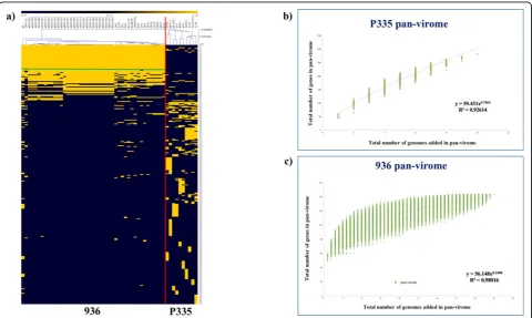
Understanding the complexity or conservation of a given phage species is vital to define the necessity of future phage genome sequencing projects and these aspects can be well resolved by the application of computational meth-odologies involving comparative genomics and pan-genome analysis on the available P335 and 936 sequences mentioned above. The relatively recent concept of pan-genome (or pan-virome if it applies to viral pan-genomes) ana-lysis has enjoyed considerable application as a means to describe genomics of bacterial species, facilitated by the increasing number of Next Generation Sequencing (NGS) projects that have been undertaken. Pan-genome analysis considers a species as a single entity composed of the entire set of genes present in each representative (also called pan-genome), which can be further divided into two classes of genes, i.e. those commonly present in all (core-genome) and the unique genes or those shared between a few members (dispensable genome) [48]. The P335 pan-virome, which is based on the sequenced representatives of this phage species, displays a considerable level of Figure 1Schematic representation of the known morphotypes of phages infecting LAB.1Arepresents the prolate-headedSiphoviridae
genetic diversity (Figure 2a). The open state (indicated by the“exponential”appearance of the graph that has not yet reached a plateau) of the pan-virome function indicates that this diversity has not yet been fully captured by the currently sequenced P335 genomes (Figure 2b), as it pre-dicts that with the addition of new P335 genomes a con-siderable number of new genes will be added to the P335 pan-virome. In contrast, given the high degree of conser-vation of phages belonging to the 936 species (Figure 2a) and the almost“closed state”(indicated by the plateaued appearance of the graph reflecting that newly sequenced genomes of this phage species do not contain genes that had not been found in previously sequenced phages of this species) of the pan-virome function achieving a plateau status (Figure 2c), sequencing of additional 936 phage gen-omes is not likely to uncover new genes, although phages with new combinations of previously found genes may still be out there.
Host-encoded receptors of LAB-infecting phages
The primary interaction between phages and their hosts is based on the recognition of a host-encoded receptor by a structure at the distal end of the phage tail known as the receptor binding protein (RBP). The molecular players involved in this initial physical connection
between LAB phages and their hosts have been the subject of intense scrutiny, particularly over the past decade. While there are multiple levels at which phages may interact with their hosts involving various different host and phage structures, these may be simplified into groups based on their receptor material: protein or car-bohydrate (or (lipo)teichoic acid).
The lactococcal 936 and P335 phages are believed to recognise cell surface-located saccharidic moieties [49,50], which are part of a so-called pellicle or cell wall polysaccharide (CWPS) [51]. The CWPS of three lacto-coccal strains (MG1363, 3107 and SMQ388) have been defined as a phospho-polysaccharide [51-53] composed of repeating subunits of a phospho-penta/hexasaccharide linked by phosphodiester bonds (Figure 3) [51]. Intrigu-ingly, within these repeating structures is a conserved tri-saccharide component that is believed to be a common receptor for phages p2, TP901-1 and 1358 of the 936, P335 and 1358 species, respectively [53]. The operon encoding the biosynthetic machinery for this CWPS has been identified as a 20 - 30 kb genomic region and muta-tions in genes within this operon render the host strain insensitive to infection by 936-type phages [54]. Genetic diversity within this cluster of genes has been associated
with potential diversity in the biochemical structure of the CWPS of lactococcal strains and comparative geno-mic analysis of this cluster within sequenced strains has led to the identification of three CWPS-specifying (geno) types (type A, B and C) based on genetic elements that are specific to that group [10]. Furthermore, a link has been established between the CWPS type of the lactococ-cal host and the phylogenetic grouping of 936 phages RBPs [10]. Thus, it is possible to predict the sensitivity of particular lactococcal hosts to particular subgroups of 936 phages. This is the first such molecular tool for defining relationships between collections of phages and strains. Further exploration of the biochemical character-istics and compositional structure of the CWPS of lacto-coccal strains will undoubtedly provide key information regarding the saccharidic components that act as recep-tors for these phages. It is likely that many other species of lactococcal phages and indeed those of other LAB gen-era that are still underrepresented in current phage/host studies, employ similar saccharidic receptors. As the number of phage/host genome sequences becoming available increases, it may be possible, through compara-tive genomics and mutational analyses, to identify the operons/genes involved and thus to develop predictive tools, such as the PCR-based method described for lacto-coccal phages and hosts, for a wider range of LAB hosts and phages in order to provide a risk assessment of phage infection.
In the dairy industry, the development of bacterioph-age-insensitive mutants (BIMs) [24,55] of important fer-mentation strains is a crucial short-term measure used to counter the phage problem. For decades this has been a practice that has been performed without any significant understanding of the underlying reasons for the associated phage-resistance. Perhaps now it is possi-ble to suggest that mutations in/diversification of the CWPS-specifying gene cluster may be responsible for at least some of the elements underpinning the acquired phage-resistance in lactococci. While knowledge of the potential reason for the phage-resistance in BIMs is
unlikely to herald the end of the ‘BIMs’ approach to avert phage-mediated problems it will most likely facili-tate a knowledge-based strategy to the design of the next generation of robust, technologically appropriate and stable BIMs.
In contrast to saccharide-recognizing phages, the lact-cococcal c2 phages recognise a protein component on the surface of their host cell [56]. The protein involved is termed the“Phage InfectionProtein” (PIP) and is a large membrane-associated protein analogous to the YueB receptor for the Bacillus subtilisphage, SPP1 [57]. This gene encoding this protein appears to be univer-sally present in lactococcal genomes, thus accounting for the typically broad-host range of these phages. As expected, removal of the genetic material encoding the predicted membrane-spanning domains of PIP prevents phage infection of the resulting PIP-lactococcal strain, a fact that may be harnessed to prevent proliferation in the dairy fermentation setting, assuming that the growth and technological characteristics of the PIP-strain are not negatively affected [58]. This could be achieved by genetic modification (GM) as previously described [58] or by means of a non-GM method through the isolation of BIMs of lactococcal strains resistant to c2 phages after exposure of the parent culture to the phage. The resulting BIMs (or many of) presumably possess a dele-tion/alteration of the PIP-encoding gene. Once it is established that such a deletion does not affect the BIM’s ability to be used as a starter culture, such strains can be applied as “natural derivatives” of the parent strain.
more than 90% reduction of phage infection [60]. While LL-H has served as an excellent model system for the interactions ofLb. delbrueckiiphages and hosts, a signifi-cant knowledge gap still remains in understanding the interactions betweenLactobacillusstrains and infecting phages given the genetic diversity found within members of this bacterial genus and their viral parasites. While this poses a significant scientific challenge, it also presents an exciting opportunity and novel area of phage-host inter-actions that deserves research attention.
The role of divalent cations in the phage infection process
The dairy industry and its associated starter culture tech-nologists have long held the view that calcium is required for phage infection, at least by some phages. It is for this reason that phage-inhibitory medium (PIM) was devel-oped incorporating phosphate in a whey-based bulk star-ter medium to“mop up”excess divalent cations during the propagation of the starter culture prior to inoculation of the milk [61] (For a review, see [62]). While this was a rational approach, it is not always successful, a phenom-enon that could not be fully explained until quite recently. Thus, the discovery by the Cambillau group [63] describing the activation of the baseplate of a 936 species phage, p2, in the presence of calcium or other divalent cations was particularly apposite. In this study, the crystal structure of the baseplate of p2 was analysed and it was observed that in the absence of calcium the receptor binding region of the baseplate was flipped upwards facing the capsid rather than the host cell, an orientation that seems counterintuitive. However, upon calcium addition, the baseplate realigns itself through a 200° downward movement to face the host. This intriguing baseplate activation may represent one possible explana-tion for the success of the 936 phages as the dominant species in the dairy industry as it will only be activated in a calcium-rich environment, such as milk, while remain-ing in a “closed” yet stable state until that situation occurs (Figure 4).
For the P335 phages, there is a mixed requirement for
divalent cations. For example, TP901-1 and jLC3 do
not require calcium for phage infection while Tuc2009 infection positively correlates to the concentration of calcium present in the medium [64]. The finding that TP901-1 does not require calcium for infection is in agreement with its permanent“infection-ready” confor-mation with the baseplate facing downwards in the pre-sence or abpre-sence of calcium (Figure 5).
These observations represent the first crucial evidence explaining why phage-inhibitory media do not success-fully prevent all phage infections, and in fact provide an exquisite example of the role of structural biology in addressing a long-standing conundrum relating to the
variable performance of PIM when used in commercial practice.
To further ascertain the requirement for divalent cations in the infection process representative members of six additional species of lactococcal phages, as well as some previously characterised phages of the P335 and 936 spe-cies have been assessed (or to verify) for their requirement for calcium. Plaque assays incorporating a range of cal-cium chloride concentrations from 0 - 10 mM and in the presence of the chelating agent EDTA were performed. In this study, only phage 1706 [65] was found to require cal-cium for plaque formation, although it should be noted that plaque visibility was significantly improved for phage 949 [66] and members of the 936 phage species in the pre-sence of calcium (unpublished data). Phage 1706 was unable to produce plaques with medium containing less than 10 mM calcium chloride. The c2 phages as well as Q54 [67] and P087 [68] infect their host with comparable efficiency in the presence or absence of calcium, or in media containing EDTA (unpublished data).
Identification of phage-encoded receptor binding proteins of LAB-infecting phages
Phage genome sequencing and subsequent bioinformatic analysis has led to the presumptive identification of RBP-encoding genes [38]. The isolation of chimeric phages con-taining a“swapped”receptor binding domain allowed the identification of the gene responsible for host recognition inS. thermophilusphages DT1 and MD4 [38]. Employing a similar strategy, the RBPs of the lactococcal P335 species phages, TP901-1 and Tuc2009 have also been identified [37] as were those of the lactococcal 936 phages, sk1 and bIL170 with confirmation by immunogold labelling elec-tron microscopy [49]. Furthermore, immunogold staining has also been central to the identification of the genetic determinants of c2 phages responsible for the interaction with their protein receptor, PIP, i.e.l10andorf31of phages c2 and bIL67, respectively, [46,47].
Most recently, chimeric phage development has been applied to identify the RBP ofLeuconostoc pseudomesenter-oidesphages, P793 and LN04 [69]. Bioinformatic analysis and domain exchange have not only permitted the identifi-cation of the RBP, but also the specific protein domain that is required for this interaction to occur. It is now well established that the amino-terminal portion of these pro-teins may be well-conserved within a group or species of phages, while the carboxy-terminus may vary considerably, and it is these unique or variable regions that dictate the specific interactions with the host [37,38,49]. Given the hypervariability associated with these host-specificity domains, it is perhaps unsurprising that the host range of these phages is generally quite limited and specific.
Figure 4The crystal structure of the“heads-down”conformations ofL. lactisphage p2 baseplate.(A)Side view in ribbon representation of the“heads-down”conformations ofL. lactisphage p2 baseplate.(B)View from top (ORF15, green; ORF16, pink; ORF18 (RBP), blue).
lactococcal 936 and P335 phages which has considerably enhanced our understanding of the mechanisms of interaction with the receptor and in specific cases it has led to the identification of the saccharidic target com-pound on the host cell surface [70]. The 936 phage p2 was the first lactococcal phage RBP to be analysed in this manner and this work revealed three domains of the RBP structure, namely the head (receptor binding domain), neck and shoulder domains [71]. This was fol-lowed by studies of chimeric RBPs of the 936 phages bIL170 and p2, which showed that while there is little sequence homology between the head domains of these two phages, a structural similarity is retained [72,73]. This supports genomic studies which suggest that while sequence similarity may not be maintained, gene order and the architecture of functional modules is conserved [27,74]. The observation of structural conservation in the absence of sequence conservation has been observed not only between members of the 936 species but also between the 936 and the P335 phage TP901-1 [73]. For example, it is now possible to predict which genes of P335 or 936 phages may encode the initiator complex or baseplate components such as the distal tail protein (Dit), the tail-associated lysin (Tal) and baseplate/RBP components once the RBP itself has been defined, even in the absence of sequence similarity. Such elements may then be exploited for structural or functional analy-sis [12,70,75-78]. Indeed, the finding of structural
conservation goes far beyond lactococcal phage species as similarities have been observed between the head domains of phage p2 and those of mammalian viruses including adenoviruses and reoviruses [71]. Using the information derived from such structural studies, propo-sals for assembly pathways of the phage tail and its baseplate components of P335 and 936 phages have been established based on Western blot analysis [79] and mass spectrometry [80], respectively. It is now pro-posed that Dit acts as a central hub around which the baseplate is formed and to which the Tal is attached to act as the initial puncturing device (For extensive reviews on this topic, see [11,78,81]).
Structural biology studies such as these represent one of the major areas of success in phage research in recent years and have transformed our previously primitive view of phage-host interactions and filled many of the knowledge gaps that were difficult to resolve by other methods including the identification of the saccharide binding sites.
Phage-host Co-evolution
immunity against phages and incoming foreign DNA that can also include plasmids [86]. Spacers of short DNA segments are acquired from the infecting phage and provide immunity against subsequent exposures to the same phage. Noteworthy however, is the relative ease with which these systems may be overcome by phages as only a single nucleotide substitution is required to bypass the immunity system [20].
The justified attention that has been paid to CRISPR-mediated phage-resistance has unearthed many interesting findings. Among these is the fact that CRISPR systems may account for the relative ease with which BIMs of S. thermophilusare generated in comparison to their non-CRISPR containing lactococcal counterparts. non- CRISPR-mediated BIMs may occur at relatively high frequencies compared to non-CRISPR-mediated BIMs, which in the latter case may require host genome mutation or IS ele-ment repositioning. For decades, it has been known that it is more difficult to generate spontaneous BIMs ofL. lactis compared toS. thermophilus; however, with the current knowledge of CRISPR systems in lactic streptococci, it is now possible to explain this phenomenon.
In addition to CRISPR defence systems, co-evolution studies of phages that have overcome abortive infection (Abi) systems have also been described, most abundantly for lactococcal phages, and through the isolation of these so-called escape mutant phages, it has been possible to identify the genes targeted by various Abi systems, including AbiQ, AbiT, AbiV [21,22,88,89]. The molecular targets of these abortive resistance systems are dispersed throughout the relevant phage genomes depending on the mechanism of the individual Abi in question. Sequence analysis of such escape mutant phages provides information which is vital to develop a basic
understand-ing of themodus operandi of Abi systems. In recent
years, the finding of novel Abi systems has slowed dra-matically in comparison to the 1980’s and 1990’s; how-ever, research attention has since shifted towards mechanistic studies, which will be invaluable in develop-ing the next generation of knowledge-based defences against phages in the dairy industry and beyond.
WhileS. thermophilusstrains appear to rely quite heav-ily on CRISPR systems to combat phage attack, lactococci on the other hand engage in“stacking”of phage-resistance systems encoded both on their plasmids and on their
type II R/M system to be defined, ScrfI, was found on the genome of the lactococcal strain UC503 [83,91] and this initiated the search for other such systems. Akin to Abi systems, the identification of novel lactococcal R/M sys-tems has lost momentum in recent years although their more recent identification in other genera, e.g. bifidobac-teria [92], is notable. In addition to host-encoded phage-resistance systems, prophage-encoded systems have been characterised over the past decade predominantly includ-ing DNA injection blockinclud-ing systems epitomised by the lac-tococcal superinfection exclusion system Sie2009encoded by the temperate lactococcal phage Tuc2009 [93]. Since the identification of this system, several others were described in both L. lactisandS. thermophilus[94,95]. The structures of phage-resistance proteins, including those of theS. thermophilussuperinfection exclusion
sys-tem, LtpTP-J34 and the Abi system AbiQ, have been
resolved [89,96]. These data shed new light on matters which were hitherto not understood and provide a unique angle from which we can begin to understand the subtle-ties of the relationship between phage and host.
Conclusions
phase, etc.). Additionally, post-translational modification of host proteins should also be considered as another level of host-response in cases where global shut-down of the host is observed post-infection as suggested by Lavigne and colleagues [98].
Perhaps, it is evident that while significant advances have been made in defining the initial interactions between LAB phages and their hosts, much remains to be discovered in aspects relating to the mechanics of DNA injection and replication, which may represent the next generation of LAB phage research in the post-genomics era.
Competing interests
The authors declare that they have no competing interests.
Acknowledgements
DvS is a recipient of a Science Foundation Ireland (SFI) Principal Investigator award (Ref. No. 08/IN.1/B1909). FB received the financial support of Science Foundation Ireland (SFI) under Grant Number SFI/12/RC/2273. We are very grateful to Sylvain Moineau and Denise Tremblay of Université Laval, Quebec, Canada for supplying phages Q54, P087, 949 & 1706 and their respective host strains used in the divalent cation dependency work referred to in this manuscript.
Declarations
This article has been published as part ofMicrobial Cell FactoriesVolume 13 Supplement 1, 2014: Proceedings of the 11th International Symposium on Lactic Acid Bacteria. The full contents of the supplement are available online at http://www.microbialcellfactories.com/supplements/13/S1. Publication charges for this supplement were funded by the Stichting Symposium on Lactic Acid Bacteria. The articles have undergone the journal’s standard peer review process for supplements. EJ is employed by Chr Hansen A/S, is a shareholder in Chr Hansen A/S; and has applied for patents relating to the content of an article which he did not edit but which forms part of this supplement. MK is an employee of Nizo. The other Supplement Editors declare that they have no competing interests.
Authors’details
1School of Microbiology, University College Cork, Cork, Ireland.2Alimentary Pharmabiotic Centre, University College Cork, Cork, Ireland.
Published: 29 August 2014
References
1. McKay LL, Baldwin KA:Plasmid distribution and evidence for a proteinase plasmid inStreptococcus lactisC2-1.Appl Microbiol1975,29:546-548. 2. Kempler GM, McKay LL:Characterization of plasmid deoxyribonucleic acid
inStreptococcus lactissubsp.diacetylactis: Evidence for plasmid-linked citrate utilization.Appl Environ Microbiol1979,37:316-323.
3. Walsh PM, McKay LL:Recombinant plasmid associated cell aggregation and high-frequency conjugation ofStreptococcus lactisML3.J Bacteriol
1981,146:937-944.
4. Klaenhammer TR, Sanozky RB:Conjugal transfer fromStreptococcus lactis
ME2 of plasmids encoding phage resistance, nisin resistance and lactose-fermenting ability: evidence for a high-frequency conjugative plasmid responsible for abortive infection of virulent bacteriophage.
J Gen Microbiol1985,131:1531-1541.
5. Kuipers OP, Beerthuyzen MM, Deruyter PGGA, Luesink EJ, Devos WM:
Autoregulation of nisin biosynthesis inLactococcus lactisby signal-transduction.J Biol Chem1995,270:27299-27304.
6. Kleerebezem M, Beerthuyzen MM, Vaughan EE, de Vos WM, Kuipers OP:
Controlled gene expression systems for lactic acid bacteria: transferable nisin-inducible expression cassettes forLactococcus, Leuconostoc, and
Lactobacillusspp.Appl Environ Microbiol1997,63:4581-4584.
7. Cotter PD:Bioengineering: a bacteriocin perspective.Bioengineered2012,
3:313-319.
8. Cotter PD, Ross RP, Hill C:Bacteriocins - a viable alternative to antibiotics?
Nat Rev Microbiol2013,11:95-105.
9. Crowley S, Mahony J, van Sinderen D:Broad-spectrum antifungal-producing lactic acid bacteria and their application in fruit models.Folia Microbiol (Praha)2013,58:291-299.
10. Mahony J, Kot W, Murphy J, Ainsworth S, Neve H, Hansen LH, Heller KJ, Sorensen SJ, Hammer K, Cambillau C,et al:Investigation of the relationship between lactococcal host cell wall polysaccharide genotype and 936 phage receptor binding protein phylogeny.Appl Environ Microbiol2013,79:4385-4392.
11. Mahony J, van Sinderen D:Structural aspects of the interaction of dairy phages with their host bacteria.Viruses2012,4:1410-1424.
12. Bebeacua C, Bron P, Lai L, Vegge CS, Brondsted L, Spinelli S, Campanacci V, Veesler D, van Heel M, Cambillau C:Structure and molecular assignment of lactococcal phage TP901-1 baseplate.J Biol Chem2010,
285:39079-39086.
13. Delavenne E, Mounier J, Deniel F, Barbier G, Le Blay G:Biodiversity of antifungal lactic acid bacteria isolated from raw milk samples from cow, ewe and goat over one-year period.Int J Food Microbiol2012,
155:185-190.
14. Crowley S, Mahony J, van Sinderen D:Current perspectives on antifungal lactic acid bacteria as natural bio-preservatives.Trends Food Sci & Technol
2013,33:93-109.
15. Ainsworth S, Zomer A, Mahony J, van Sinderen D:Lytic infection of
Lactococcus lactisby bacteriophages Tuc2009 and c2 triggers alternative transcriptional host responses.Appl Environ Microbiol2013,79:4786-4798. 16. Liu M, Bayjanov JR, Renckens B, Nauta A, Siezen RJ:The proteolytic system
of lactic acid bacteria revisited: a genomic comparison.BMC Genomics
2010,11:36.
17. Brussow H, Fremont M, Bruttin A, Sidoti J, Constable A, Fryder V:Detection and classification ofStreptococcus thermophilusbacteriophages isolated from industrial milk fermentation.Appl Environ Microbiol1994,
60:4537-4543.
18. Fallico V, Ross RP, Fitzgerald GF, McAuliffe O:Novel conjugative plasmids from the natural isolateLactococcus lactissubspecies cremoris DPC3758: a repository of genes for the potential improvement of dairy starters.
J Dairy Sci2012,95:3593-3608.
19. Whitehead HR, Cox GA:The occurrence of bacteriophage in cultures of lactic streptococci: A preliminary note.NZ J Dairy Sci & Technol1935,
16:319-320.
20. Deveau H, Barrangou R, Garneau JE, Labonte J, Fremaux C, Boyaval P, Romero DA, Horvath P, Moineau S:Phage response to CRISPR-encoded resistance in Streptococcus thermophilus.J Bacteriol2008,190:1390-1400. 21. Labrie SJ, Tremblay DM, Moisan M, Villion M, Magadan AH, Campanacci V,
Cambillau C, Moineau S:Involvement of the major capsid protein and two early-expressed phage genes in the activity of the lactococcal abortive infection mechanism AbiT.Appl Environ Microbiol2012,
78:6890-6899.
22. Haaber J, Rousseau GM, Hammer K, Moineau S:Identification and characterization of the phage gene sav, involved in sensitivity to the lactococcal abortive infection mechanism AbiV.Appl Environ Microbiol
2009,75:2484-2494.
23. Murphy J, Mahony J, Ainsworth S, Nauta A, van Sinderen D:Bacteriophage orphan DNA methyltransferases: insights from their bacterial origin, function, and occurrence.Appl Environ Microbiol2013,79:7547-7555. 24. Coffey A, Ross RP:Bacteriophage-resistance systems in dairy starter
strains: molecular analysis to application.Antonie Van Leeuwenhoek2002,
82:303-321.
25. Deveau H, Labrie SJ, Chopin MC, Moineau S:Biodiversity and classification of lactococcal phages.Appl Environ Microbiol2006,72:4338-4346. 26. Ali Y, Kot W, Atamer Z, Hinrichs J, Vogensen FK, Heller KJ, Neve H:
Classification of lytic bacteriophages attacking dairyLeuconostocstarter strains.Appl Environ Microbiol2013,79:3628-3636.
27. Brussow H, Desiere F:Comparative phage genomics and the evolution of
Siphoviridae: insights from dairy phages.Mol Microbiol2001,39:213-222. 28. Desiere F, Lucchini S, Bruttin A, Zwahlen MC, Brussow H:A highly
conserved DNA replication module fromStreptococcus thermophilus
phages is similar in sequence and topology to a module from
Lactococcus lactisphages.Virology1997,234:372-382.
33. Rohwer F, Edwards R:The phage proteomic tree: a genome-based taxonomy for phage.J Bacteriol2002,184:4529-4535.
34. Deasy T, Mahony J, Neve H, Heller KJ, van Sinderen D:Isolation of a virulentLactobacillus brevisphage and its application in the control of beer spoilage.J Food Prot2011,74:2157-2161.
35. Chibani-Chennoufi S, Dillmann ML, Marvin-Guy L, Rami-Shojaei S, Brussow H: Lactobacillus plantarumbacteriophage LP65: a new member of the SPO1-like genus of the familyMyoviridae.J Bacteriol2004,186:7069-7083. 36. Munsch-Alatossava P, Alatossava T:The extracellular phage-host
interactions involved in the bacteriophage LL-H infection of
Lactobacillus delbrueckiissp.lactisATCC 15808.Front Microbiol2013,4.
37. Vegge CS, Vogensen FK, Mc Grath S, Neve H, van Sinderen D, Brondsted L:
Identification of the lower baseplate protein as the antireceptor of the temperate lactococcal bacteriophages TP901-1 and Tuc2009.J Bacteriol
2006,188:55-63.
38. Duplessis M, Moineau S:Identification of a genetic determinant responsible for host specificity inStreptococcus thermophilus
bacteriophages.Mol Microbiol2001,41:325-336.
39. Mills S, Griffin C, O’Sullivan O, Coffey A, McAuliffe OE, Meijer WC, Serrano LM, Ross RP:A new phage on the‘Mozzarella’block: Bacteriophage 5093 shares a low level of homology with other
Streptococcus thermophilusphages.Int Dairy Journal2011,21:963-969. 40. Villion M, Moineau S:Bacteriophages ofLactobacillus.Front Biosci
(Landmark Ed)2009,14:1661-1683.
41. del Rio B, Binetti AG, Martin MC, Fernandez M, Magadan AH, Alvarez MA:
Multiplex PCR for the detection and identification of dairy bacteriophages in milk.Food Microbiol2007,24:75-81.
42. Labrie S, Moineau S:Multiplex PCR for detection and identification of lactococcal bacteriophages.Appl Environ Microbiol2000,66:987-994. 43. Mahony J, Deveau H, Mc Grath S, Ventura M, Canchaya C, Moineau S,
Fitzgerald GF, van Sinderen D:Sequence and comparative genomic analysis of lactococcal bacteriophages jj50, 712 and P008: evolutionary insights into the 936 phage species.FEMS Microbiol Lett2006,261:253-261. 44. Castro-Nallar E, Chen HL, Gladman S, Moore SC, Seemann T, Powell IB,
Hillier A, Crandall KA, Chandry PS:Population genomics and phylogeography of an Australian dairy factory derived lytic bacteriophage.Gen Biol Evol2012,4:382-393.
45. Mahony J, Martel B, Tremblay DM, Neve H, Heller KJ, Moineau S, van Sinderen D:Identification of a new P335 subgroup through molecular analysis of lactococcal phages Q33 and BM13.Appl Environ Microbiol
2013,79:4401-4409.
46. Lubbers MW, Waterfield NR, Beresford TP, Le Page RW, Jarvis AW:
Sequencing and analysis of the prolate-headed lactococcal bacteriophage c2 genome and identification of the structural genes.
Appl Environ Microbiol1995,61:4348-4356.
47. Schouler C, Ehrlich SD, Chopin MC:Sequence and organization of the lactococcal prolate-headed bIL67 phage genome.Microbiol1994,140(Pt 11):3061-3069.
48. Tettelin H, Masignani V, Cieslewicz MJ, Donati C, Medini D, Ward NL, Angiuoli SV, Crabtree J, Jones AL, Durkin AS,et al:Genome analysis of multiple pathogenic isolates ofStreptococcus agalactiae: implications for the microbial“pan-genome”.Proc Natl Acad Sci USA2005,
102:13950-13955.
49. Dupont K, Vogensen FK, Neve H, Bresciani J, Josephsen J:Identification of the receptor-binding protein in 936-species lactococcal bacteriophages.
Appl Environ Microbiol2004,70:5818-5824.
50. Spinelli S, Campanacci V, Blangy S, Moineau S, Tegoni M, Cambillau C:
Modular structure of the receptor binding proteins ofLactococcus lactis
Lactococcus lactiscell wall pellicle by phage 1358 receptor binding protein.J Virol2014.
54. Dupont K, Janzen T, Vogensen FK, Josephsen J, Stuer-Lauridsen B:
Identification ofLactococcus lactisgenes required for bacteriophage adsorption.Appl Environ Microbiol2004,70:5825-5832.
55. Mills S, Coffey A, McAuliffe OE, Meijer WC, Hafkamp B, Ross RP:Efficient method for generation of bacteriophage insensitive mutants of
Streptococcus thermophilusyoghurt and mozzarella strains.J Microbiol Methods2007,70:159-164.
56. Valyasevi R, Sandine WE, Geller BL:A membrane protein is required for bacteriophage c2 infection ofLactococcus lactissubsp.lactisC2.
J Bacteriol1991,173:6095-6100.
57. Sao-Jose C, Baptista C, Santos MA:Bacillus subtilisoperon encoding a membrane receptor for bacteriophage SPP1.J Bacteriol2004,186:8337-8346. 58. Mooney DT, Jann M, Geller BL:Subcellular location of phage infection
protein (Pip) inLactococcus lactis.Can J Microbiol2006,52:664-672. 59. Raisanen L, Draing C, Pfitzenmaier M, Schubert K, Jaakonsaari T, von
Aulock S, Hartung T, Alatossava T:Molecular interaction between lipoteichoic acids andLactobacillus delbrueckiiphages depends on D-alanyl and alpha-glucose substitution of poly(glycerophosphate) backbones.J Bacteriol2007,189:4135-4140.
60. Raisanen L, Schubert K, Jaakonsaari T, Alatossava T:Characterization of lipoteichoic acids asLactobacillus delbrueckiiphage receptor components.J Bacteriol2004,186:5529-5532.
61. Richardson GH, Cheng CT, Young R:Lactic bulk culture system utilizing a whey-based bacteriophage inhibitory medium and pH control .1. Applicability to American Style Cheese.J Dairy Sci1977,60:378-386. 62. Marco MB, Moineau S, Quiberoni A:Bacteriophages and dairy
fermentations.Bacteriophage2012,2:149-158.
63. Sciara G, Bebeacua C, Bron P, Tremblay D, Ortiz-Lombardia M, Lichiere J, van Heel M, Campanacci V, Moineau S, Cambillau C:Structure of lactococcal phage p2 baseplate and its mechanism of activation.Proc Natl Acad Sci USA2010,107:6852-6857.
64. Veesler D, Spinelli S, Mahony J, Lichiere J, Blangy S, Bricogne G, Legrand P, Ortiz-Lombardia M, Campanacci V, van Sinderen D, Cambillau C:Structure of the phage TP901-1 1.8 MDa baseplate suggests an alternative host adhesion mechanism.Proc Natl Acad Sci USA2012,109:8954-8958. 65. Garneau JE, Tremblay DM, Moineau S:Characterization of 1706, a virulent
phage fromLactococcus lactiswith similarities to prophages from other
Firmicutes.Virology2008,373:298-309.
66. Samson JE, Moineau S:Characterization ofLactococcus lactisphage 949 and comparison with other lactococcal phages.Appl Environ Microbiol
2010,76:6843-6852.
67. Fortier LC, Bransi A, Moineau S:Genome sequence and global gene expression of Q54, a new phage species linking the 936 and c2 phage species ofLactococcus lactis.J Bacteriol2006,188:6101-6114.
68. Villion M, Chopin MC, Deveau H, Ehrlich SD, Moineau S, Chopin A:P087, a lactococcal phage with a morphogenesis module similar to an
Enterococcus faecalisprophage.Virology2009,388:49-56.
69. Kot W, Hammer K, Neve H, Vogensen FK:Identification of the receptor-binding protein in lyticLeuconostoc pseudomesenteroides
bacteriophages.Appl Environ Microbiol2013,79:3311-3314.
70. Tremblay DM, Tegoni M, Spinelli S, Campanacci V, Blangy S, Huyghe C, Desmyter A, Labrie S, Moineau S, Cambillau C:Receptor-binding protein of
Lactococcus lactisphages: identification and characterization of the saccharide receptor-binding site.J Bacteriol2006,188:2400-2410. 71. Spinelli S, Desmyter A, Verrips CT, de Haard HJ, Moineau S, Cambillau C:
suggests a common ancestor gene with bacterial and mammalian viruses.Nat Struct Mol Biol2006,13:85-89.
72. Siponen M, Spinelli S, Blangy S, Moineau S, Cambillau C, Campanacci V:
Crystal structure of a chimeric receptor binding protein constructed from two lactococcal phages.J Bacteriol2009,191:3220-3225. 73. Ricagno S, Campanacci V, Blangy S, Spinelli S, Tremblay D, Moineau S,
Tegoni M, Cambillau C:Crystal structure of the receptor-binding protein head domain fromLactococcus lactisphage bIL170.J Virol2006,
80:9331-9335.
74. Chandry PS, Moore SC, Boyce JD, Davidson BE, Hillier AJ:Analysis of the DNA sequence, gene expression, origin of replication and modular structure of theLactococcus lactislytic bacteriophage sk1.Mol Microbiol
1997,26:49-64.
75. Bebeacua C, Tremblay D, Farenc C, Chapot-Chartier MP, Sadovskaya I, van Heel M, Veesler D, Moineau S, Cambillau C:Structure, adsorption to host, and infection mechanism of virulent lactococcal phage p2.J Virol2013,
87:12302-12312.
76. Collins B, Bebeacua C, Mahony J, Blangy S, Douillard FP, Veesler D, Cambillau C, van Sinderen D:Structure and functional analysis of the host recognition device of lactococcal phage Tuc2009.J Virol2013,
87:8429-8440.
77. Sciara G, Blangy S, Siponen M, Mc Grath S, van Sinderen D, Tegoni M, Cambillau C, Campanacci V:A topological model of the baseplate of lactococcal phage Tuc2009.Journal of Biological Chemistry2008,
283:2716-2723.
78. Veesler D, Cambillau C:A common evolutionary origin for tailed-bacteriophage functional modules and bacterial machineries.Microbiol Mol Biol Rev2011,75:423-433.
79. Mc Grath S, Neve H, Seegers JFML, Eijlander R, Vegge CS, Brondsted L, Heller KJ, Fitzgerald GF, Vogensen FK, van Sinderen D:Anatomy of a lactococcal phage tail.J Bacteriol2006,188:3972-3982.
80. Shepherd DA, Veesler D, Lichiere J, Ashcroft AE, Cambillau C:Unraveling lactococcal phage baseplate assembly by mass spectrometry.Molecular & Cellular Proteomics2011,10.
81. Spinelli S, Veesler D, Bebeacua C, Cambillau C:Structures and host-adhesion mechanisms of lactococcal siphophages.Front Microbiol2014,5:3. 82. Josephsen J, Vogensen FK:Identification of three different
plasmid-encoded restriction/modification systems inStreptococcus lactissubsp.
cremorisW56.FEMS Microbiol Lett1989,59:161-166.
83. Fitzgerald GF, Daly C, Brown LR, Gingeras TR:ScrFI : a new sequence-specific endonuclease fromStreptococcus cremoris.Nucleic Acids Res1982,
10:8171-8179.
84. Forde A, Daly C, Fitzgerald GF:Identification of four phage resistance plasmids fromLactococcus lactissubsp.cremorisHO2.Appl Environ Microbiol1999,65:1540-1547.
85. Horvath P, Coute-Monvoisin AC, Romero DA, Boyaval P, Fremaux C, Barrangou R:Comparative analysis of CRISPR loci in lactic acid bacteria genomes.Int J Food Microbiol2009,131:62-70.
86. Horvath P, Romero DA, Coute-Monvoisin AC, Richards M, Deveau H, Moineau S, Boyaval P, Fremaux C, Barrangou R:Diversity, activity, and evolution of CRISPR loci inStreptococcus thermophilus.J Bacteriol2008,
190:1401-1412.
87. Millen AM, Horvath P, Boyaval P, Romero DA:Mobile CRISPR/Cas-mediated bacteriophage resistance inLactococcus lactis.PLoS One2012,7:e51663. 88. Samson JE, Belanger M, Moineau S:Effect of the abortive infection
mechanism and type III toxin/antitoxin system AbiQ on the lytic cycle of
Lactococcus lactisphages.J Bacteriol2013,195:3947-3956.
89. Samson JE, Spinelli S, Cambillau C, Moineau S:Structure and activity of AbiQ, a lactococcal endoribonuclease belonging to the type III toxin-antitoxin system.Mol Microbiol2013,87:756-768.
90. Seegers JF, van Sinderen D, Fitzgerald GF:Molecular characterization of the lactococcal plasmid pCIS3: natural stacking of specificity subunits of a type I restriction/modification system in a single lactococcal strain.
Microbiol2000,146(Pt 2):435-443.
91. Davis R, van der Lelie D, Mercenier A, Daly C, Fitzgerald GF:ScrFI restriction-modification system ofLactococcus lactissubsp.cremoris
UC503: cloning and characterization of two ScrFI methylase genes.Appl Environ Microbiol1993,59:777-785.
92. O’Connell Motherway M, O’Driscoll J, Fitzgerald GF, Van Sinderen D:
Overcoming the restriction barrier to plasmid transformation and
targeted mutagenesis inBifidobacterium breveUCC2003.Microb Biotechnol2009,2:321-332.
93. McGrath S, Fitzgerald GF, van Sinderen D:Identification and characterization of phage-resistance genes in temperate lactococcal bacteriophages.Mol Microbiol2002,43:509-520.
94. Mahony J, McGrath S, Fitzgerald GF, van Sinderen D:Identification and characterization of lactococcal-prophage-carried superinfection exclusion genes.Appl Environ Microbiol2008,74:6206-6215.
95. Sun X, Gohler A, Heller KJ, Neve H:Theltpgene of temperate
Streptococcus thermophilusphage TP-J34 confers superinfection exclusion toStreptococcus thermophilusandLactococcus lactis.Virol2006,
350:146-157.
96. Bebeacua C, Lorenzo Fajardo JC, Blangy S, Spinelli S, Bollmann S, Neve H, Cambillau C, Heller KJ:X-ray structure of a superinfection exclusion lipoprotein from phage TP-J34 and identification of the tape measure protein as its target.Mol Microbiol2013,89:152-165.
97. Fallico V, Ross RP, Fitzgerald GF, McAuliffe O:Genetic response to bacteriophage infection inLactococcus lactisreveals a four-strand approach involving induction of membrane stress proteins,
D-alanylation of the cell wall, maintenance of proton motive force, and energy conservation.J Virol2011,85:12032-12042.
98. Lavigne R, Lecoutere E, Wagemans J, Cenens W, Aertsen A, Schoofs L, Landuyt B, Paeshuyse J, Scheer M, Schobert M, Ceyssens PJ:A multifaceted study ofPseudomonas aeruginosashutdown by virulent podovirus LUZ19.MBio2013,4:e00061-00013.
doi:10.1186/1475-2859-13-S1-S1
Cite this article as:Mahonyet al.:Progress in lactic acid bacterial phage research.Microbial Cell Factories201413(Suppl 1):S1.
Submit your next manuscript to BioMed Central and take full advantage of:
• Convenient online submission
• Thorough peer review
• No space constraints or color figure charges
• Immediate publication on acceptance
• Inclusion in PubMed, CAS, Scopus and Google Scholar
• Research which is freely available for redistribution

![Figure 3 Figure adapted from Chapot-Chartier et alpolysaccharide of., 2010 [51] and Farenc et al., 2014 [53] highlighting the structure of the cell wall L](https://thumb-us.123doks.com/thumbv2/123dok_us/9156865.1911080/5.595.60.539.89.205/figure-figure-adapted-chapot-chartier-alpolysaccharide-highlighting-structure.webp)


