R E S E A R C H
Open Access
Cardiogenol C can induce Mouse Hair Bulge
Progenitor Cells to Transdifferentiate into
Cardiomyocyte-like Cells
Winifred WY Yau
1, Mei Kuen Tang
1, Elve Chen
1, YaoYao
1, Ivan WC Wong
1, Henry SS Lee
1, Kenneth KH Lee
1,2*Abstract
Background:Hair bulge progenitor cells (HBPCs) are multipotent stem cells derived from the bulge region of mice vibrissal hairs. The purified HBPCs express CD34, K15 and K14 surface markers. It has been reported that HBPCs could be readily induced to transdifferentiate into adipocytes and osteocytes. However, the ability of HBPCs to transdifferentiate into cardiomyocytes has not yet been investigated.
Methodology/Principal Findings:The cardiomyogenic potential of HBPCs was investigated using a small cell-permeable molecule called Cardiogenol C. We established that Cardiogenol C could induce HBPCs to express transcription factors GATA4, Nkx2.5 and Tbx5, which are early specific markers for pre-cardiomyogenic cells. In prolonged cultures, the Cardiogenol C-treated HBPCs can also express muscle proteins, cardiac-specific troponin I and sarcomeric myosin heavy chain. However, we did not observe the ability of these cells to functionally contract. Hence, we called these cells cardiomyocyte-like cells rather than cardiomyocytes. We tried to remedy this
deficiency by pre-treating HBPCs with Valproic acid first before exposing them to Cardiogenol C. This pretreatment inhibited, rather than improved, the effectiveness of Cardiogenol C in reprogramming the HBPCs. We used comparative proteomics to determine how Cardiogenol C worked by identifying proteins that were differentially expressed. We identified proteins that were involved in promoting cell differentiation, cardiomyocyte development and for the normal function of striated muscles. From those differentially expressed proteins, we further propose that Cardiogenol C might exert its effect by activating the Wnt signaling pathway through the suppression of Kremen1. In addition, by up-regulating the expression of chromatin remodeling proteins, SIK1 and Smarce1 would initiate cardiac differentiation.
Conclusions/Significance:In conclusion, our CD34+/K15+HBPCs could be induced to transdifferentiate into cardiomyocyte-like cells using a small molecule called Cardiogenol C. The process involves activation of the Wnt signaling pathway and altered expression of several key chromatin remodeling proteins. The finding is clinically significant as HBPCs offer a readily accessible and autologous source of progenitor cells for cell-based therapy of heart disease, which is one of major killers in developed countries.
Introduction
The hair follicle is a structure that constantly undergoes cyclic self-renewal of anagen (growth), catagen (regres-sion) and telogen (resting) stages for the replacement of natural hair loss [1]. Studies over the past two decades have been documented the presence of a progenitor cell population residing in the hair bulge region, near where
the arrector pili muscle attaches to the outer hair root sheath [2,3]. It was elucidated that hair bulge progenitor cells (HBPCs) were derived from neural crest cells that migrated to the bulge during embryonic development [4,5]. These neural crest cells that are multipotent have the capability to differentiate into various cell types in the embryo, including neurons, schwann cells, glial cells, sensory neurons, melanocytes, endocrine cells, chondro-cytes and smooth muscles [5-9]. It has been reported that there are cardiac neural crest-derived cells residing in the heart, as a rare population of dormant * Correspondence: kaholee@cuhk.edu.hk
1
Stem Cell and Regeneration Thematic Research Programme, School of Biomedical Sciences, Chinese University of Hong Kong, Shatin, Hong Kong Full list of author information is available at the end of the article
multipotent stem cells that can be induced to differenti-ate into cardiomyocytes when given the appropridifferenti-ate sti-mulation [10]. However, it would be impractical to harvest cardiac neural crest cells as a source of progeni-tor cells for the therapeutic repair of damaged heart tis-sues. Therefore, it is useful to identify a reservoir of these progenitor cells, which are abundant and readily accessible. HBPCs are readily accessible since they reside on the outer root sheath of the hair follicle and contain a rich source of neural crest-derived progenitor cells, but their ability to transdifferentiate into cardiomyocytes has never been investigated. In this context, it is impor-tant to establish a method for directing HBPCs to trans-differentiate into cardiomyocytes. There are several known chemicals that can induce embryonic and bone marrow-derived mesenchymal stem cells into cardio-myocytes-like cells, such as dimethyl sulfoxide and 5-azacytidine [11-17]. Although the induction mechanisms are not yet fully understood, it has been reported that the structure of azacytidine is similar to cytidine. 5-azacytidine can induce demethylation of cytosine and activate the expression of myogenic gene MyoD1 which in turn facilitates the differentiation of bone marrow stem cells into cardiomyocyte-like cells [16]. Wu et al. synthesized a novel small molecule from a class of dia-minopyrimidine compounds, called Cardiogenol C that could specifically induce embryonic stem cells to differ-entiate into the cardiomyocytes [18]. They reported that up to 90% of the Cardiogenol C treated cells positively expressed GATA4, Mef2 and Nkx2.5, which are essen-tial transcription factors involved in cardiogenesis. To date, Cardiogenol C has not been applied to induce adult stem cells type to differentiate into cardiomyo-cytes. Moreover, it is still not known how this molecule works or the proteins that it targets.
In the present study, we first investigated the multipo-tency of HBPCs and then tested the ability of Cardio-genol C to induce HBPCs to transdifferentiate into cardiomyocytes. In addition, we used comparative pro-teomics to understand how Cardiogenol C worked by identifying differentially expressed proteins that were directly or indirectly influenced by Cardiogenol C.
Materials and methods Ethics Statement
All experimental procedures have been approved by the animal ethics committee, The Chinese University of Hong Kong with approval number (09-245) in DH/ HA&P/8/2/1 Pt.7.
Isolation of hair bulge explants
Adult female ICR mice (8-10 weeks old) were sacrificed by cervical dislocation and anagen staged vibrissal hair follicles were extracted from the whisker pads according
to methods reported by Sieber-Blum et al. [5]. Briefly, the whisker pads were isolated and sterilized in 70% ethanol for 1 min and then washed 3 times in dissecting medium (DMEM/F12, 0.5% FBS, Invitrogen). Under the dissecting microscope, the dermis and adipose tissues were carefully removed from the vibrissal hair follicle using sharp tungsten needles. The follicle was then cut at cross-sectioned at levels above the cavernous sinus and below the attachment for the arrestor pili muscle (Figure 1B). After the hair bulge region was isolated, it was then plated onto a collagen-coated 35-mm organ culture dish containing 0.5 ml culture medium. The cul-ture medium is composed of the Glasgow Minimal Essential Medium (GMEM; Sigma-Aldrich, St. Louis, USA), supplemented with 10% USDA-approved embryo-nic stem-cell qualified fetal bovine serum (ESQ-FBS, Invitrogen), and penicillin-streptomycin (0.01 mg/ml). The explants were maintained in 5% CO2 at 37 °C
inside a humidified cell incubator. The culture medium was changed every three days.
Production, isolation and purification of CD34+HBPCs
After seven days culture, cells have migrated out from all around the hair bulge explant (Figure 1C). The explant was then removed using the tungsten needles and the cells that have attached to the culture plate were rinsed with PBS and digested with 0.25% trypsin solution (0.03% EDTA, 10 mM HEPES in Hank’s balanced salt solution) for 2 min. The reaction was then
stopped with GMEM plus 1% ESQ-FBS and the cell sus-pension was further centrifuged at 1,500 rpm for 3 min. These cells were resuspended and seeded onto two 60-mm culture dishes in GMEM with 10% ESQ-FBS, 5% CO2at 37°C inside the humidified cell incubator. It has
been reported the HBPCs expressed cell surface marker CD34, therefore we employed Dynal CD34 Progenitor Cell Selection System (Invitrogen) to select CD34+ HBPCs out from our cell cultures. Briefly, 4 × 10⁷/100 μl of CD34-coated magnetic beads (Dynabeads, Invitro-gen) were first washed with 1 ml of isolation buffer (PBS without Ca2+ and Mg2+, 0.1% BSA and 2 mM EDTA, pH 7.4). The tube was placed in a magnetic stand (Dynal MPC-S Magnetic Particle Concentrator; Invitrogen) and then the supernatant was aspirated. The tube was then removed from the magnetic stand, and the washed magnetic beads resuspended in 100 μl of isolation buffer, ready for use. The primary hair bulge cultures were trypsinized and the cells were suspended at 1 × 10⁸ cells/ml. The appropriated cell density of 1 ml of the crude hair bulge cells suspension was mixed with 100μl of pre-washed magnetic beads. The mixture was then incubated at 4°C for 30 min with gentle tilting and rotation. The tube was then filled with isolation buffer (to the level of the magnets) and the cell-bead complexes were resuspended. The tube was placed in the magnetic stand for 2 min and then the supernatant was discarded. The bead-bound cells were washed and resuspended in 100μl of isolation buffer. The suspen-sion was further centrifuged for 10 min at 400 × g to remove excess detached beads. Finally, the purified CD34+ HBPCs pellet was resuspended and cultured in GMEM plus 10% ESQ-FBS.
Testing the multipotency of the CD34+HBPCs
CD34+HBPCs were assessed for their ability to transdif-ferentiate into adipocytes, osteocytes and cardiomyocytes. Purified HBPCs (1 × 10⁵ cells/ml), in normal culture medium, were plated onto four-well culture plates con-taining 13-mm glass coverslips. After incubation at 37°C overnight, the HBPCs were treated with adipogenic indu-cing medium composing of GMEM, 1 mg/ml insulin (Sigma-Aldrich), 100 μM dexamethasone Aldrich), 100 mM 3-isobutyl-1-methylxanthine (Sigma-Aldrich) and 7.5% ESQ-FBS. After three weeks culture, the presence of adipocytes was determined using Oil Red O staining. For osteogenic induction, we used medium containing GMEM, 10 mMb-glycerophosphate (Sigma-Aldrich), 50μM ascorbic acid 2-phosphate, 1μM dexa-methasone and 7.5% ESQ-FBS. After 3-weeks culture, the presence of osteocytes was identified using Alizarin Red S staining, which detected the presence of mineralized calcium-deposits. For cardiogenic induction, we used GMEM plus 5μM Cardiogenol C (Calbiochem, CA,
USA) and 7.5% ESQ-FBS. The cultures were harvested at different day-intervals after induction for immunohisto-chemistry, semi-quantitative RT-PCR analysis, western blot analysis and comparative proteomic.
Immunohistochemistry
Briefly, Cardiogenol C treated and untreated CD34+ HBPCs that have been cultured on coverslips were fixed in 10% formalin overnight. The samples washed 3 times with PBS and permeabilized with 2 M HCl with 0.5% (v/v) Triton X-100 for 30 min. These samples were then blocked with 3% BSA (Amresco) in PBS for 1 hr, and incubated with primary antibody (1:100) overnight at room temperature with gentle agitation. Primary antibo-dies used were mouse monoclonal antiboantibo-dies against CD34 (Lab Vision, CA, USA), K14 (Calbiochem, CA, USA), activeb-catenin (Millipore, CA, USA), GATA4 (Santa Cruz Biotechnology, CA, USA), sarcomeric myo-sin heavy chain (Developmental Studies Hybridoma Bank, Iowa, IA, USA), Cardiac-specific troponin I (Lab Vision) and Islet1 (R & D Systems). In addition, rabbit monoclonal anti-K15 (Epitomics, CA, USA) and goat polyclonal anti-Nkx-2.5 antibodies (Santa Cruz Biotech-nology, CA, USA) were also used. The cells were washed three times with PBST (PBS buffer with 0.05% Tween 20) for 20 min to remove unbound primary antibody. After-wards, the appropriate secondary antibody (1:300) was added for 1 hr at room temperature in the dark with gen-tle shaking. The secondary antibodies used were FITC-conjugated donkey anti-mouse immunoglobulin G (IgG) and Cy2-conjugated donkey anti-goat IgG (Jackson ImmunoResearch). Unbound secondary antibody was removed by washing with PBST and then PBS. The sam-ples were counterstained with the nuclear stained dye DAPI (Molecular Probes, Eugene, OR, USA) in 50% (v/v) glycerol and mounted onto slides. The samples were then examined and recorded under a confocal microscopy (Confocal Laser Scanning Biological Microscope FV1000; Olympus, Japan) with fixed exposure settings for all the samples. Image analysis was performed using a FV10-ASW software (version 1.6a, Olympus). Three replicates of each sample were analyzed.
Semi-quantitative RT-PCR analysis
polymerase (Bio-Firm, Hong Kong), 1μl of forward and reverse primers (10 μM; 1st BASE, Singapore), and DEPC-treated water was added up to a final volume of 25 μl. The primers, listed in Table 1 were designed using Primer3 software (version 0.4.0, Rozen and Ska-letsky; http://frodo.wi.mit.edu/ April 13, 2008). The reaction mixture was then placed in a PTC-100 thermal cycler with a heated lid (MJ Research, Watertown, MA, USA) operated under the following amplification condi-tions: initial denaturation at 95°C for 2 min, followed by a total of 35 cycles of denaturation at 95°C for 1 min, annealing at 55°C for 1 min, and extension at 72°C for 1 min. There was a final extension at 72°C for 5 min. The PCR products were analyzed by 1.5% agarose (USB) gel electrophoresis and stained with ethidium bromide (0.5 μg/ml). The expected bands in the gels were then examined under ultraviolet (UV) light, using a FluorChem 8000 imaging system (Alpha Innotech, San Leandro, CA). The intensity of the band for each sample was normalized using housekeeping b-actin gene. Each experiment was repeated three times.
Western blot analysis
Briefly, Cardiogenol C treated and untreated of HBPCs were trypsinized and harvested by centrifugation at 1,500 rpm for 3 min. The protein lysate samples were then prepared from the cell pellets that were solubilized in 200 μl of lysis buffer made up of 8 M urea, 2% CHAPS (Sigma-Aldrich), 2 M thiourea (Sigma-Aldrich),
Table 1 Primers for semi-quantitative RT-PCR analysis
Gene Primer sequences (5’to 3’) Product size (bp)
b-actin Sense 5’-TGT TAC CAA CTG GGA CGA C-3’ 573
Anti-sense
5’-AAG GAA GGC TGG AAA AGA G-3’
CD34 Sense 5’-TAG CCC AGT CTG AGG TTA GG-3’ 205
Anti-sense
5’-AGC AGA ACT CCA GAG GTG AC-3’
Dkk1 Sense 5’-TGT CTA GGG GGT CGA ATG TA-3’ 300
Anti-sense
5’-CAC AGA GCC CCA AAT GTA TC-3’
Etv6 Sense 5’-GTC TCC TTC AGC AGA AAC CA-3’ 424
Anti-sense
5’-CTG ATC CCA ACT GTG TGT CA-3’
GATA4 Sense 5’-GTG GTG GGT TTT TCT TTG TG-3’ 443
Anti-sense
5’-CAT AGT CAC CAA GGC TGC TT-3’
Hes6 Sense 5’-AGG GTA GCA GCT TTC AGG AT-3’ 354
Anti-sense
5’-CCT GAG CTG TCT CCA CCT TA-3’
Keratin 5 Sense 5’-GGA CCA GTC AAC ATC TCT GT-3’ 378
Anti-sense
5’-TGC CTA AAA GAA GCA GTC TC-3’
Keratin 14 Sense 5’-GAA TGG TTC TTC AGC AAG AC-3’ 386
Anti-sense
5’-ATG AGG AGA ATT GGG AAG AT-3’
Kremen1 Sense 5’-GCA TCC ATT TCA ACT TCA CC-3’ 431
Anti-sense
5’-CGT ACA CAG TCC ATC CTT CC-3’
Mthfr Sense 5’-CGT AGA GCA ACT TAG GGA GA-3’ 314
Anti-sense
5’-CCC ATG AGA AGA ACT AGC AG-3’
Nestin Sense 5’-GAA TCA GAT CGC TCA GAT CC-3’ 487
Anti-sense
5’-GCA CGA CAC CAG TAG AAC TGG-3’
Nkx-2.5 Sense 5’-ATC CTA AAC CTG GAG CAG CA-3’ 501
Anti-sense
5’-AGA TCT TGA CCT GCG TGG AC-3’
Osteocalcin Sense 5’-GCA GCT TGG TGC ACA CCT AG-3’ 405
Anti-sense
5’-ACC TTA TTG CCC TCC TGC TT-3’
Phc1 Sense 5’-AGC AGA CTT TGC AGA GAA GA-3’ 212
Anti-sense
5’-TAT ACT GGC TGG GGA TCC TG-3’
Plod2 Sense 5’-GCC AGA AGG TGA GAT TAC TG-3’ 397
Anti-sense
5’GCA CAA CAG GGT ACT TGT CT-3’
PPAR-g Sense 5’-ACT GTC GGT TTC AGA AGT GC-3’ 258
Anti-sense
5’-GAT CTC TTG CAC GGC TTC TA-3’
Sik1 Sense 5’-GGA GAG GAA AGG AGA CAC TT-3’ 296
Anti-sense
5’-GGA TAC TGA GTG CTT GCT TC-3’
Table 1 Primers for semi-quantitative RT-PCR analysis
(Continued)
Sox2 Sense 5’-GCC CTG CAG TAC AAC TCC AT-3’ 363
Anti-sense
5’-GTC ATT TGC TGT GGG TGA TG-3’
Srms Sense 5’-TCC CTA TGA AGG AAT GAC CA-3’ 354
Anti-sense
5’-AGC CTG TGT CAG TGA AGG AG-3’
Tbx5 Sense 5’-CAA AGA CAG GTC TTG CGA TT-3’ 426
Anti-sense
5’-GGT GAG TTT GAG CTT CTG GA-3’
Tcf3 Sense 5’-CTT ATC CCC CAA CTA CGA TG-3’ 315
Anti-sense
5’-ATA GGA GTC GGG AGG TCT CT-3’
Wnt11 Sense 5’-AGG ATC CCA AGC CAA TAA AC-3’ 330
Anti-sense
5’-GGA TCC CAC CTT CTC ATT CT-3’
YY1 Sense 5’-GCG GCA AGA AGA GTT ACC TG-3’ 418
Anti-sense
5’-GGT GTG CAG ATG CTT TCT CA-3’
Ezh2 Sense 5’-AGG CTA ATT GGG ACC AAA AC-3’ 306
Anti-sense
40 mM dithiothreitol (Sigma-Aldrich), 1% (v/v) Nonidet P-40 (USB, USA), 0.01% (v/v) TBP, Bezonase®nuclease (Novagen, USA) and a cocktail of protease inhibitors (consisting of leupeptin, pepstatin, aprotinin, PMSF and EDTA-Na2; Roche Molecular Biochemicals, USA). After
incubation on ice for 2 hr, the cell lysate samples were centrifuged at 12,000 rpm at 4°C for 15 min. The super-natant, which contained the proteins, was transferred into Eppendorf tubes. The concentration of protein for each sample was determined using a Bio-Rad Protein Assay Kit. After SDS-PAGE, the proteins were trans-ferred using a Trans-Blot Semi-Dry Transfer Cell (Bio-Rad) set at 90 mA for 60 min. The blots were stained with Ponseau S (Sigma-Aldrich) to confirm the presence of the proteins. The blots were then blocked with 5% skimmed milk and 1:1,000 primary antibodies added to the blots overnight at 4°C with agitation. Primary anti-bodies used were mouse monoclonal antianti-bodies against
b-catenin (Millipore, CA, USA), Ezh2 (Abnova), Kre-men1 (Clone 252525, R&D Systems), Phc1 (Abnova) and tubulin-a (LabVision). The blots were then washed with TBST (TBS buffer with 0.05% Tween 20) and probed with the appropriate HRP-conjugated sec-ondary antibody solution (1:10,000, Zymed), and incu-bated for 1 hr with gentle agitation. Finally, the blots were washed and developed using an ECL Western blotting detection kit (GE Healthcare), according to manufacturer’s instructions. There were three repli-cates of each sample. The staining was viewed and analyzed using a FluorChem 8000 imaging system (Alpha Innotech Corporation, San Leandro, CA). The band intensity measurement for each protein band was recorded and normalized against measurements house-keeping protein tubulin-a. All procedures were per-formed in triplicate and results were expressed as the mean value.
Cell proliferation assay
The effects of Cardiogenol C on HBPCs proliferation were determined by MTT assay. Briefly, 200 μl of CD34+ HBPCs (1 × 10⁴ cells/ml) was seeded into a 96-well plate. The cells were allowed to adhere and then treated with Cardiogenol C. At set time intervals between 1-5 days, 20μl of 12 mM 3-(4,5-dimethylthia-zol-2-yl)-2, 5-diphenyltetrazolium bromide (MTT) solution in medium without the phenol red was added to the cultures and incubated for 4 hr at 37°C. The supernatants were then discarded and 200μl of DMSO solution was added. The plates were placed on an orbi-tal shaker for 15 min to dissolve formazan crysorbi-tals and then measured on a microplate reader (Bio-Rad) set at 490 nm. There were three replicates for each time point analyzed.
Scanning electron microscopy
Briefly, treated and untreated HBPCs cultured on cover-slips were washed with PBS and fixed in 2.5% glutaral-dehyde dissolved in 0.1 M freshly prepared Sorensen’s Phosphate Buffer (PB, pH 7.4) for 4 hr. The samples were post-fixed with 1% aqueous osmium tetraoxide for 15 min and washed 3 times in PB for 10 min. The sam-ples were then dehydrated through a graded series of ethanol, critical point dried and coated with palladium-gold. The coated specimens were examined under a JSM-6301F scanning electron microscope (Joel, Tokyo, Japan).
Transmission electron microscopy
The treated and untreated HBPCs were fixed in freshly prepared 2.5% glutaraldehyde in 0.1 M phosphate buffer (pH 7.4) for 4 h. After rinsing in phosphate buffer, the cells were post-fixed in 1% osmium tetraoxide for 30 min. The cultures were then washed with MilliQ-water, dehydrated through a graded series of ethanol, cleared in propylene oxide, and then embedded in Epon 812 embedding resin. The embedded cultures were sec-tioned with an UltraCut R microtome (Leica, Austria), double-stained with uranyl acetate and lead citrate, and then examined using a transmission electron microscope (Hitachi H-7100FA, Hitachi, Tokyo, Japan).
Proteomics: sample preparations for two dimensional gel electrophoresis (2-DE)
Comparative proteomic analysis was performed as we previously reported [19,20]. Briefly, 100 μg of total pro-teins from Cardiogenol C treated and untreated CD34+ HBPCs were used in each 2-DE. The samples were first washed in ice-cold saline and then lyzed in the presence of 7 M Urea, 2 M thiourea, 0.01% TBP, 4% CHAP, 0.01% NP 40 and a mixture of protease inhibitors (Roche Molecular Biochemicals, Mannheim, Germany). After 2 hr incubation at 4°C, the supernatants were harvested by centrifugation at 13,000 rpm for 15 min. The total protein concentration of the samples was determined using a protein assay kit (Bio-Rad, Hercules, USA).
Proteomics: two dimensional gel electrophoresis
for 20000 vhr. Vertical sodium dodecyl sulphate polya-crylamide gel electrophoresis (SDS-PAGE) was used for the second dimension, using 10% polyacrylamide slab gels (18 × 16 cm). Briefly, the gel strips were removed from the IPGphor IEF system and equilibrated for 30 min in 6 M urea, 30% w/v glycerol, 2% w/v SDS, 0.05 M Tris-HCl, pH 6.8 with 2% w/v DTT. They were then treated with 2% iodoacetamide for 30 min. The gel strips were embedded on the cathode side of a pre-pre-pared SDS-PAGE gel and 0.2% agarose was poured into the cathode side to seal the gel strip. The second dimen-sion electrophoresis was performed in an ISO-DALT apparatus (Hoefer Scientific Instruments, San Francisco, USA). A tris-tricine dissociating buffer system was used and the gel was run at 60 mA constant current over-night. The gels were then fixed in 40% methanol con-taining 10% acetic acid for 1 hr and followed by a second fixative containing 50% ethanol. The fixed gels were further sensitized with 0.02% sodium thiosulphate for 10 min. After sensitization, the gels were stained with silver nitrate (Sigma-Aldrich Co., St Louis, USA) and developed. The molecular mass of the protein spots was determined by co-running the samples with stan-dard protein markers, covering the range of 14.4 - 116 kDa (Fermanta International Inc, Burlington, Canada). The pI values were determined according to the infor-mation provided by the supplier of the IPG strips. The silver stained 2-DE gels of Cardiogenol C treated and untreated HBPCs were scanned using an Agfa DUOS-CAN densitometer. The distribution of the protein spots in the 2-DE gels was recorded, compared and quantified using the ImageMaster 2 D Elite software (Amersham Biosciences, Freiburg, Germany). The data were normal-ized with respect to the total density of the gel image. Three replicates of each sample were analyzed.
Proteomics: in-gel digestion and MALDI-TOF analysis Protein spots were isolated from the silver stained gels using a spot picker (EXQuest™ Spot Cutter). Each iso-lated spot was destained in 500 μl of 15 mM potassium ferricyanide and 50 mM sodium thiosulfate for 10 min. The sample was then further washed 3 times for 15 min each in 500μl of 50% acetonitrile/25 mM NH4 bicarbo-nate at pH 8.0. The spot was soaked in 100% acetoni-trile for 5 min to dehydrate the gel, the acetoniacetoni-trile was removed when the gel turned opaque white and the gel was finally dried in a Speed-Vac Evaporator (UniEquip, Martinsried, Germany). For enzyme digestion, the gel spot was rehydrated in cold trypsin (15 μg/mL, Pro-mega, Madison) made up in 25 mM ammonium bicar-bonate, pH 8.0. After the gel had swelled and cleared, it was incubated at 37°C for 16 - 24 h. The peptide was then extracted using 50% acetonitrile and 5% trifluoroa-cetic acid. The extract peptides (0.5 μl) were then mixed
with 1 μl of fresh cyano-matrix solution (consisting of an 8 mg a-cyano-4-hydroxycinnamic acid in 50% aceto-nitrile/0.1% TFA) on a MALDI plate. The protein sam-ple was analyzed in a time-of-flight mass spectrometer (Voyager-DE™PRO Biospectrometry™, Applied Biosys-tems) using an accelerating voltage of 20 kV. For data-base search, known contamination peaks such as autoproteolysis and keratin were extracted prior to a protein mass fingerprint search with MASCOT software (Matrix Science, London, UK) in CBInr database. Up to one missed tryptic cleavage was considered and a mass accuracy of 100 ppm was used for all tryptic-mass searches. Protein identification was confirmed by using MS-Fit software http://prospector.ucsf.edu.
Results
Isolation and Purification of CD34+HBPCs
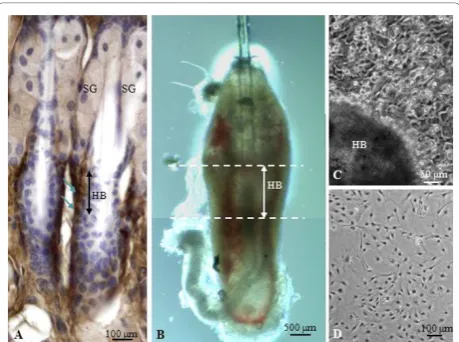
It has been reported that cell surface marker CD34 is specifically expressed by HBPCs isolated from the hair mouse bulge [21]. We performed immunohistological staining to determine where CD34+cells were normally distributed in the vibrissa. CD34+ HBPCs were evident in the bulge region of the outer root hair sheath, inferior to the sebaceous glands (Figure 1A). We carefully microdissected and isolated the bulge area from the vibrissa follicles and explanted them onto organ culture dishes (Figure 1B). We observed cells migrating out from the bulge explants after seven days culture. Colo-nies of cells were found grown around the bulge region (Figure 1C) which were trypsinized and seeded onto the 60 mm plate (Figure 1D). The cells from the primary hair bulge culture was then harvested and purified using magnetic beads coated with CD34 monoclonal antibody. We also confirmed that these cells expressed other HBPC cell surface markers K15 and K14 (Figure 2A, B). Moreover, semi-quantitative RT-PCR revealed that these
Figure 2 Characterization of CD34+ HBPCs. (A-B) Immuno-fluorescent staining showed HBPCs specifically expressed
cells expressed K5, Snail, Sox2, K14, CD34 and Nestin (Figure 2C). Dermal fibroblasts, isolated from adjacent to the hair bulge, did not express any of the HBPC sur-face markers (Figure 2D). This confirms that our HBPCs were derived from cells that have migrated out from bulge explants and not from connective tissue cells that have contaminated the bulge explants during isolation.
Establishing the multipotency of CD34+HBPCs
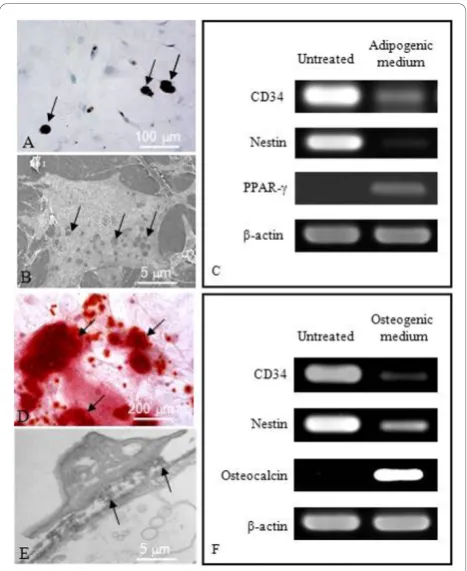
The multipotency of HBPCs was assessed for their abil-ity to transdifferentiate into adipocytes and osteocytes. The HBPCs were cultured in the presence of adipogenic or osteogenic-inducing media. We established that the HBPCs could be readily induced to differentiate into adipocytes after culturing 21 days that they were posi-tively stained with Oil Red O solution (Figure 3A). Under scanning electron microscopy, the cytoplasm of induced HBPCs clearly show the presence of empty vacuoles which originally contained storage of lipids (Figure 3B). Semi-quantitative RT-PCR analysis revealed that, following adipogenic-inducing medium treatment, CD34 and Nestin (HBPC markers) were down-regulated whereas PPAR-g(adipocyte marker) expression was up-regulated (Figure 3C). Similarly, HBPCs could be induced to transdifferentiate into osteocytes by osteo-genic-inducing medium (Figure 3D). Transmission elec-tron microscopy revealed that the induced HBPCs could secrete bone matrix-like materials into the interstitial space (Figure 3E). Semi-quantitative RT-PCR analysis showed that CD34 and Nestin expression were down-regulated while osteocalcin (osteocyte marker) expres-sion was up-regulated (Figure 2F).
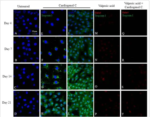
We also investigated the ability of HBPCs to transdif-ferentiate into cardiomyocytes using small molecule, Car-diogenol C. Semi-quantitative RT-PCR analysis revealed that Cardiogenol C could activate the expression of tran-scription factors GATA4, Tbx5 and homeodomain pro-tein Nkx2.5, which are all early pre-cardiac cell markers that are indispensible for initiating cardiomyogenesis (Figure 4A). Immunofluorescent staining further con-firmed that Cardiogenol C induced expressions of cardiac marker Nkx2.5 (Figure 4B) and GATA4 (Figure 5A-D). In addition, western blot analysis revealed that GATA4 expression was initiated from day 4 culture onwards in Cardiogenol C-treated HBPCs (Figure 5E). Immunofluor-escent staining showed the Cardiogenol C-treated HBPCs also progressively expressed Cardiac-specific tro-ponin I (Figure 6E-H) and sarcomeric myosin heavy chain proteins (Figure 6I-L). However, we did not observe any contracting cells in the cardiogenol C-treated cultures. In this context, we called these cells cardiomyo-cyte-like cells rather than cardiomyocytes. Huangfu et al. reported that treating fibroblasts with Valproic acid, a histone deacetylase inhibitor, enabled the fibroblasts to be more efficiently reprogrammed to become induced pluripotent stem cells [22]. Hence, we treated our HBPCs simultaneously with Valproic acid and Cardiogenol C. The mixture did not improve cardiomyocyte transdif-ferentiation. In fact, the presence of Valporic acid inhib-ited the process (Figure 6M-T). We also investigated the effects of Cardiogenol C on cell division. MTT assay
revealed that Cardiogenol C significantly inhibited cell proliferation (Figure 7).
Comparative proteomic analysis
We used comparative proteomics to elucidate how Cardiogenol C was able to induce HBPCs to become cardiomyocyte-like cells. Two-dimensional gel electro-phoresis was performed and the protein profile of HBPCs treated with Cardiogenol C for four days was compared with untreated HBPCs. We identified 18 silver-stained protein spots that were differentially expressed from 3 independent experiments. Twelve of the proteins were up-regulated by Cardiogenol C treat-ment (Figure 8), while 6 of the proteins were down-regulated (Figure 9). MALDI-TOF MS analysis revealed that the up-regulated proteins included: 1) COP9 sig-nalosome complex subunit 6, 2) emerin, 3) methylene-tetrahydrofolate reductase (Mthfr), 4) myosin light polypeptide 3, 5) myosin light polypeptide 6, 6) procol-lagen-lysine, 2-oxoglutarate 5-dioxygenase 2 precursor (Plod2), 7) protein C-ets-1, 8) salt-inducible kinase 1 (SIK1), 9) SWI/SNF related protein Smarce1, 10) tran-scription cofactor HES-6, 11) tripartite motif-contain-ing protein 54, and 12) troponin C (summarized in Table 2). The down-regulated proteins were included:
1) cell division protein kinase 6 (Cdk6), 2) growth/dif-ferentiation factor 8 precursor (GDF-8), 3) Kremen protein 1 precursor (Kremen1), 4) tight junction pro-tein ZO-1, 5) transcription factor ETV6, and 6) Tyro-sine protein kinase Srms. The observed pI and molecular mass of each proteins identified on the 2DE gel matched closely with the theoretical values pro-vided in the bioinformatic database. Their functions were also summarized in the Table 2 and 3.
We next performed semi-quantitative RT-PCR analysis to determine whether some of the differentially-expressed proteins identified were also affected at the transcriptional level. We established that Hes6, Mthfr, Plod2 and SIK1 transcriptions were up-regulated following Cardiogenol C treatment (Figure 10A); whereas, ETV6, GDF-8, Kremen1 and Srms transcriptions were down-regulated (Figure 10B). These results were the same as those observed in the compare proteomic analyses.
Cardiogenol C activates Wnt/beta-catenin signaling Kremen1 was one of the proteins found down-regu-lated in our comparative proteomic analysis. This pro-tein normally acts as a receptor for Dickkopf (DKK) protein and both cooperate together to block Wnt/b -catenin signaling [23,24]. Hence, we decided to investi-gate whether the presence of Cardiogenol C could acti-vate the Wnt/b-catenin pathway. Western blot analyses revealed that there were significant increase in the Kre-men1 and b-catenin following Cardiogenol C treatment (Figure 11A). It has been reported that Wnt-11 is one of the potential activator of the Wnt/b-catenin signal-ing dursignal-ing cardiogenesis [25]. Transcriptional factor, Lef1, participates in Wnt/b-catenin signaling by med-iating in the phosphorylation of b-catenin [26]. We established that Dkk1 and Kremen1 expression were down-regulated; whereas, Lef1 and Wnt11 expression were up-regulated by semi-quantitative RT-PCR analy-sis (Figure 11B). Immunofluorescent staining revealed that b-catenin was detected in the cytoplasm and nucleus of Cardiogenol C-treated HBPCs at Day 3 but not in untreated cultures (Figure 12A-B). Recently, Islet1 has been reported to be a downstream target of
b-catenin in cardiac progenitor cells [27]. Therefore, we examined whether Cardiogenol C could induce HBPCs to express Islet1. We established that the Car-diogenol C-treated cells expressed Islet1 after 3 days culture (Figure 12C-D).
Cardiogenol C suppresses genes involved in chromatin remodeling
SIK1 was also one of the proteins that we found up-regulated in the comparative proteomic analysis. SIK1 has been identified as a class II Histone deactylases (HDACs) kinase that is specifically expressed in the
Figure 4Cardiomyogenic induction of HBPCs by Cardiogenol C. (A) Semi-quantitative RT-PCR analysis revealed that Cardiogenol C could induce HBPCs to express cardiac-specific markers GATA4, Tbx5 and Nkx2.5.b-actin served as an internal control. (B-C)
mouse embryonic heart [28]. SIK1 is known to phos-phorylate cytoplasmic class II HDACs to trigger their translocation into the nucleus and activate MEF2-dependent transcription [28,29]. This suggests that chromatin remodeling is also involved in Cardiogenol C-induced cardiogenesis. Recent studies revealed that the Polycomb gene complex may competitively antago-nize nucleosome remodeling by the SWI/SNF-family complex [30]. Hence, we examined the effects of Cardiogenol C on the polycomb group gene complex. Semi-quantitative RT-PCR analysis revealed that poly-homeotic-like 1 (Phc1), Zeste homolog 2 (Ezh2) and transcription factor YY1 expression were significantly down-regulated following Cardiogenol C treatment (Fig-ure 13A). Moreover, western blot analysis confirmed that Phc1 and Ezh2 expressions were inhibited by Car-diogenol C (Figure 13B-C).
Discussion
Previous studies on HBPCs have mostly been related to hair regeneration and re-epithelialisation of burn wound, chronic wound and ulcerated skins [4,31-34]. In the present study, we have demonstrated that the
HBPCs, isolated from mouse vibrissa, are multipotent and can potentially provide a source of autologous pro-genitor cells for cardiac repair. These HBPCs expressed K15, a specific marker for hair bulge stem cells, and also expressed neural crest stem cell markers Nestin and Snail [32]. Furthermore, these cells expressed cell sur-face markers K5, K14 and CD34 which confirm these cells were originated from the bulge region and not from adjacent connective tissue which do not express these markers [35]. Our HBPCs also expressed Sox2 which is a key transcription factor involved in maintain-ing pluripotency and self-renewal in embryonic stem cells [36-38]. Since HBPCs express the pluripotent mar-ker Sox2, we investigated the developmental potential of these cells. These cells were able to transdifferentiate into adipocytes and osteocytes when chemically induced. To investigate the ability of HBPCs to transdifferentiate into cardiac cells, we used a small cell permeable mole-cule called Cardiogenol C. This molemole-cule was first reported to be able to induce embryonic stem cells to differentiate into beating cardiomyocytes [18]. We found that Cardiogenol C-treated HBPCs can be induced to express Nkx2.5 and GATA4, two early markers for pre-cardiac cells [39]. These genes are evolutionary highly conserved and indispensable for normal heart develop-ment [39,40]. In mature Cardiogenol C-treated cultures, we established that the cells can also express cardiac-specific troponin I and sarcomeric myosin heavy chain. In contrast to findings reported by Wu et al., who observed beating cardiomyocytes following Cardiogenol C-treated of embryonic stem cells, we could not find cardiomyocytes capable of contracting in our genol C-treated HBPCs [18]. In this context, Cardio-genol C cannot be used to produce fully functional cardiomyocytes by HBPCs - despite its ability to induce expression of key cardiac transcriptional factors Nkx2.5, GATA4, Tbx5 and Islet1.
Recently, Huangfu et al. revealed that Valporic acid (a histone deacetylase inhibitor) could be used to enhance the reprogramming of somatic cells into induced pluri-potent stem cells by more than 100-fold [41]. We there-fore decided to use Valporic acid, in combination with our Cardiogenol C, to induce a more comprehensive transdifferentiation of our HBPCs - producing cardio-mycytes that were capable of spontaneous contraction. However, we found that the HBPCs were not responsive to the Valporic acid treatment. Our results imply that HBPCs are only capable of transdifferenting into cardio-myocyte-like cells when induced by Cardiogenol C. We believe that this limited response may be attributed to the developmental plasticity of our HBPCs verses embryonic stem cells (i.e. multipotent verses pluripotent, respectively). Liu et al. recently reported that hair follicle stem cells from the bulge region could differentiate into
smooth contractile muscle cells using a tissue-specific promoter [42]. In this study, our isolated CD34+ HBPCs behave like mesenchymal stem cells capable of differen-tiating into various mesenchymal lineages, such as adipocytes and osteocytes. Though HBPCs can only transdifferentiate into cardiomyocyte-like cells, they may still be potentially useful once a method for stimulating these cells to contract has been established.
In this study, we used comparative proteomic approach to elucidate how Cardiogenol C was able to induce HBPCs to transdifferentiate into cardiomyocyte-like cells. We found a number of differentially expressed proteins in our treated HBPCs. Kremen1 expression was significantly down-regulated in the Cardiogenol C-treated cells. It has been reported that Kremen1 and Kremen2 are two dick-kopf homolog 1 (DKK1) transmembrane receptors which regulate the canonical Wnt/b-catenin signaling pathway.
Figure 6Effects of Valproic acid on cardiomyogenic induction by Cardiogenol C. Cardiogenol C-treated and untreated HBPCs were stained with Cardiac Specific Troponin I and sarcomeric myosin heavy chain (MF20) antibodies. The results revealed that Cardiogenol C-treated HBPCs expressed cardiac specific troponin I (G-H) and sarcomeric myosin heavy chain (J-L) progressively. HBPCs treated with Valproic acid alone (M-P) or Valproic acid plus Cardiogenol C (Q-T) did not induce GATA4 or cardiac specific troponin I expressions (M-T).
Figure 8Comparative proteomic analysis: Identification of protein up-regulated by Cardiogenol C. The silver stained 2-DE gel showing proteins differentially up-regulated in HBPCs by Cardiogenol C.
The binding of DKK1 to the Kremen receptors antagonize the canonical Wnt/b-catenin signaling by blocking Wnt co-receptors LRP5/6 [23,24]. Both canonical and nonca-noncial Wnt signaling pathways are essential regulators for coordinating cardiac specification and morphogenesis.
Canonical Wnt/b-catenin signaling regulates early car-diogenesis by enhancing the proliferation of cardiac pro-genitors and differentiation of cardiomyocytes [43,44].
b-catenin is thought to interact with members of the LEF-1/TCF family of transcription factors to mediate in
Table 2 Comparative proteomic analysis: proteins up-regulated by Cardiogenol C in HBPCs
Proteins identified Observed MW(kDa)/
pI
Theoretical MW (kDa)/
pI
Accession number
Folds increase
Reported functions
COP9 signalosome complex subunit 6
41/5.4 35.9/5.5 O88545 1.7 A complex involved in various cellular and developmental processes. It is an essential regulator of the ubiquitin conjugation pathway
Emerin 27/4.6 29.4/4.9 O08579 2.3 Emerin may be required for the stability and normal function of rigorously moving nuclei in skeletal muscle and heart
Methylenetetrahydro folate reductase (Mthfr)
70/4.9 74.7/5.2 Q9WU20 2.4 Folate is important for normal heart development. It is a key enzyme in folate and homocysteine metabolism
Myosin light polypeptide 6 14/4.7 16.8/4.6 Q60605 1.4 Regulatory light chain of myosin Myosin light polypeptide 3 19/4.9 21.8/5.0 P08590 1.6 Regulatory light chain of myosin
Procollagen-lysine,2-oxoglutarate 5- dioxygenase 2 precursor (Plod2)
80/6.5 84.5/6.3 Q9R0B9 4.0 Essential for the stability of the intermolecular collagen cross-links; highly expressed in heart, lung, kidney, eye, ovary, and placenta
Protein C-ets-1 55/4.8 50.2/5.0 P27577 1.7 Transcription factor. Binds to DAXX. Interacts with UBE2I. Essential for normal coronary and myocardial development in chicken embryos
Salt-inducible kinase (SIK1) 81/6.4 85.0/6.4 Q60670 3.8 Transient role during the earliest stages of myocardial cell differentiation and/or primitive chamber formation and may also be important for the earliest stages of skeletal muscle growth and/or differentiation. Potential role in G2/M cell cycle regulation SWI/SNF related protein
Smarce1
49/4.5 46.6/4.9 O54941 1.9 Involved in transcriptional activation and repression of select genes by chromatin remodeling (alteration of DNA-nucleosome topology)
Transcription cofactor HES-6 23/5.3 24.4/5.2 Q9JHE6 1.4 Up-regulate the transcription of ASCL1 and TCF3. Promotes cell differentiation
Tripartite motif- containing protein 54
36/5. 40.3/5.1 Q9BYV2 1.6 It may bind and stabilize microtubule during myotube formation
Troponin C 17/4.2 18.4/4.0 P19123 1.4 Troponin is the central regulatory protein of striated muscle contraction. The binding of calcium to Tn-C abolishes the inhibitory action of Tn on actin filaments
Table 3 Comparative proteomic analysis: proteins down-regulated by Cardiogenol C in HBPCs
Proteins identified Observed MW (kDa)/pI
Theoretical MW (kDa)/pI
Accession number
Folds decrease
Reported functions
Cell division protein kinase 6 (Cdk6)
36/6.2 37.0/6.2 Q64261 1.7 Probably involved in the control of the cell cycle. Interacts with D-type G1 cyclins
Growth/ differentiation factor 8 (GDF-8)
39/6.6 42.9/6.6 O08689 2.2 Acts specifically as a negative regulator of skeletal muscle growth
Kremen protein 1 precursor (Kremen1)
63/6.5 51.7/6.7 Q99N43 3.7 Receptor for Dickkopf protein. Cooperation with Dickkopf to block Wnt/ beta-catenin signaling
Tight junction protein ZO-1
130/6.4 194.7/6.2 P39447 1.4 The N-terminal may be involved in transducing a signal required for tight junction assembly, while the C-terminal may have specific properties of tight junctions. The alpha domain might be involved in stabilizing junctions
Transcription factor ETV6
60/6.5 56.4/6.8 P97360 3.6 Transcription repressor; binds to the DNA sequence 5’CCGGAAGT-3’
Tyrosine protein kinase Srms
Wnt signaling [26]. b-catenin also modulates the expression of Islet1 in cardiac progenitor cells which is required for cardiogenesis [27]. The noncanonical Wnt signaling pathway, which is independent ofb-catenins, involves protein kinase C and Jun amino-terminal kinase also regulates cardiac differentiation [45]. Wnt11
in the noncanonical pathway was reported to enhance cardiomyocytes differentiation in various stem cell populations [25,46,47]. In our semi-quantitative RT-PCR studies, we found Lef1 and Wnt11 expression were up-regulated by Cardiogenol C. Furthermore, our immunofluorescent staining results revealed that b -catenin was present in both the nucleus and cytoplasm. Therefore, it appears that Cardiogenol C could activate Wnt/b-catenin signaling to induce cardiogenesis. The results of our MTT cell proliferation assay confirmed that Cardiogenol C-treatment significantly decreased HBPCs proliferation. Nevertheless, we cannot explain why Cardiogenol C induced an increase inb-catenin yet a decrease in cell proliferation, as activation of the Wnt signaling pathway is normally associated with increased cell proliferation. This paradox may be required to be investigated in the future.
Besides cardiac-inducing transcription factors, epige-netic factors may also play a contributory role in cardio-myocyte differentiation. This idea is supported by reported findings that 5-azacytidine, an unspecific DNA methyltransferase inhibitor, can induce cardiogenesis [48]. This reagent prevents methylation at cytosine, which makes CpG islands in the promoter sequen-ces of genes involved in cardiac differentiation. The unmethylated sequence allows the binding of transcrip-tion initiatranscrip-tion machinery. Moreover, several chromatin remodeling proteins, such as methyltransferase Smyd1, SWI/SNF protein Baf60c, HDAC5 and HDAC9, have also been implemented in cardiomyocytes differentiation [49-51]. In this context, we identified two chromatin remodeling proteins, SIK1 and Smarce1, which were up-regulated by Cardiogenol C in our comparative proteo-mic analysis. SIK1 is a kinase of class II HDACs. It stimu-lates cardiac-specific transcription factor Mef2 via phosphorylation of HDACs [29]. Smarce1 is a compo-nent of the SWI/SNF complex. It can interact specifically with transcription factor REST to repress neuronal genes. Therefore, up-regulation of Smarce1 might facilitate the repression of neuronal- and neural crest-related genes (e.g. Nestin and Snail) in our Cardiogenol C-trea-ted HBPCs. Recently, the polycomb group complex proteins have been identified as essential in the mainte-nance of embryonic and adult stem cells, by silencing genes that are necessary for stem/progenitor cells to dif-ferentiate into various tissue types [52-54]. Therefore, we examined whether the polycomb group proteins were also involved in cardiac differentiation induced by Cardiogenol C. We found that Cardiogenol C sup-pressed Phc1, Ezh2 as well as YY1 expression. Ezh2 con-tains SET domain and belongs to polycomb repressor complex 2; while Phc1 and YY1 contain zinc-finger domain and are components of PRC1 maintenance
Figure 10RT-PCR analysis of differentially expressed proteins. Semi-quantitative RT-PCR analysis was performed to confirm that some of differentially expressed proteins identified in the comparative proteomics were also differentially transcribed. (A) The transcription levels of Hes6, Mthfr, Plod2 and SIK1 were found up-regulated in the Cardiogenol C-treated samples. (B) ETV6, GDF-8, Kremen1 and Srms expression were down-regulated by Cardiogenol C.b-actin served as an internal control.
complex [54,55]. These findings lead us to speculate that up-regulation of SIK1 as well as down-regulation of polycomb group proteins may silence genes that normally represses cardiac differentiation.
We have also identified several more proteins that were down-regulated by Cardiogenol C. Cdk6 was inhibited by Cardiogenol C. This protein is a vertebrate cdc-2 related kinase. It interacts with the G-type cyclins in the early G1 phase and functions as a retinoblastoma protein (Rb) kinase that phosphorylates the Rb protein. Phosphorylated Rb releases its binding partner tran-scription activator E2F. The free E2F in turn stimulates the transcription of genes essential for DNA replication, which initiates the cell cycle into the S phase [56]. Indeed, it has also been reported that cdk6 expression must be suppressed in order to allow proper osteoblasts and osteoclasts differentiation [55,57,58]. Therefore, it would be expected that mitogenic cdk6 expression would be inhibited so that the HBPCs could exit the cell cycle to initiate differentiation. Myostatin (also known as GDF-8) expression was also suppressed in response to Cardiogenol C treatment. Morissette et al. reported that myostatin was a negative regulator involved in controlling the growth of striated muscles in the heart [59]. Therefore, it was not surprising to observe the decreased myostatin expression when
Figure 12 b-catenin and Islet1 expression in HBPCs after Cardiogenol C treatment. (A-B) Immunofluorescent staining revealed the presence of theb-catenin (white arrows) in the Cardiogenol C-treated HBPCs but not in the untreated HBPCs. (C-D). Immunofluorescent staining revealed HBPCs expressed Islet1 (white arrows) following Cardiogenol C-treatment but not in the untreated cells.
Cardiogenol C-treated HBPCs transdifferentiate into cardiomyocyte-like cells.
In conclusion, we demonstrated for the first time that HBPCs can be induced to transdifferentiate into cardi-omyocyte-like cells using Cardiogenol C. With more research into understanding the developmental proper-ties of HBPCs, these readily accessible cells may in the future provide an abundant potential source of pro-genitor cells for the therapeutic treatment of heart diseases.
Acknowledgements
This study was supported by GRF grant (RGC Ref. No. CUHK469809) and Direct Grant (Project ID: 2041396) to KKH Lee.
Author details
1
Stem Cell and Regeneration Thematic Research Programme, School of Biomedical Sciences, Chinese University of Hong Kong, Shatin, Hong Kong.
2
Key Laboratory for Regenerative Medicine, Ministry of Education, China.
Authors’contributions
WWWY conducted the Cardiogenol C studies and the draft of the manuscript; MKT performed comparative proteomics analysis, histology and also help prepare the manuscript; EC performed the RT semi-quantitative assay, MTT assay; YY carried out the western blot assay; I WC isolated and characterized the mouse hair bulge progenitor cells, comparative proteomics, SEM and TEM studies; HSSL performed the immunohistological staining and KKH L designed and coordinated the experiments, proof read the manuscript. All authors read and approved the final manuscript. All of the authors declare that they have no competing interests.
Received: 10 August 2010 Accepted: 19 January 2011 Published: 19 January 2011
References
1. Barth JH, Messenger AG:Measurement of hair growth and investigation of hair disease.InDiseases of the Hair and Scalp..3 edition. Edited by: Dawwber R. Oxford: Blackwell Scientific; 1997:564-579.
2. Cotsarelis G, Sun TT, Lavker RM:Label-retaining cells reside in the bulge area of pilosebaceous unit - implications for follicular stem cells, hair cycle, and skin carcinogenesis.Cell1990,61:1329-1337.
3. Ohyama M, Terunuma A, Tock CL, Radonovich MF, Pise-Masison CA,et al:
Characterization and isolation of stem cell-enriched human hair follicle bulge cells.J Clin Invest2006,116:249-260.
4. Oshima H, Rochat A, Kedzia C, Kobayashi K, Barrandon Y:Morphogenesis and renewal of hair follicles from adult multipotent stem cells.Cell2001,
104:233-245.
5. Sieber-Blum M, Grim M, Hu YF, Szeder V:Pluripotent neural crest stem cells in the adult hair follicle.Dev Dyn2004,231:258-269.
6. Rao MS:Multipotent and restricted precursors in the central nervous system.Anat Rec2000,257(4):137-48.
7. Richardson MK, Sieber-Blum M:Pluripotent neural crest cells in the developing skin of the quail embryo.Dev Biol1993,157(2):348-58. 8. Etchevers HC, Vincent C, Le Douarin NM, Couly GF:The cephalic neural
crest provides pericytes and smooth muscle cells to all blood vessels of the face and forebrain.Development2001,128:1059-1068.
9. Lee G, Kim H, Elkabetz Y, Al Shamy G, Panagiotakos G, Barberi T, Tabar V, Studer L:Isolation and directed differentiation of neural crest stem cells derived from human embryonic stem cells.Nat Biotechnol2008,
25(12):1468-75.
10. Tomita Y, Matsumura K, Wakamatsu Y, Matsuzaki Y, Shibuya I,et al:Cardiac neural crest cells contribute to the dormant multipotent stem cell in the mammalian heart.J Cell Biol2005,170:1135-1146.
11. Morley P, Whitfield JF:The differentiation inducer, dimethyl sulfoxide, transiently increases the intracellular calcium ion concentration in various cell types.J Cell Physiol1993,156:219-225.
12. Skerjanc IS:Cardiac and skeletal muscle development in P19 embryonal carcinoma cells.Trends Cardiovasc Med1999,9:139-143.
13. Makino S, Fukuda K, Miyoshi S:Cardiomyocytes can be generated from marrow stromal cells in vitro.J Clin Invest1999,103:697-705.
14. Fukuda K:Reprogramming of bone marrow mesenchymal stem cells into cardiomyocytes.CR Biol2002,325:1027-1038.
15. Rangappa S, Fen C, Lee EH,et al:Transformation of adult mesenchymal stem cells isolated from the fatty tissue into cardiomyocytes.Ann Thorac Surg2003,75:775-779.
16. Choi SC, Yoon J, Shim WJ, Ro YM, DS Lim:5-Azacytidine induces cardiac differentiation of P19 embryonic stem cells.Exp Mol Med2004,
36:515-523.
17. Taylor SM, Jones PA:Changes in phenotype expression in embryonic and adult cells treated with 5-azacytidine.J Cell Physiol1982,111:187-194. 18. Wu X, Ding S, Ding Q, Gray NS, Schultz PG:Small molecules that induce
cardiomyogenesis in embryonic stem cells.J Am Chem Soc2004,
126:1590-1591.
19. Tang MK, Wang CM, Shan SW, Chui YL, Ching AK,et al:Comparative proteomic analysis reveals a function of the novel death receptor-associated protein BRE in the regulation of Prohibitin and p53 expression and proliferation.Proteomics2006,6:2376-2385. 20. Liu Y, Tang MK, Cai DQ, Li M, Wong WM,et al:Cyclin I and p53 are
differentially expressed during the terminal differentiation of the postnatal mouse heart.Proteomics2007,7:23-32.
21. Trempus CS, Morris RJ, Bortner CD, Cotsarelis G, Faircloth RS,et al:
Enrichment for living murine keratinocytes from the hair follicle bulge with the cell surface marker CD34.J Invest Dermatol2003,
120:501-511.
22. Huangfu D,et al:Induction of pluripotent stem cells by defined factors is greatly improved by small-molecule compounds.Nat Biotechnol2008,
26:795-797.
23. Davidson G, Mao B, del Barco Barrantes I, Niehrs C:Kremen proteins interact with Dickkopf1 to regulate anteroposterior CNS patterning.
Development2002,129(24):5587-96.
24. Mao B, Wu W, Davidson G, Marhold J, Li M,et al:Kremen proteins are dickkopf receptors that regulate Wnt/beta-catenin signaling.Nature2002,
417:664-667.
25. Pandur P, Läsche M, Eisenberg LM, Kühl M:Wnt-11 activation of a non-canonical Wnt signalling pathway is required for cardiogenesis.Nature 2002,418:636-41.
26. Hsu SC, Galceran J, Grosschedl R:Modulation of transcriptional regulation by LEF-1 in response to Wnt-1 signaling and association with beta-catenin.Mol Cell Biol1998,18(8):4807-18.
27. Lin L, Cui L, Zhou W, Dufort D, Zhang X, Cai CL, Bu L, Yang L, Martin J, Kemler R,et al:Beta-catenin directly regulates Islet1 expression in cardiovascular progenitors and is required for multiple aspects of cardiogenesis.Proc Natl Acad Sci USA2007,104:9313-9318.
28. Berdeaux R, Goebel N, Banaszynski L, Takemori H, Wandless T,et al:SIK1 is a class II HDAC kinase that promotes survival of skeletal myocytes.Nat Med2007,13:597-603.
29. van der Linden AM, Nolan KM, Sengupta P:KIN-29 SIK regulates chemoreceptor gene expression via an MEF2 transcription factor and a class II HDAC.EMBO J2007,26(2):358-70.
30. Shirai M, Osugi T, Koga H, Kaji Y, Takimoto E, Komuro I, Hara J, Miwa T, Yamauchi-Takihara K, Takihara Y:The Polycomb-group gene Rae28 sustains Nkx2.5/Csx expression and is essential for cardiac morphogenesis.J Clin Invest2002,110(2):177-84.
31. Singer AJ, Clark RA:Cutaneous wound healing.N Engl J Med1999,
341:738-746.
32. Morris RJ, Liu Y, Marles L,et al:Capturing and profiling adult hair follicle stem cells.Nat Biotechnol2004,22:411-417.
33. Greco V, Chen T, Rendl M, Schober M, Pasolli HA, Stokes N, Dela Cruz-Racelis J, Fuchs E:A two-step mechanism for stem cell activation during hair regeneration.Cell Stem Cell2009,4(2):155-69.
34. Ito M, Liu Y, Yang Z, Nguyen J, Liang F, Morris RJ, Cotsarelis G:Stem cells in the hair follicle bulge contribute to wound repair but not to homeostasis of the epidermis.Nat Med2005,11(12):1351-4.
35. Fuchs E, Segre JA:Stem cells: a new lease on life.Cell2000,100(1):143-55. 36. Avilion AA, Nicolis SK, Pevny LH, Perez L, Vivian N, Lovell-Badge R:
37. Lefebvre V, Dumitriu B, Penzo-Mendez A, Han Y, Pallavi B:Control of cell fate and differentiation by Sry-related high-mobility-group box (Sox) transcription factors.Int J Biochem Cell Biol2007,39:2195-2214.
38. Takahashi K, Yamanaka S:Induction of pluripotent stem cells from mouse embryonic and adult fibroblast cultures by defined factors.Cell2006,
126:663-667.
39. Durocher D, Charron F, Warren R, Schwartz RJ, Nemer M:The cardiac transcription factors Nkx2-5 and GATA-4 are mutual cofactors.EMBO J 1997,16:5687-5696.
40. Akazawa H, Komuro I:Cardiac transcription factor Csx/Nkx2-5: Its role in cardiac development and diseases.Pharmacol Ther2005,107:252-268. 41. Huangfu D,et al:Induction of pluripotent stem cells from primary
human fibroblasts with only Oct4 and Sox2.Nat Biotechnol2008,
26:1269-1275.
42. Liu JY, Peng HF, Andreadis ST:Contractile smooth muscle cells derived from hair-follicle stem cells.Cardiovasc Res2008,79:24-33.
43. Nakamura T, Sano M, Zhou S, Schneider MD:A Wnt- andβ -catenin-dependent pathway for mammalian cardiac myogenesis.Proc Natl Acad Sci USA2003,100(10):5834-5839.
44. Kwon C, Arnold J, Hsiao EC, Taketo MM, Conklin BR, Srivastava D:Canonical Wnt signaling is a positive regulator of mammalian cardiac progenitors.
Proc Natl Acad Sci USA2007,104(26):10894-9.
45. Sheldahl LC, Park M, Malbon CC, Moon RT:Protein kinase C is differentially stimulated by Wnt and Frizzled homologs in a G-protein-dependent manner.Curr Biol1999,9:695-698.
46. Terami H, Hidaka K, Katsumata T, Iio A, Morisaki T:Wnt11 facilitates embryonic stem cell differentiation to Nkx2.5-positive cardiomyocytes, Biochem. Biophys.Res Commun2004,325:968-975.
47. Koyanagi M, Haendeler J, Badorff C, Brandes RP, Hoffmann J, Pandur P, Kühl AMZeiher, Dimmeler S:Non-canonical Wnt signaling enhances differentiation of human circulating progenitor cells to cardiomyogenic cells.J Biol Chem2005,280:16838-16842.
48. Xu C, Police S, Rao N, Carpenter MK:Characterization and enrichment of cardiomyocytes derived from human embryonic stem cells.Circ Res2002,
91:501-508.
49. Gottlieb PD, Pierce SA, Sims RJ, Yamagishi H, Weihe EK,et al:Bop encodes a muscle-restricted protein containing MYND and SET domains and is essential for cardiac differentiation and morphogenesis.Nat Genet2002,
31:25-32.
50. Chang S, McKinsey TA, Zhang CL, Richardson JA, Hill JA, Olson EN:Histone deacetylases 5 and 9 govern responsiveness of the heart to a subset of stress signals and play redundant roles in heart development.Mol Cell Biol2004,24:8467-8476.
51. Lickert H, Takeuchi JK, Von Both I, Walls JR, McAuliffe F,et al:Baf60c is essential for function of BAF chromatin remodelling complexes in heart development.Nature2004,432:107-112.
52. Boyer LA, Plath K, Zeitlinger J, Brambrink T, Medeiros LA,et al:Polycomb complexes repress developmental regulators in murine embryonic stem cells.Nature2006,441:349-353.
53. Bracken AP, Dietrich N, Pasini D, Hansen KH, Helin K:Genome-wide mapping of polycomb target genes unravels their roles in cell fate transitions.Genes Dev2006,20:1123-1136.
54. Shao Z,et al:Stabilization of chromatin structure by PRC1, a Polycomb complex.Cell1999,98:37-46.
55. Shan SW, Tang MK, Chow PH, Morato M:Induction of growth arrest and polycomb gene expression by reversine allows C2C12 cells to be reprogrammed to various differentiated cell types.Proteomics2007,
7:4303-4326.
56. Ekholm SV, Reed SI:Regulation of G (1) cyclin-dependent kinases in the mammalian cell cycle.Curr Opin Cell Biol2000,12:676-684.
57. Ogasawara T, Katagiri M, Yamamoto A, Hoshi K, Takato T,et al:Osteoclast differentiation by RANKL requires NF-kappaB-mediated downregulation of cyclin-dependent kinase 6 (Cdk6).J Bone Miner Res2004,19:1128-1136.
58. Ekholm SV, Reed SI:Regulation of G(1) cyclin-dependent kinases in the mammalian cell cycle.Curr Opin Cell Biol2000,12:676-684.
59. Morissette MR, Cook SA, Foo S, McKoy G, Ashida N,et al:Myostatin regulates cardiomyocyte growth through modulation of akt signaling.
Circ Res2006,99:15-24.
doi:10.1186/1477-5956-9-3
Cite this article as:Yauet al.:Cardiogenol C can induce Mouse Hair Bulge Progenitor Cells to Transdifferentiate into Cardiomyocyte-like Cells.Proteome Science20119:3.
Submit your next manuscript to BioMed Central and take full advantage of:
• Convenient online submission
• Thorough peer review
• No space constraints or color figure charges
• Immediate publication on acceptance
• Inclusion in PubMed, CAS, Scopus and Google Scholar
• Research which is freely available for redistribution