M E T H O D O L O G Y
Open Access
Protocol: Chromatin immunoprecipitation (ChIP)
methodology to investigate histone modifications
in two model diatom species
Xin Lin, Leïla Tirichine
*and Chris Bowler
*Abstract
In this report we describe a chromatin immunoprecipitation (ChIP) protocol for two fully sequenced model diatom speciesPhaeodactylum tricornutumandThalassiosira pseudonana. This protocol allows the extraction of satisfactory amounts of chromatin and gives reproducible results. We coupled the ChIP assay with real time quantitative PCR. Our results reveal that the two major histone marks H3K4me2 and H3K9me2 exist inP. tricornutumandT. pseudonana.As in other eukaryotes, H3K4me2 marks active genes whereas H3K9me2 marks transcriptionally inactive transposable elements. Unexpectedly however,T. pseudonanahousekeeping genes also show a relative enrichment of H3K9me2. We also discuss optimization of the procedure, including growth conditions, cross linking and sonication. Validation of the protocol provides a set of genes and transposable elements that can be used as controls for studies using ChIP in each diatom species. This protocol can be easily adapted to other diatoms and eukaryotic phytoplankton species for genetic and biochemical studies.
Keywords:Phaeodactylum tricornutum,Thalassiosira pseudonana, Histone modifications, Chromatin immunoprecipitation, Epigenomics
Background
Diatoms are a group of eukaryotic phytoplankton with a wide distribution and a large diversity in marine and fresh water ecosystems. It is estimated that there are between 10,000 and 100,000 extant diatom species [1]. Diatoms play essential roles in global biogeochemical cycles because they are believed to be responsible for 20% of global carbon fixation and 40% of marine primary productivity [2]. Diatoms capture CO2 through photosynthesis and act as a critical buffer against global warming by sequestering organic carbon in the ocean interior. They can live under different conditions in all oceans from the poles to the tropics, and are often the first group of phytoplankton to benefit from sporadic nutrient upwelling events, indicating their intrinsic adaptability to changing environments.
Completed whole genome sequences from three species, the centric diatom Thalassiosira pseudonana,
and the pennate diatoms Phaeodactylum tricornutum and Fragilariopsis cylindrus are now available [3,4], (http://genome.jgi-psf.org/Fracy1/Fracy1.home.html). Additional pennate species, the toxic Pseudo-nitzschia multiseries, Fistulifera sp. and Seminavis variabilis are also being sequenced and will provide an additional source for comparative genomics. T. pseudonana is widely distributed in marine environments and is of significant ecological importance. P. tricornutum on the other hand is considered to be of little ecological rele-vance but is the model system for pennate diatoms be-cause of a long history of physiological experiments and the availability of a wide range of tools for reverse genet-ics [5,6]. Furthermore, a digital gene expression database (http://www.diatomics.biologie.ens.fr/EST3/index.php) is available for both species.
The whole genome sequences have revealed a wealth of information about diatom genes. It was shown for example that diatoms have acquired genes both from their endosym-biotic ancestors and by horizontal gene transfer from prokaryotes. But while DNA is the substrate for mutations upon which natural selection can act, DNA sequence in * Correspondence:tirichin@biologie.ens.fr;cbowler@biologie.ens.fr
Ecole Normale Supérieure, Institut de Biologie de l’ENS, IBENS, Inserm, U1024, CNRS, UMR 8197, Génomique, Environnementale et Evolutive Section 3 CNRS UMR8197, 46 rue d’Ulm, Paris 75005, France
itself may not explain adequately their ability to adapt to changing environments, and more flexible mechanisms based on epigenetic processes could provide additional control. These changes include DNA methylation and histone tail post-translational modifications that alter chro-matin structure. Study of diatom epigenomes can therefore provide a more in depth look at the regulatory mechanisms underlying their natural phenotypic adaptability to environ-mental changes.
Several tools for studying chromatin have been deve-loped. Among them, chromatin immuno-precipitation (ChIP) has become a powerful tool to detectin vivo interac-tions between a DNA-associated protein and genomic DNA. ChIP combined with microarray or massively parallel sequencing is used to study gene regulatory networks active during development and/or in response to the environ-ment. ChIP is also a valuable tool for mapping genome wide epigenetic modifications such as histone marks, and has been used to characterize several eukaryotic genomes [7,8]. However, no ChIP protocol has been reported for marine phytoplankton.T. pseudonana andP. tricornutum were therefore chosen to set up a ChIP protocol in diatoms using two histone marks known to characterize active and repressive chromatin states in other organisms.
The principle of a ChIP procedure includes: (1) Cross linking of DNA and protein with formaldehyde to cova-lently combine DNA and attached proteins in vivo, (2) Fragmentation of the fixed chromatin to an average size of 500 bp ranging from 200 to 1000 bp, (3) Chromatin extraction by a succession of extraction buffers, (4) Immu-noprecipitation with specific antibodies, (5) Purification of immune complexes after immunoprecipitation and reverse crosslinking, and (6) Analysis of bound DNA by PCR which involves comparison of the intensity of PCR signals from the precipitated template with positive and negative controls. Standard PCR on immunoprecipitated DNA from a specific genomic region provides a direct assessment of protein association with that region, whereas quantitative PCR can assess not only whether a protein binds to that region, but also further compare the relative abundance at different genomic regions.
The protocol described in this work was optimized for each of the steps described above. It is an adaptation of ChIP protocols used for yeast andArabidopsis [9,10]. It is therefore not new in its principles but takes into con-sideration features inherent to diatoms such as their sili-ceous frustule, the cell wall and the chloroplast.
Materials and methods
Reagents:
Formaldehyde 36.5% (Fluka cat. No. 200018) Glycine (Sigma cat. No. 2412)
Sucrose (Sigma cat. No. S-0389)
EDTA (Sigma cat. No. E-4884)
2-MERCAPTOETHANOL (Sigma cat. No. 125 k0165) SDS (EUROMEDEX cat. No. EU660)
Triton X 100 (Amnersham bioscience)
Magnesium chloride (Sigma cat. No. 7786-30-3) Sodium chloride (Sigma cat. No. 7647-14-5) Sodium Acetate (Sigma cat. No. 127-09-3) Lithium chloride (Sigma cat. No. 7447-41-8) Sodium deoxycholate (Sigma cat. No. D-6750) IGEPAL CA-630 (Sigma cat. No. 043 K0654) NaHCO3(Prolabo cat. No. 2 778.293)
Complete protease cocktail Inhibitor (Roche cat. No. 11873 580 001)
Proteinase K (Ambion cat. No. AM2546) RNase A (Fermantas cat. No. EN0531)
Dynabeads protein G (Invitrogen cat. No. 100.04D) Dynabeads protein A (Invitrogen cat. No. 100.02D) Ethanol
Phenol (CAROLO ERBA Cat. no. 108-95-2) Chloroforms (Prolab cat. No. 22 716.296) Glycogen (Fermentas cat. No. R0561)
SYBERWGreen master mix (Life Technologies)
Equipment
Sonicator (BioruptorWUCD-200 from Diagenode) Vortex
Rotating wheel Incubator
Freezers (−80 and−20°C) Falcon tubes (50 ml)
Eppendorf tubes (1.5 and 2 ml) Siliconized eppendorf
Magnet Dynal
Solutions
Extraction buffer I:0.4 M sucrose, 1 mini tablet Roche per 50 ml, 10 mM MgCl2, 5 mM
2-MERCAPTOETHANOL, 10 mM Tris–HCl pH 8 Extraction buffer II:0.25 M sucrose, 10 mM Tris–HCl pH8, 10 mM MgCl2, 1% triton, 1 mini tablet Roche diluted in 1 ml (for 10 ml), 5 mM
2-MERCAPTOETHANOL
Extraction buffer III:1.7 M sucrose, 1 minitablet diluted in 1 ml (for 10 ml), 0.15% Triton X-100, 2 mM MgCl2, 5 mM 2-MERCAPTOETHANOL, 10 mM Tris HCl pH8
Nuclei lysis buffer:50 mM Tris HCl pH 8, 10 mM
EDTA, 1 mini tablet of protease inhibitors diluted in 1 ml (for 10 ml), 1% SDS
ChIP dilution buffer:1% triton, 1.2 mM EDTA,
Low salt wash buffer:150 mM NaCl, 0.1% SDS, 20 mM Tris–HCl pH8, 2 mM EDTA, 1% Triton X-100 CRITICAL keep at 4°C.
High salt wash buffer:500 mM NaCl, 0.1% SDS, 1%
Triton X-100, 20 mM Tris–HCl pH8, 2 mM EDTA CRITICAL keep at 4°C.
LiCl wash buffer:0.25 M LiCL, 1% IGEPAL CA-630,
10 Mm Tris–HCl pH8, 1 mM EDTA, 1% sodium deoxycholate CRITICAL keep at 4°C.
TE buffer:10 Mm Tris–HCl pH8, 1 Mm EDTA
CRITICAL keep at 4°C.
Elution buffer:1% SDS, 0.1 M NaHCO3CRITICAL
keep at 4°C.
CRITICAL: All the buffers should be prepared fresh and when required, the addition of the protease inhibi-tors, should be done directly before using the buffer.
Protocol
Harvesting cells and cross-linking
1. GrowP. tricornutumculture in 400 ml artificial sea water under the standard conditions until cell density reaches around 1 million cells/ml.
2. Add 11.27 ml of 36.5% of formaldehyde to the culture to get final 1% concentration in the whole medium.
3. Stop fixation by adding 2 M glycine (final concentration is 0.125 M) for 5 min at room temperature.
4. Wash the cells with PBS solution twice by centrifugation at 4000 rpm for 5 min at 4°C.
Comment: Fixed pellet can be stored at−80°C for sev-eral months.
Chromatin extraction and sonication
5. Add approximately 5 ml of Extraction buffer I to 50 ml culture pellet.
6. Leave the falcon tubes on ice for 5 min.
7. Spin the solution at 4000 rpm for 20 min at 4°C. 8. Gently remove supernatant and resuspend the pellet
in 1 ml of Extraction Buffer II. 9. Spin at 10,000 rpm for 10 min at 4°C.
10. Remove supernatant and resuspend pellet in 300μl of Extraction Buffer III.
11. In a clean eppendorf, add 300μl of Extraction Buffer III. Take the 300μl solution (resuspended pellet) from last step and carefully layer it on top of the clean 300μl of Extraction Buffer III.
12. Spin at 13,000 rpm for 1 hour at 4°C. 13. Remove the supernatant and resuspend the
chromatin pellet in 300μl (or 200μl if small pellet)
of Nuclei Lysis Buffer. Resuspend the pellet by pipetting up and down and vortexing (keep solution cold between vortexing).
14. Sonicate the chromatin solution for 9 cycles 30 seconds ON and 1 minute OFF for each cycle on full power. Keep 5μl for DNA extraction to check sonication efficiency.
CRITICAL Supernatant is usually less than 300μl following sonication due to liquid loss.
15. Check the sonicated chromatin after reverse cross-linking and recovering the DNA on 1% agarose gel. The DNA fragment should be around 200 bp-1000 bp.
Comment: Sonicated chromatin can be frozen at
−80°C for 3 months or can be used directly for immunoprecipitation.
Immunoprecipitation and reverse crosslinking
16. Spin at full speed the chromatin solution for 5 minutes at 4 degrees to pellet debris. Remove supernatant to a new tube.
CRITICAL It is important to remove 20μl for Total DNA Control for INPUT.
17. Measure out the remaining volume of sonicated chromatin and bring volume up to 3 ml with ChIP Dilution Buffer. The point here is to dilute the 1% SDS to 0.1% SDS with ChIP dilution buffer. 18. Split the chromatin solution into 3 tubes (1 ml
each).
Tube 1. With DNA A beads labeled H3K4me2 Tube 2. With DNA A beads labeled H3K9me2 Tube 3. With DNA A beads labeled No
Antibody (mock)
19. For each IP, mix 45μl beads A and 45μl DNA beads G in siliconized tubes. Wash DNA beads twice with ChIP Dilution buffer and resuspend beads in 90μl Chip dilution buffer. Split the 90μl resuspended beads into 30 and 60μl volumes. 20. Add 5μl of your antibody to the siliconized
eppendorf tube that contains mixed 60μl beads for Ig capturing. Add the diluted chromatin solutions into the tubes that contain 30μl mixed beads for preclearing. Leave tubes with gentle rotation at 4°C for 2 hours.
Comment: Using precleared chromatin and siliconized eppendorf tubes can significantly decrease negative control (mock) noise background. 21. Wash the beads-Ig complexes once by 1 ml ChIP
beads-Ig complexes. Leave tubes with gentle rotation at 4°C overnight.
22. Wash the DNA beads-Ig-antigen complexes 4 times using Low Salt Wash Buffer, High Salt Wash Buffer, LiCl Wash Buffer and TE Buffer in sequence. Use 1 ml of each buffer per wash and wash twice per sample. One wash is quick without agitation and the other one is at 4°C with gentle rotation for 5 min.
Comment: Put all the wash solutions on ice prior to immune complex collection.
23. Elute immune complexes by adding 250μl of Elution Buffer to the washed beads. Vortex briefly for mixing and incubate at 65°C for 15 min (mix tubes during incubation). Put tubes on the magnet Dynal and carefully transfer the eluate to another eppendorf tube (not siliconized tube) and repeat elution once again. Combine them to obtain final 500μl of elute.
25. Add 20μl 5 M NaCl to the eluate and reverse crosslink at 65°C overnight.
Comment: Do not forget to get your total DNA out of the freezer and reverse cross-link with other samples (Add 500μl of Elution buffer + 20μl 5 M NaCl).
DNA recovery
26. Add 10μl of 0.5 M EDTA, 20μl Tris–HCl 1 M (pH 6.5), 2μl of 10 mg/ml proteinase K, and 1μl of 10 mg/ml RNase to the eluate and incubate for one hour at 45°C.
27. Recover DNA by phenol and chloroform (phenol; phenol/chloroform 1:1; chloroform) extraction (equal volume) and precipitate DNA with EtOH (2 vol 1/2) and 1/10 vol NaAc (3 M pH 5.3). Add 2μl glycogen (20 mg/ml) to ethanol precipitation step. Incubate at - 80°C for 15 min or 2 hours at−20°C. 28. Centrifuge at 4°C at 13,000 rpm for 30 min and
wash pellets with 400μl 70% ethanol. Dry the pellet at room temperature.
29. Resuspend the pellet in 50μl of distilled water or 30μl to concentrate DNA.
Quantitative PCR
For both diatoms, specific primers were designed for two genes and a set of TEs (Table 1). The input DNA pulled out from ChIP was diluted 10 times before q-PCR. Quan-titative PCR was performed using a Roche LightCyclerW 480 machine on 1μl of IP, input and mock DNA which were mixed to 5 μl LightCyclerW DNA Master SYBR Green I 2X, 3μl forward/reverse primers 1μM, and 1μL H2O. The PCR program was performed as follows:
10 min at 95°C; 45 cycles of 95°C for 15 seconds and 60°C for 1 min.
Data analysis
The Ct value (number of cycles required for the fluorescent signal to cross the threshold) is recorded in the experimental report after analysis by Roche LightCyclerW 480 software. The Ct values of the duplicates should show minimal variability, indicating that samples were properly handled (ideally, it should be below 0.2). Ct values were used for performing the calculation which consists on Table 1 Primer sequences used for QPCR analysis
Primer name Sequence
Tp H4 Promoter fw AGCCTGATGGAGAGAGTGGA
Tp H4 Promoter rev TACATCCAGGACCTCCGTTC
Tp H4 body fw TCGTAGAAAACGGTCCCATC
Tp H4 body rev CCTCCACTTGGAAGAAGCAG
Tp Phyto promoter fw CGATGTTGGTTGAGTGTTGG
Tp Phyto promoter rev GCGATGTGCTCTTTTTGACA
Tp Phyto body fwd TTTGGATTCCGTGAGAAAGG
Tp Phyto body rev GTCTCGTGCACTCATCTCCA
Tp EU432485 fw CCAGAGCTCGACAAACATGA
Tp EU432485 rev TCGTTTTCCCTACGTGGAAC
Tp EU432490 fw AGGAACTCGGAGACAAAGCA
Tp EU432490 rev ATGTGCCCTCTTCAACAACC
Tp EU432492 fw GCTCTGTCGTCGGAAAACTC
Tp EU432492 rev AGGACAGCCTGCGTAGAAAA
Tp EU432500 fw TGATGCAACAGGACGAAGAG
Tp EU432500 rev GCATTGTTGGCCTTGTACCT
Pt H4 promoter fw GTTGGTCGTCCATCGTTAGC
Pt H4 promoter rev CCGTGGACGTTCTTGGTAGT
Pt H4 body fw AATTACCAAGCCCGCTATCC
Pt H4 body rev GTTTCTGTGAAACGGCAGGT
Pt PHY promoter fw CTTGCCATGTCTTTGCAGTG
Pt PHY promoter rev GTCAACACGCAATCAAGCAC
Pt PHY body1 fw CAGCGACGGAAATGGACTAC
Pt PHY body1 rev TTAGCAAGCAAGTGCGTCAG
Pt BKB4022 fw CGAAGCTACTATGCCGGAAG
Pt BKB4022 rev AAGGACACGAGAGTCGAGGA
PTC30 fw CGGACTTCACCGAAGACAAT
PTC30 rev GAATGGCTTTGGCATCATCT
PTC66 fw AGCGATGGAACATTGGTTTATC
PTC66 rev AACGTATCGTGAGCCTGACC
PTC25 fw GCCTACCCCATGAAAACTGA
PTC25 rev AGGCTCACTCTGCCACTGAT
SCF Fw CAGCCTGAGGCGAAAGATAC
evaluating the fold difference between experimental sample and normalized input.
ΔCt (normalized to the input samples) value for each sample.ΔCt [normalized ChIP] = (Ct [ChIP] (Ct [Input] -Log2 (Input Dilution Factor).
Where Input Dilution Factor = (fraction of the input chromatin saved)-1× Input dilution factor before q PCR. Here the fraction of Input chromatin saved is 20μl and the fraction for each IP is 90μl. The IP fraction is 4.5 times the input fraction. For QPCR runs Input was diluted 10 times which makes the final dilution factor of the Input fraction (Input Dilution Factor) = 4.5 × 10 = 45. Then the equation above is as follows:ΔCt [normalized ChIP] = (Ct [ChIP] -(Ct [Input] - Log2 (45). Finally, the percentage (Input %) value for each sample is calculated as follows: Input % = 100/2 ΔCt [normalized ChIP]. The “Input %” value represents the enrichment of certain histone modification on specific region.
Peptide competition assay and western blot
The antibody was pre-incubated with the peptide prior to use in immunoblotting assays. Different amounts (depends on different peptides and antibody) of peptides were added to a 10 ml BSA solution containing antibody and incubated under gentle agitation for 4 h at room temperature and an additional 1 hour at 4°C before the immunoblotting assays. Calibration curve were performed for each antibody to determin the amount of antibody to use (data not shown). The antibodies and peptides used in this work include H3K4me2 (Millipore Ref: 07–030), H3K9me2 (Millipore, Ref: 17–681), H3K4me1 peptide (Abcam Ref: ab1340), H3K4me2 peptide (Abcam Ref: ab7768), H3K4me3 peptide (Abcam Ref: ab1342), H3K9me1 peptide (Millipore, Ref: 12–569), H3K9me2 peptide (Millipore, Ref: 12–430), H3K9me3 peptide (Millipore, Ref: 12–568). The references of H3K9me2 antibodies that were discarded because of the lack of specificity are Millipore Ref: 17–648 and Millipore ref: 05–1249. Different concentrations of antibody and pep-tide concentration were compared. The nuclear enriched protein used for immunoblotting was extracted following the chromatin extraction protocol with minor modifica-tions: the culture was not fixed by formaldehyde and soni-cation was not needed.
Results and discussion
Chromatin extraction and processing
ChIP is a very powerful technique for revealing association of specific DNA regions with proteins of interest. However, it is not a trivial technique, and highly specific antibodies against the protein of interest or a particular histone modi-fication are required. Furthermore, false-negative signals may originate from inefficient antibody binding, and the beads used in ChIP can bind non-specific sites and cause background noise in negative controls. The starting
materials for immunoprecipitation are also critical in terms of number of cells in a given volume, and sample dilution can effectively decrease background noise. To avoid satur-ation of antibody in ChIP assays, calibrsatur-ation curves should be built before precipitation with the antibody to determine the optimal amount of antibody to use.
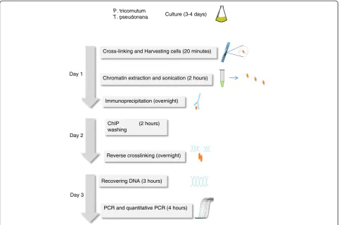
The protocol described herein (Figure 1) has taken into consideration these issues as well as the particularities of diatoms. Diatoms have the unique ability to precipitate sol-uble silicic acid into a finely patterned cell wall built from amorphous silica, and they also possess photosynthetic chloroplasts. Chromatin extraction buffers modified from Arabidopsis ChIP protocols were used for P. tricornutum and T. pseudonana which have plant features, such as chloroplasts and a cell wall. It is important in this protocol to use artificial sea water instead of natural sea water in order to control the different components of the medium such as silica. Extraction of chromatin from diatom species can be difficult because of the silica based rigid cell wall [11], which can interfere with chromatin extraction because it binds DNA. In this study, we have chosen species that have different requirements for silicon.P. tricornutumhas a facultative requirement for silicic acid, whereasT. pseudo-nana needs a silicon source to grow. A growth medium without silicic acid was used for P. tricornutum while T. pseudonana grew in medium containing a relatively high concentration of silica (~ 88 μM) reflecting the amount found in natural environment.
To preserve cell integrity, diatom cells were fixed in the growth medium prior to any handling. Formaldehyde was used for cross linking. Cross linking is a time dependent procedure and our trials have established 10 minutes as an optimal time for both P. tricornutumand T. pseudonana. Excessive cross linking might reduce antigen accessibility and sonication efficiency. For a different diatom species, we recommend 5 minutes for a start, proceed with sonication, reverse cross link and run a gel to see how much DNA is recovered and whether the size is optimal. If not, we recommend trying longer or shorter times for cross linking. Sonication time was determined after trying three dif-ferent times, 7 cycles of 25 seconds ON and 1 minute OFF, 9 and 12 cycles with 30 seconds ON and 1 minute OFF. Nine cycles gave the best range of DNA sizes which is between 220 and 1000 bp with a maximum of DNA fragments at 500 bp (Figure 2). Different dilutions of 200 ml sample containing 5 million cells per ml were used for sonication as high cell density can interfere with the efficiency of sonication. Four times dilution gave the best range of fragment sizes (Figure 2).
Peptide competition assay
Antibodies for two histone marks H3K4me2 and H3K9me2 were used for validation of the ChIP protocol. A peptide competition assay was performed to confirm the specific band reactivity of the antibodies. This is an impor-tant issue especially for genome wide studies as unspecific antibody binding will lead to non-specific signals increasing background noise and the occurrence of false positives. Both antibodies were pre-incubated with three different concentrations of the corresponding peptides (see Methods section) prior to immunobloting. For the H3K4me2 histone mark, the peptide competition assay did not detect the presence of a band for peptide H3K4me2 while a band was seen for the two other peptides, indicating the absence of competition with the other modifications of lysine 4 (Figure 3A). Three different H3K9me2 antibodies from Millipore were tested in this assay (see methods section for references). Two of them were discarded because of a lack of specificity while the third one gave better results in terms of specificity and noise (Figure 3B).
H3K4me2 is enriched in genes inP. tricornutumandT. pseudonana
Quantitative real time transcriptase polymerase chain reaction (Q-PCR) coupled with the ChIP protocol Cross-linking and Harvesting cells (20 minutes)
Chromatin extraction and sonication (2 hours)
Immunoprecipitation (overnight)
Recovering DNA (3 hours)
PCR and quantitative PCR (4 hours) P. tricornutum
T. pseudonana
Day 1
ChIP (2 hours) washing
Reverse crosslinking (overnight) Day 2
Day 3
Culture (3-4 days)
Figure 1Outline of the ChIP QPCR protocol.Timing for each step is indicated between parentheses.
7cycles 25 s
original 4x 2x
9cycles 30 s
4x
200 bp
10x dilution 7 cycles 25S
1000 bp
described herein was used to investigate the enrichment of H3K4 and H3K9 dimethylation on two genes, histone H4 and diatom phytochrome (Dph), and four transpos-able elements (TEs) in P. tricornutum and T. pseudo-nana. A clear enrichment of genes in H3K4me2 shown by a set of primers spanning promoter and gene body was demonstrated by Q-PCR (Figure 4A). Both histone H4 and Dph show significant differences in the enrich-ment in H3K4me2 between immunoprecipitated sample and mock which is the no antibody control. However, no significant differences were observed for the five TEs chosen in this study. Likewise, in T. pseudonana, the Dph and histone H4 show enrichment in H3K4me2 in both promoter and gene body, whereas the four chosen TEs (C12, C4, C19 and G1) show no enrichment in H3K4me2 (Figure 4B). Additional genes and TEs were tested and showed similar results (data not shown). Altogether, our data show a conservation of H3K4me2 location between the two diatom species and multicel-lular organisms, because genome wide studies in plants and mammals have indeed shown the presence of H3K4me2 on genes [12-14].
H3K9me2 shows a different enrichment profile inP. tricornutumandT. pseudonana
ChIP analysis of H3K9me2 in P. tricornutum revealed that TEs contain a significant enrichment for this mark. We particularly focused on two previously characterized diatom-specific copia-like retrotransposable elements known as Blackbeard (Bkb) and Surcouf (Scf) [15] Bkb was particularly enriched in H3K9me2 whileScfwas the
least enriched. Intermediate levels were observed among the remaining TEs (Figure 5A). On the other hand, the two protein-coding genes histone H4 and Dph were clearly depleted of H3K9me2 in both promoter and gene body regions (Figure 5A). Both genes were shown by us and others to be transcriptionally active ([5], unpub-lished results). It was previously shown thatBkband Scf are also transcriptionally inactive in normal growth con-ditions [15]. This is consistent with the primary function of H3K9me2 in repressing TEs in order to maintain gen-ome stability. In P. tricornutum, the ChIP assay shows that repressed TEs are marked by H3K9me2 while active genes are depleted in this mark which is similar to what has been observed in plants and mammals [16,17].
In T. pseudonana, a similar pattern of enrichment of TEs by H3K9me2 was observed (Figure 5B). The four
A
B
1 2 3 4
1 2 3 4
Figure 3Analysis of antibody specificity using peptide competition assays on western blots ofP. tricornutumnuclear extracts.(A)Upper lane contains from left to right 1μg of H3K4me2 antibody alone or with 0.25μg of one of the different modified peptides, H3K4me1, H3K4me2 and H3K4me3. Lower lane contains H4 antibody as internal loading control. (B) Upper lane contains 1μg of H3K9me2 antibody alone or with 0.25μg of one of the different modified peptides, H3K9me1, H3K9me2 and H3K9me3. Lower lane contains H4 antibody as internal loading control.
A
B
0 1 2 3 4 5 6 7 8 9% Input
Mock
H3K4me2
0 1 2 3 4 5 6
% Input
H3K4me2
Mock
Transposons Genes
Transposons Genes
tested TEs were highly enriched for H3K9me2, indica-ting a conserved profile for this mark among both diatoms. Surprisingly, some genes also showed a signifi-cant enrichment for H3K9me2, particularly at promoter regions (Figure 5B). This unusual association of H3K9me2 with active genes can be due to an intrinsic feature of the centric diatom T. pseudonana which is believed to have diverged from its distantly related pennate diatom P. tricornutum90 million years ago. Comparative genomics and analysis of molecular divergence has shown indeed that both genomes are as different as those of mammals and fish [4]. Furthermore, combinatorial patterns of anta-gonistic chromatin marks are known to occur [18-21]. In Drosophila S2 cells, clusters of transcriptionally active genes were reported to be enriched in H3K9me2 [22]. Similarly, differentiated mouse ES cells were reported to contain large domains of H3K9me2 [23]. A hypothesis is that combinatorial chromatin marks can poise genes for transcription, creating more flexible chromatin states ready to adjust for subtle changes in the microenvironment of
regulated genes [22,24]. The presence of chromatin marks known to be silent on euchromatin regions or active genes is intriguing and future analysis will be particularly impor-tant for elucidating this question.
Conclusions
The ChIP protocol described herein provides reproducible results with two different diatom species grown in different media. The quality of the procedure monitored by the no antibody control is high as no or insignificant background noise was observed. This protocol is also rapid, and can be completed within 3 days. The quantity and quality of eluted DNA from immunoprecipitation are also satisfactory. Using the described ChIP method combined with real time quantitative PCR, we have demonstrated the existence in diatoms of two types of histone modifications, H3K4me2 and H3K9me2. The H3K4me2 mark is associated with transcriptionally active genes in P. tricornutum and T. pseudonana. This result is consistent with the distribution of H3K4me2 in plants and mammals. H3K9me2 binds TEs whereas no association with genes was detected in P. tricornutum. In T. pseudonana, H3K9me2 also correlated significantly with TEs, although it also appears to bind protein-coding genes. This is different from the distribution pattern of H3K9me2 inP. tricornutum. The differences of H3K9me2 distribution pattern betweenP. tricornutumand T. pseudonana may be inherent to the genetic and/or epigenetic background of the two species which belong to pennate and centric diatoms, respectively. Gene enrichment with H3K9me2 could be further confirmed by a genome wide study of H3K9me2 distribution. Our results show that our experimental and data analysis approach are indeed highly sensitive to detect differences between the two species if they do occur. The ChIP protocol we describe can be combined with microarray or massively parallel sequencing for further genome-wide studies. This protocol has been indeed successfully combined with Illumina sequencing for studies of global histone modifications inP. tricornutum (unpublished results). Furthermore, our ChIP assay can likely be easily adapted to other eukaryotic phyto-plankton species for in vivo protein DNA interaction studies.
Acknowledgements
Funding for this work was provided by the Agence Nationale de Recherche (France) and a European Research Council Advanced Grant award to CB.
Received: 24 October 2012 Accepted: 5 December 2012 Published: 7 December 2012
References
1. Round FE, Crawford RM, Mann DG:The diatoms: biology and morphology of the genera. London, UK: Cambridge University Press; 1990.
2. Nelson DM, Treguer P, Brzezinski MA, Leynaert A, Queguiner B: Production and dissolution of biogenic silica in the ocean–revised global estimates, comparison with regional data and relationship to biogenic sedimentation.Global Biogeochem Cycles1995,9:359–372.
A
B
0 2 4 6 8 10 12 14 16 18% Input
Mock
H3K9me2
0 5 10 15 20 25 30 35
% Input
H3K9me2
Mock
Transposons Genes Transposons Genes
3. Armbrust EV, Berges JA, Bowler C, Green BR, Martinez D, Putnam NH, Zhou S, Allen AE, Apt KE, Bechner M,et al:The genome of the diatom Thalassiosira pseudonana: ecology, evolution, and metabolism. Science2004,306:79–86.
4. Bowler C, Allen AE, Badger JH, Grimwood J, Jabbari K, Kuo A, Maheswari U, Martens C, Maumus F, Otillar RP,et al:The Phaeodactylum genome reveals the evolutionary history of diatom genomes.Nature2008, 456:239–244.
5. Siaut M, Heijde M, Mangogna M, Montsant A, Coesel S, Allen A, Manfredonia A, Falciatore A, Bowler C:Molecular toolbox for studying diatom biology in Phaeodactylum tricornutum.Gene2007,406:23–35. 6. De Riso V, Raniello R, Maumus F, Rogato A, Bowler C, Falciatore A:Gene
silencing in the marine diatom Phaeodactylum tricornutum.Nucleic Acids Res2009,37:e96.
7. Gendrel AV, Lippman Z, Martienssen R, Colot V:Profiling histone modification patterns in plants using genomic tiling microarrays. Nature methods2005,2:213–218.
8. Grably M, Engelberg D:A detailed protocol for chromatin immunoprecipitation in the yeast Saccharomyces cerevisiae. Methods mol biol2010,638:211–224.
9. Saleh A, Alvarez-Venegas R, Avramova Z:An efficient chromatin immunoprecipitation (ChIP) protocol for studying histone modifications in Arabidopsis plants.Nat Protoc2008,3:1018–1025.
10. Nelson JD, Denisenko O, Bomsztyk K:Protocol for the fast chromatin immunoprecipitation (ChIP) method.Nat Protoc2006,1:179–185. 11. Kim BH, Ramanan R, Cho DH, Choi GG, La HJ, Ahn CY, Oh HM, Kim HS:
Simple, rapid and cost-effective method for high quality nucleic acids extraction from different strains of Botryococcus braunii.PLoS One2012, 7:e37770.
12. Liu CL, Kaplan T, Kim M, Buratowski S, Schreiber SL, Friedman N, Rando OJ: Single-nucleosome mapping of histone modifications in S. cerevisiae. PLoS biol2005,3:e328.
13. Mikkelsen TS, Ku M, Jaffe DB, Issac B, Lieberman E, Giannoukos G, Alvarez P, Brockman W, Kim TK, Koche RP,et al:Genome-wide maps of chromatin state in pluripotent and lineage-committed cells.Nature2007, 448:553–560.
14. Zhang X, Bernatavichute YV, Cokus S, Pellegrini M, Jacobsen SE: Genome-wide analysis of mono-, di- and trimethylation of histone H3 lysine 4 in Arabidopsis thaliana.Genome Biol2009,10:R62.
15. Maumus F, Allen AE, Mhiri C, Hu H, Jabbari K, Vardi A, Grandbastien MA, Bowler C:Potential impact of stress activated retrotransposons on genome evolution in a marine diatom.BMC Genomics2009,10:624. 16. Zhou J, Wang X, He K, Charron JB, Elling AA, Deng XW:Genome-wide
profiling of histone H3 lysine 9 acetylation and dimethylation in Arabidopsis reveals correlation between multiple histone marks and gene expression.Plant Mol Biol2010,72:585–595.
17. Tachibana M, Sugimoto K, Nozaki M, Ueda J, Ohta T, Ohki M, Fukuda M, Takeda N, Niida H, Kato H, Shinkai Y:G9a histone methyltransferase plays a dominant role in euchromatic histone H3 lysine 9 methylation and is essential for early embryogenesis.Genes Dev2002,16:1779–1791. 18. Bapat SA, Jin V, Berry N, Balch C, Sharma N, Kurrey N, Zhang S, Fang F,
Lan X, Li M,et al:Multivalent epigenetic marks confer microenvironment-responsive epigenetic plasticity to ovarian cancer cells.Epigenetics: off j DNA Methylation Soc2010,5:716–729.
19. Bernstein BE, Mikkelsen TS, Xie X, Kamal M, Huebert DJ, Cuff J, Fry B, Meissner A, Wernig M, Plath K,et al:A bivalent chromatin structure marks key developmental genes in embryonic stem cells.Cell2006,
125:315–326.
20. Roudier F, Ahmed I, Berard C, Sarazin A, Mary-Huard T, Cortijo S, Bouyer D, Caillieux E, Duvernois-Berthet E, Al-Shikhley L,et al:Integrative epigenomic mapping defines four main chromatin states in Arabidopsis.EMBO J2011, 30:1928–1938.
21. Weishaupt H, Sigvardsson M, Attema JL:Epigenetic chromatin states uniquely define the developmental plasticity of murine hematopoietic stem cells.Blood2010,115:247–256.
22. Riddle NC, Minoda A, Kharchenko PV, Alekseyenko AA, Schwartz YB, Tolstorukov MY, Gorchakov AA, Jaffe JD, Kennedy C, Linder-Basso D,et al:
Plasticity in patterns of histone modifications and chromosomal proteins in Drosophila heterochromatin.Genome Res2011,21:147–163.
23. Wen B, Wu H, Shinkai Y, Irizarry RA, Feinberg AP:Large histone H3 lysine 9 dimethylated chromatin blocks distinguish differentiated from embryonic stem cells.Nat Genet2009,41:246–250.
24. Lanzuolo C, Lo Sardo F, Orlando V:Concerted epigenetic signatures inheritance at PcG targets through replication.Cell cycle2012,11:1300.
doi:10.1186/1746-4811-8-48
Cite this article as:Linet al.:Protocol: Chromatin immunoprecipitation (ChIP) methodology to investigate histone modifications in two model diatom species.Plant Methods20128:48.
Submit your next manuscript to BioMed Central and take full advantage of:
• Convenient online submission
• Thorough peer review
• No space constraints or color figure charges
• Immediate publication on acceptance
• Inclusion in PubMed, CAS, Scopus and Google Scholar
• Research which is freely available for redistribution