R E S E A R C H A R T I C L E
Open Access
Mutational spectrum of the
SPG4
(SPAST) and
SPG3A
(ATL1) genes in Spanish patients with
hereditary spastic paraplegia
Victoria Álvarez
1*, Elena Sánchez-Ferrero
1,2, Christian Beetz
2, Marta Díaz
1, Belén Alonso
1, Ana I Corao
1,
Josep Gámez
3, Jesús Esteban
4, Juan F Gonzalo
4, Samuel I Pascual-Pascual
5, Adolfo López de Munain
6,
Germán Moris
7, Renne Ribacoba
8, Celedonio Márquez
9, Jordi Rosell
10, Rosario Marín
11, Maria J García-Barcina
12,
Emilia del Castillo
13, Carmen Benito
13, Eliecer Coto
1, the group for the study of the genetics of Spastic Paraplegia*
Abstract
Background:Hereditary Spastic Paraplegias (HSP) are characterized by progressive spasticity and weakness of the lower limbs. At least 45 loci have been identified in families with autosomal dominant (AD), autosomal recessive (AR), or X-linked hereditary patterns. Mutations in theSPAST(SPG4) andATL1(SPG3A) genes would account for about 50% of the ADHSP cases.
Methods:We defined theSPASTandATL1mutational spectrum in a total of 370 unrelated HSP index cases from Spain (83% with a pure phenotype).
Results:We found 50SPASTmutations (including two large deletions) in 54 patients and 7ATL1mutations in 11 patients. A total of 33 of theSPASTand 3 of theATL1were new mutations. A total of 141 (31%) were familial cases, and we found a higher frequency of mutation carriers among these compared to apparently sporadic cases (38% vs. 5%). Five of theSPASTmutations were predicted to affect the pre-mRNA splicing, and in 4 of them we demonstrated this effect at the cDNA level. In addition to large deletions, splicing, frameshifting, and missense mutations, we also found a nucleotide change in the stop codon that would result in a larger ORF.
Conclusions:In a large cohort of Spanish patients with spastic paraplegia,SPASTandATL1mutations were found in 15% of the cases. These mutations were more frequent in familial cases (compared to sporadic), and were associated with heterogeneous clinical manifestations.
Background
The hereditary spastic paraplegias (HSP) are character-ized by progressive spasticity and weakness of the lower limbs due to axonal degeneration in the pyramidal tract. The disease is classified as “pure” when spasticity is the only clinical finding, and as “complicated” when other clinical features (dementia, cerebellar ataxia, epilepsy, peripheral neuropathy) are also present [1]. HSP is fre-quently familial and at least 45 loci have been identified in families with autosomal dominant (AD), autosomal recessive (AR), or X-linked inheritance [2-4]. This
genetic heterogeneity partly explains the differences in disease severity, age at onset, rate of progression, and degree of disability between families. However, intrafa-milial heterogeneity is also frequent.
SPG4 (OMIM#604277) is the most common form of HSP, accounting for approximately 40% of the familial and 6-15% of the sporadic cases. TheSPAST/SPG4 gene encodes spastin, a member of the AAA (ATPase asso-ciated with various cellular activities) family of proteins, implicated in the remodeling of protein complexes upon ATP hydrolysis and the coordination of axonal microtu-bule interactions with the tubular endoplasmic reticu-lum network [5,6]. To date, >240SPASTmutations have been reported (http://www.hgmd.cf.ac.uk/ac/index.php; date of consultation August 2, 2010), mainly in patients
* Correspondence: victoria.alvarez@sespa.princast.es
1
Laboratory of Molecular Genetics -Genetic Unit, Hospital Universitario Central de Asturias, Oviedo, Spain
Full list of author information is available at the end of the article
with pure HSP [7-12]. Most of theSPASTmutations are single-nucleotide changes or small deletions/insertions, but large deletions and duplications have also been reported [13-16]. This suggested that both,
haploinsuffi-ciency and “toxic” gain of function could explain the
pathogenic mechanism ofSPASTmutations [5,17-19].
SPG3A (OMIM#606439) is the second most frequent
form of ADHSP. TheATL1/SPG3A gene encodes
atlas-tin, a protein localized in the endoplasmic reticulum and the Golgi and implicated in vesicle trafficking and neurite outgrowth [20,21]. To date, >25ATL1mutations have been reported (http://www.hgmd.cf.ac.uk/ac/index. php; date of consultation August 2, 2010), mainly mis-sense changes that supported a gain of function
patho-genic mechanism. ATL1 mutations accounted for
approximately 10% of the ADHSP families, and have
been mainly found in pure HSP [22,23].ATL1
muta-tions are also frequent in early onset (childhood or ado-lescence) cases [24-26].
This is the first report of the mutational spectrum of the SPASTand ATL1 genes in a large cohort of unre-lated HSP patients from Spain. Few studies on cohorts >200 patients have been published, and the parallel screening of both genes was rarely reported.
Methods
Patients
This study was approved by the Ethical Committee of Hospital Universitario Central Asturias (HUCA), and all the participants (patients and controls) signed an informed consent. A total of 370 non-related patients (index cases) were recruited through the Neurology Departments of several Hospitals from Spain. HSP was diagnosed by quali-fied neurologists on the basis of Harding’s criteria. Based on the clinical, radiological, and biochemical findings, cases with diseases that mimicked spastic paraplegia were excluded [27]. A total of 141 patients (38%) had a family history of HSP with a dominant inheritance pattern, and were thus classified as ADHSP cases. The absence of family history of the disease was established in 229 patients (62%) after interview on their first and second degree relatives. These cases were classified as“apparently” sporadic or with an uncertain inheritance pattern. In 177 of these the two parents were alive and did not have symp-toms consistent with HSP, while in 52 the inheritance pat-tern could not be established because no clinical data were available from relatives.
SPASTandATL1sequencing
DNA was obtained from blood leukocytes and the 17 coding exons ofSPAST(plus at least 50 bp of the flank-ing intronic sequences) were polymerase chain reaction (PCR) amplified (Additional file 1, Table S1). PCR frag-ments were purified and the two strands were sequenced
using Big Dye chemistry in an ABI3130 system (Applied Biosystems, Ca, USA). These sequences were compared
with theSPASTreference sequence (ENSG00000021574
for the genomic; ENST00000315285 for the transcript; http://www.ensembl.org). In patients who were negative for SPAST mutations and had ADHSP (n = 88) or patients without a family history of HSP and onset age ≤20 years (n = 99), we amplified and sequenced the 13
coding exons of ATL1 (reference sequences
ENSG00000198513 for the genomic, and
ENST00000358385 for the transcript; http://www. ensembl.org). All the DNA sequence variants were named following the guidelines of the Human Genome Variation Society (http://www.hgvs.org/mutnomen).
Controls screening
All the new putative mutations (not previously reported as SPASTor ATL1 mutations or polymorphisms) were screened in 400 controls. These were healthy individuals aged 21-65 years recruited through the Blood Bank of HUCA. The new nucleotide changes were genotyped through PCR-RFLP, single strand conformation analysis (SSCA), or denaturing high performance liquid chroma-tography (DHPLC), as reported [28]. Each PCR fragment containing a putative mutation gave a characteristic RFLP, SSCA or DHPLC pattern, and we could thus determine its presence/absence in the controls. When a new nucleotide change was not found among the 400 controls we determined, when it was possible, the car-rier status of all the available affected relatives to con-firm the segregation of the mutation with the disease.
MLPA analysis
The multiplex ligation dependent probe amplification (MLPA) assay with the Salsa Kit P165 HPA (MRC Hol-land, Amsterdam) was used to determine the existence
of genome copy number aberrations in the SPASTand
ATL1 genes in ADHSP patients who were negative for
sequencing mutations [13]. To investigate the conse-quences of largeSPASTdeletions at the transcript level, we isolated the mRNA from leukocytes in 10 ml of blood (Trizol reagent, Invitrogen, Carlsbad, CA, USA) and synthesised the cDNA (Quantitec Reverse Tran-scripcion kit, Quiagen, Hilden, Germany). The cDNA was amplified with primers that matched exons flanking the deletion (conditions available upon request), and the PCR products were purified and sequenced.
Analysis ofSPASTtranscripts
To determine the effect of some SPASTmutations on
Statistical analysis
The analysis of variance (SPSS 17.0 software) was used to compare the mean onset age and disease duration
between patients with mutations in SPAST and ATL1
and patients without mutations.
Results
Patients characteristics
We studied a total of 370 unrelated HSP index cases, with a mean onset age of 28 (± 21) years (range 1-77 years), and a mean disease duration of 17 (± 15) years. A total of 321 patients (87%) had a pure HSP, while 49 (13%) were complicated cases with peripheral neuropa-thy (the most frequent finding), cerebellar or cerebral atrophy, mental retardation, nystagmus, or dysarthia.
A total of 44 of the 141 patients (31%) with ADHSP had aSPASTmutation, and 10 mutations were found among the 229 (5%) cases apparently sporadic or with an uncer-tain inheritance pattern. However, in three of these patients the mutation (p.Leu426Val, p.Lys503insArg, and p.Met390Val) wasde novobecause none of the two parents was mutation carrier (the paternity was confirmed). We
found 10ATL1mutation carriers among 88 ADHSP cases
(11%), and the only apparently sporadic proband had ade novomutation (p.Gln154Glu) (Additional file 2, Figure S1).
SPASTmutations
We found 48SPASTmutations in 52 patients (Table 1).
These were missense changes in amino acids conserved between species (n = 22), nonsense mutations (n = 7), small frameshifting insertions/deletions (n = 13), nucleo-tide changes in the intronic splicing consensus sequence (n = 5), and a sense mutation in the stop codon (Figure 1). A total of 15 mutations had been previously reported, while 33 (69%) were novel.
SPASTmissense mutations out of the AAA domain
Most of the missense mutations were in the AAA domain. Two novel missense changes (p. Pro293Leu, and p. Asp613Ala) were outside of the AAA domain, where missense mutations have been rarely found. The two amino acids were evolutionary conserved. The p. Pro293Leu proband was a 51-year-old man with pro-gressive gait disorder starting at the age of 31, who has been confined to a wheelchair due to his spasticity (pure form) for the last two years. His 46-year-old sister, and one of his daughters (18 years old) also carried this mutation. They both presented brisk reflexes and Babinski’s sign, but no spasticity and an almost normal gait. The p.Asp613Ala was found in a 45 years old man with a pure phenotype affecting only the lower limbs and the first symptoms at the age of 36 years. He did not report a family history of the disease, although no relatives were studied.
We also found a new SPAST missense variant (p.
Ile328Leu) in a 15 years old male with complicated spastic paraplegia starting at the age of 18 months. This putative mutation was also found in his father’s grand-mother (asymptomatic), but the proband’s father was not available for study. Although this variant was not found among the controls and involved a conserved amino acid, a bioinformatics analysis (SIFT v2. program, Sorting Intolerant From Tolerant; http://sift.jcvi.org/) indicated a non significant change in the protein func-tion, and was thus classified as a change of uncertain pathogenic effect.
G (exon 3) affected
splicing",1,0,1,0,0pc,0pc,0pc,0pc>SPASTc.583C>G (exon 3) affected splicing
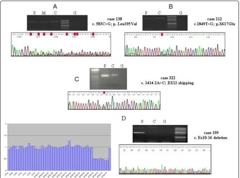
The c.583C>G (p.Leu195Val) was found in a 19 years old male with an onset at the age of 2 years and a phe-notype complicated with neuropathy. He was from a family with ADSPH in which we confirmed the segrega-tion of the disease with this mutasegrega-tion. This has been previously reported as a missense mutation, and the bioinformatic analysis (SIFT v2. program) indicated that this change would affect the protein function. However, the c.583C>G was four nucleotides from the 3’end of exon 3, and was also predicted to reduce the score of the splicing consensus site (Human Splicing Finder v. 2.4; http://www.umd.be/SSF). This mutation could thus result in an aberrant mRNA sequence. To confirm this, we isolated the mRNA from the patient’s blood
leuko-cytes and amplified the SPASTcDNA with primers for
exons 2 and 7. The last 4 nucleotides of exon 3 were missed in the transcript, that would thus be translated into an aberrant protein (Figure 2A).
SPASTsense mutation
We found a novel mutation in theSPAST stop codon,
c.1849T>G (p.X617Glu) in two non related patients with-out known affected relatives. The mutated transcript was predicted to be translated into a 46 amino acids longer protein. To determine the stability of the mRNA contain-ing this mutation, we synthesized the cDNA from RNA obtained from leukocytes of one of the patients and amplified and sequenced a PCR-fragment generated with
primers that matched exons 11 and the 3’untranslated
region (UTR). The two alleles gave equally intense sequence electropherogram peaks, suggesting that c.1849G did not increase the mRNA decay and was likely translated into a mutated protein (Figure 2B).
SPASTmutations affecting splicing
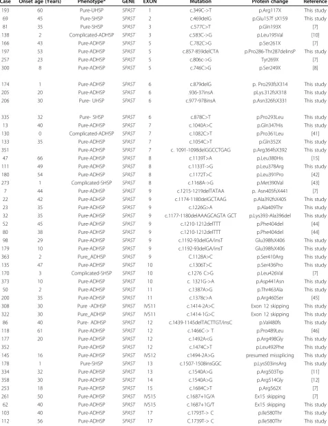
Table 1 Mutations identied in theSPASTgene in the Spanish HSP cohort
Case Onset age (Years) Phenotype* GENE EXÓN Mutation Protein change Reference
193 60 Pure-UHSP SPAST 1 c.349C->T p.Arg117X This study
69 45 Pure-SHSP SPAST 2 c.469delG p.Glu157f sX159 This study
81 35 Pure-SHSP SPAST 3 c.577C>T p.Gln193X [7]
138 2 Complicated-ADHSP SPAST 3 c.583C->G p.Leu195Val [10]
166 43 Pure-ADHSP SPAST 5 C.782C>G p.Ser261X [7]
197 53 Pure-ADHSP SPAST 5 c.857-859delCTA p.Pro286-Thr287delinsP This study
257 23 Pure-ADHSP SPAST 5 c.806c->G Tyr269X [7]
300 8 Pure-ADHSP SPAST 5 c.746C>G p.Ser249X [8]
174 1 Pure-ADHSP SPAST 6 c.879delG p. Pro293fsX314 This study
205 20 Pure-ADHSP SPAST 6 .936-37insA pLys.312fsX318 This study
206 30 Pure- UHSP SPAST 6 c.977-978insA p.Asn326fsX331 This study
335 32 Pure- SHSP SPAST 6 c.878C>T p.Pro293Leu This study
13 40 Pure-ADHSP SPAST 7 c.1040A>C p.Gln347His This study
130 0 Complicated-ADHSP SPAST 7 c.1082C>T p.Pro361Leu [41]
133 35 Pure-ADHSP SPAST 7 c.1054C>T p.Gln352X This study
351 Pure-ADHSP SPAST 7 c. 1091-1098delGGCCTGAG p.Arg364fsX392 This study
47 66 Pure-ADHSP SPAST 8 c.1139T>A p.Leu380His [15]
111 49 Pure-ADHSP SPAST 8 c.1133T->G p.Leu378Arg This study
180 54 Pure-ADHSP SPAST 8 c.1172T>C p.Leu391Pro [42]
273 1 Complicated-SHSP SPAST 8 c.1168A->G p.Met390Val [43]
7 44 Pure-ADHSP SPAST 9 c.1215-1219delTATAA p. Asn405fsX441 [7]
22 42 Pure-ADHSP SPAST 9 c.1174-1180delGCTAAG p.Ala392fsX405 This study
23 35 Pure-ADHSP SPAST 9 c.1226G>A p.Ala409Thr This study
32 35 Pure-ADHSP SPAST 9 c.1177-1180delAAAGCAGTA GCT p.Lys393-Ala396del This study
52 45 Pure-ADHSP SPAST 9 c.1210-1212delTTT p.Phe404del [44]
80 38 Pure-ADHSP SPAST 9 c.1210-1212delTTT p.Phe404del [44]
98 29 Pure-ADHSP SPAST 9 c.1192-93delGA/insT Glu398fsX406 This study
179 10 Pure-ADHSP SPAST 9 c.1192-93delGA/insT Glu398fsX406 This study
363 2 Pure_ADHSP SPAST 9 C.1128A>C p.Ser410Arg This study
135 47 Pure-ADHSP SPAST 10 c.1306T>C p.Ser436Pro This study
170 3 Complicated-SHSP SPAST 10 c.1276 C>G p.Leu426Val [7]
373 10 Pure-ADHSP SPAST 10 c. 1321G->A p.Asp441Asn This study
50 2 Pure-ADHSP SPAST 11 c.1387A>G p.Thr463Ala This study
200 35 Pure-ADHSP SPAST 11 c.1378c>A p.Arg460Ser [45]
308 30 Pure -ADHSP SPAST IVS11 c.1414-2A>C Exon 12 skipping This study
322 30 Pure_ADHSP SPAST IVS11 c.1414-1G>C Exon 12 skipping This study
86 40 Pure- ADHSP SPAST 12 c.1439-1145delTACTTGT/insC p.Val480fs This study
118 61 Pure-ADHSP SPAST 12 c.1466C-> T p.Pro489Leu [46]
177 20 Pure-ADHSP SPAST 12 c.1492A<G p.Arg498Gly This study
352 Pure-ADHSP SPAST 12 c.1474C>T p.Leu492Phe This study
145 16 Pure-ADHSP SPAST IVS12 c1494-2A>G presumed missplicing This study
178 1 Pure-SHSP SPAST 13 c.1507-1508insGGC p.Lys503insArg This study
334 32 Pure-ADHSP SPAST 13 c.1540A>G p.Arg503Trp [11]
358 30 Pure-ADHSP SPAST 14 c.1540A>G p.Arg514Gly [12]
253 18 Pure-ADHSP SPAST 15 c.1684C>T p.Arg562X [7]
261 50 Pure-ADHSP SPAST IVS15 c.1687+1G/A Ex15 skipping [7]
62 40 Pure-ADHSP SPAST IVS15 c.1687+1G/T Ex15 skipping This study
103 40 Pure-ADHSP SPAST 17 c.1793T-> C p.Ile580Thr This study
Prediction by Neural Network; http://www.fruitfly.org/ seq_tools/splice.html) indicated that the five changes would affect the pre-mRNA splicing. We could synthe-size the cDNA from four of the patients, that was ampli-fied with primers that matched exons 7 and 17, and exons 11 and 17. In all these cases two PCR-fragments were amplified, and the longer fragment corresponded to the wild type transcript while the shorter resulted from an exon skipping. In this way, we confirmed the existence of defective transcripts (Figure 2C).
LargeSPASTdeletions
We performed MLPA analysis in the 77 ADHSP cases who were negative forATL1andSPASTsequencing mutations. Two patients (2.5%) showed a significantly reduced ampli-fication signal for exons 10-16, or exons 6-7 ofSPAST. In the patient with a putative deletion of exons 10-16 we amplified the cDNA with primers that matched exons 9 and 17, and confirmed the skipping of exons 10-16 (Figure 2D). In this pedigree, the age at onset (third decade) was similar in the three affected members who were studied, and the oldest patient (an 81-year-old man) had been con-fined to a wheelchair for the previous five years.
ATL1mutations
TheATL1 exons were sequenced in a total 88 patients
with ADHSP and in 99 patients without a family history
of HSP and an onset age ≤20 years. We found seven
mutations and a variant with uncertain pathogenic effect in 11 patients (Table 2). Three were novel missense changes (p.His256Asp, p.Gln154Glu, and p.Phe413Val), while 5 had been reported. The p.Arg239Cys was a recurrent mutation found in 3 non related patients.
Mutations p.Gln154Glu, (c.460 C>G), p. His256Asp (c.766C>A), and p.Phe413Val (c.1237T>G) are located in the guanilate binding protein domains. The p.Gln154Glu mutation was found in a patient with a pure phenotype and onset of symptoms in childhood. His son showed gait problems at the age of two years. The p.His256Asp patient was a 45 years old woman with the onset of symptoms at the age of 3 years and a progressive spastic paraplegia (Additional file 2, Figure S1). In her family, we identified mutation carriers who were asymptomatic at ages of 60
and 63 years, and this could thus be classified as a variant with uncertain pathogenic effect. Mutation p.Phe413Val was found in a patient with an onset age of 6 years and a complicated phenotype (neuropathy). Her mother was affected, but she was death and we could not confirm whether carried the mutation. None of the 77 ADHSP patients studied through MLPA showed evidence for large ATL1deletions.
SPASTand ATL1polymorphisms
We found several polymorphisms in the two genes (Additional file 1, Table S3). All the new polymorphisms were intronic, or exonic silent amino acid changes with
one exception: c.844 T>A in exon 5 of SPAST (p.
Ser282Thr). This missense change affected an amino conserved between species and was not found in any of the 400 controls. Although it could be considered a putative mutation, was in a patient with the p.Ser261X mutation (located also in exon 5). Segregation analysis to determine whether the two nucleotide changes were in different chromosomes was not possible. However, the electrophoretic pattern of bands after double restric-tion enzyme digesrestric-tion (MnlI+MboI) of the exon 5
frag-ment indicated that the mutation was in cis with the
c.844 A allele (data not shown). This suggested that this was likely a rare variant, rather than an HSP mutation. Two missense polymorphisms that have been proposed as modifiers of the clinical phenotype (p.Ser44Leu and p.Pro45Leu) were not found in our patients [29].
Genotype-phenotype correlation
The phenotype associated withSPASTandATL1
muta-tions was pure in 48 (87%) and 10 (90%) of the index patients, respectively. We analysed all the available rela-tives of the 63 index cases with mutations, and identified a total of 54SPASTand 15ATL1mutation carriers. A total
of 13 mutation carriers (10 forSPASTand 3 forATL1
gene) were asymptomatic at the time of our analysis. The mean onset age of the disease (index cases and relatives) was 34.5 (± 17.72) years forSPASTmutation carriers, and 7.67 (± 5.9 years) forATL1mutation carriers (p < 0.001).
A difference was also observed betweenATL1mutation
carriers and patients without SPAST/ATL1mutations
Table 1 Mutations identied in theSPASTgene in the Spanish HSP cohort(Continued)
212 35 Pure-ADHSP SPAST 17 c.1849T-> G p.X617Glu This study
286 54 Complicated-UHSP SPAST 17 c.1849T-> G p.X617Glu This study
310 36 Pure- UHSP SPAST 17 C.1838A-> C p.Asp613Ala This study
EXON DELETIONS
199 20 Pure-ADHSP SPAST - EX10-16 deletion [14]
225 14 Pure-ADHSP SPAST - Ex 6-7 deletion This study
(7.67 ± 5.9 vs. 27.78 ± 20.238; p < 0.001). The mean dura-tion of symptoms at the time of examinadura-tion did not differ
between patients withSPAST(14.5 ± 11.1) andATL1
(18.78 ± 15.057) mutations. No difference in disease
dura-tion was observed betweenSPASTorATL1mutation
car-riers and patients without mutations.
Discussion
SPASTmutations (including large gene deletions) were found in 15% (54/370) of the patients, but this frequency increased to 31% (44/141) among patients with ADHSP. Most of theSPASTmutations were novel, and this was in agreement with previous reports that described a high rate of private mutations in this gene [30,10,15]. We
found anATL1 mutation in 6% of the patients studied
for this gene, but this frequency could be underestimated because we did not include ADHSP patients with a SPAST mutation or patients with sporadic/uncertain HSP and an onset age >20 years. However, we think that
theATL1mutation rate should be right because double
mutated patients (SPAST + ATL1) have not been
reported, andATL1mutations were rare among
non-ADHSP patients with an onset age >20 years [22,25,26]. Considering the ADHSP cases, the mutational screening ofSPASTandATL1identified a total of 54 mutation car-riers, a frequency (38%) within the range reported in other populations [10,12,31,32]. Also in agreement with previous reports, the frequency ofSPASTandATL1mutation car-riers was much lower among patients with sporadic/ uncertain HSP [12]. Four of the sporadic cases had ade novomutation. A high rate ofde novomutational events
forSPASTandATL1has been previously described, indi-cating that sporadic cases should also be screened for mutations in these genes after exclusion of other major neurological causes of spasticity [15,23,33-35].
SPASTmutations are mostly restricted to the AAA protein domain. It is thus remarkable that three of the missense mutations in our patients were out of this domain (p.Leu195Val, p.Pro293Leu, and p.Asp613Ala).
In the case of ATL1all the mutations were missense
changes in the GTPase and transmembrane domains of atlastin, and would disrupt the normal protein folding and oligomerization [20,21]. TheSPASTc.C583G variant was previously reported as a missense mutation (p. Leu195Val) [10]. However, this change was within the consensus splicing sequence of exon 3 and we confirmed its effect on pre-RNA splicing. Nucleotide changes in the last nucleotides of exons could result in both, normal and splicing defective transcripts [36-38]. Although we found an aberrant transcript we could not define whether the c.583 G resulted also in normal transcripts.
In theSPASTgene we also found one mutation in the
stop codon (c.1849T>G; X617Glu), that would result in the translation of 46 amino acid beyond the stop codon. To our knowledge, this is the first sense mutation reported in this gene. The abolishment of a stop codon and the appearance of a longer ORF has been found in other her-editary diseases, such as the British Familial Dementia caused by a sense mutation in theITM2Bgene [39].
We performed MLPA analysis in 78 patients with
ADHSP and we found two large deletions in theSPAST
gene, which accounts for 2.5% of patients. In a recent
Figure 2A) The cDNA from patient 138 (carrier of c.583C>G in exon 3 ofSPAST) was synthesised from leukocytes mRNA. The sequencing of the fragment amplified with primers that matched exons 2 and 7 showed the deletion of the last 4 nucleotides of exon 3.B)
The cDNA from patient 212 (X617Glu) was amplified with primers for exons 11 and 17 ofSPAST, and the sequence showed similar electropherogram peaks for the two alleles.C)The cDNA of patient 322 was amplified with primers for exons 7 and 17 ofSPAST, and the sequence showed the effect on splicing of the c.1414-1G>C mutation.D)MLPA of theSPASTgene in patient 199, showing the exons 10-16 deletion. The cDNA was amplified with primers that matched exons 9 and 17, and two PCR fragments were obtained. Sequencing of the smaller fragment confirmed the absence of exons 10 to 16. P = patient (cDNA); M = Patient’s mother’s (cDNA); C = Control (cDNA); G = Genomic DNA
Table 2 Mutations in theATL1gene
Case onset age (Years) Phenotype/Inheritance GEN Exon Mutation Protein Reference
57.1 1 Pure- SHSP ATL1 4 c.460 C>G p.Gln154Glu This study
97 8 Pure-ADHSP ATL1 7 c.715C>T p.Arg239Cys [47]
102 4 Pure-ADHSP ATL1 7 c.715C>T p.Arg239Cys [47]
220 3 Pure-ADHSP ATL1 7 c.715C>T p.Arg239Cys [47]
279 5 Pure-ADHSP ATL1 7 c.715C>T p.Arg239Cys [47]
64 10 Pure-ADHSP ATL1 8 c.757 G>A p.Val253Ile [25]
159 5 Pure-ADHSP ATL1 12 c.1483c>T p.Arg495Trp [25]
110 17 Pure-ADHSP ATL1 12 c.1319A->C p.Asn440Thr [25]
232 6 Pure-ADHSP ATL1 12 c.1237T->G p.Phe413Val This study
work Shoukier et al. reported a similar frequency for this type of mutation [12]. However, previous studies reported a much higher frequency (18-23%) [13,15]. Additional stu-dies are thus necessary to define the frequency of large deletions/duplications in the aetiology of HSP.
Finaly,SPASTand ATL1mutations have been
asso-ciated with variable penetrance, leading to heterogeneous HSP phenotypes in terms of onset age and clinical symp-toms (pure vs. complicated) [40,15]. This heterogeneity could be partly explained by different mutated genes. As discussed above, we observed a significantly higher
fre-quency of familial dominant HSP amongATL1patients,
compared to SPASTmutation carriers, and pure HSP
was also more frequent among the patients withATL1
mutations. However, phenotypic variability was also com-mon acom-mong mutation carriers from the same family.
Conclusions
In conclusion, we reported the mutational spectrum of theSPASTandATL1genes in a large cohort of Spanish patients with spastic paraplegia. We found a mutation in 15% of the cases, and a frequency of mutation carriers significantly higher among ADHSP compared to spora-dic cases. Thus, the genetic screening should be more relevant in patients (pure and complicated phenotypes) with a family history of the disease. However, the fact that a significant number of apparently sporadic cases had a mutation suggested that these patients should not be excluded from the genetic study. The mutational report could be of limited value to predict the pheno-type associated to these mutations, as demonstrated by the heterogeneous behavior of most of the mutations.
Note
* Manuel Amorín, Eugenia Marzo-Sola, Carlos H. Lahoz, Pia Gallano, Concepción Alonso-Cerezo, Rafael Palencia-Luaces, Eduardo Gutiérrez- Rivas, Rogelio Simón, Loreto Martorell, Eduardo López-Laso, José M. Asensi, Luis Hernández- Echebarria, Yolanda Morgado, Alonso González-Masegosa, Juan J. Garcia- Peñas, Irene Catalina-Alvarez, José L. Muñoz-Blanco, Miguel Fernán-dez-Burriel, Juan B. Espinal, Mariano Aparicio- Blanco, Jon Infante, María Vázquez-Espinar, Elena Maside
Additional material
Additional file 1: Supplementary tables and supplementary figure 1 legendl for Alvarez et al. This file contains information about PCR primers, polymorphisms detected and legend of supplementary figure.
Additional file 2: supplementary FIGURE1 for Alvarez et al. This file contains a figure showing several examples of mutations detected.
Acknowledgements
This work was supported by grants from Spanish Fondo de Investigaciones Sanitarias PI08/0915 (European FEDER founds) to V.A. E.S-F. was a fellowship from FICYT-Principado de Asturias. The authors thank the patients and family members for their participation in this study. Authors wish to thank Fundación Parkinson Asturias and Obra Social CAJASTUR for their support.
Author details
1Laboratory of Molecular Genetics -Genetic Unit, Hospital Universitario
Central de Asturias, Oviedo, Spain.2Institute for Clinical Chemistry and Laboratory Medicine, University Hospital Jena, Jena, Germany.3Neurology
Department, Hospital Universitari Vall d’Hebron. Univ. Autonoma Barcelona, Spain.4Neurology Department, Hospital 12 de Octubre, Madrid, Spain. 5Pediatric Neurology Department, University Hospital La Paz, Madrid, Spain. 6Neurology Department, Hospital Donostia-Instituto Biodonostia-Ciberned,
San Sebastián, Spain.7Neurology Department, Hospital San Agustín, Aviles,
Spain.8Neurology Department, Hospital Alvarez-Buylla, Mieres, Spain.
9Neurology Department, Hospital Universitario Virgen del Rocio, Sevilla,
Spain.10Department of Genetics, Hospital Universitari Son Dureta, Palma de Mallorca, Spain.11HGenetics Unit, Hospital Universitario Puerta del Mar,
Cádiz, Spain.12Genetics Department, Hospital de Basurto, Bilbao, Spain.
13Genetics Unit, Hospital Universitario Carlos Haya, Málaga, Spain. Authors’contributions
All the authors contributed to this work by recruiting the patients, obtaining the clinical and analytical information, or performing the laboratory work. VA designated the work and analyzed the results. VA, ES-F, CB, M., BA, and AIC performed all the genetic analysis. VA and EC wrote the manuscript. All authors have read and approved the submission of this manuscript.
Competing interests
The authors declare that they have no competing interests.
Received: 9 June 2010 Accepted: 8 October 2010 Published: 8 October 2010
References
1. Harding AE:Classification of the hereditary ataxias and paraplegias.
Lancet1983,1:1151-1155.
2. Stevanin G, Ruberg M, Brice A:Recent advances in the genetics of spastic paraplegias.Curr Neurol Neurosci Rep2008,8:198-210.
3. Salinas S, Proukakis C, Crosby A, Warner TT:Hereditary spastic paraplegia: clinical features and pathogenetic mechanisms.Lancet Neurol2008, 7:1127-38.
4. Dursun U, Koroglu C, Kocasoy Orhan E, Ugur SA, Tolun A:Autosomal recessive spastic paraplegia (SPG45) with mental retardation maps to10q24.3-q25.1.Neurogenetics2009,10:325-31.
5. Errico A, Ballabio A, Rugarli EI:Spastin, the protein mutated in autosomal dominant hereditary spastic paraplegia, is involved in microtubule dynamics.Hum Mol Genet2002,11:153-63.
6. Park SH, Zhu PP, Parker RL, Blackstone C:Hereditary spastic paraplegia proteins REEP1, spastin, and atlastin-1 coordinate microtubule interactions with the tubular ER network.J Clin Invest2010,120: 1097-110.
7. Fonknechten N, Mavel D, Byrne P, Davoine CS, Cruaud C, Bönsch D,et al: Spectrum of SPG4 mutations in autosomal dominant spasticparaplegia.
Hum Mol Genet2000,9:637-44.
8. Sauter S, Miterski B, Klimpe S, Bönsch D, Schöls L, Visbeck A,et al:Mutation analysis of the spastin gene (SPG4) in patients in Germany with autosomal dominant hereditary spastic paraplegia.Hum Mutat2002, 20:127-32.
9. Patrono C, Scarano V, Cricchi F, Melone MA, Chiriaco M, Napolitano A,et al: Autosomal dominant hereditary spastic paraplegia:DHPLC-based mutation analysis of SPG4 reveals eleven novel mutations.Hum Mutat
2005,25:506.
10. Crippa F, Panzeri C, Martinuzzi A, Arnoldi A, Redaelli F, Tonelli A,et al:Eight novel mutations in SPG4 in a large sample of patients with hereditary spastic paraplegia.ArchNeurol2006,63:750-5.
11. Depienne C, Tallaksen C, Lephay JY, Bricka B, Poea-Guyon S, Fontaine B,
their spectrum is different from that observed in familial cases.J Med Genet2006,43:259-65.
12. Shoukier M, Neesen J, Sauter SM, Argyriou L, Doerwald N, Pantakani DV,
et al:Expansion of mutation spectrum, determination of mutation cluster regions and predictive structural classification of SPAST mutations in hereditary spastic paraplegia.Eur J Hum Genet2009, 17:187-94.
13. Beetz C, Nygren AO, Schickel J, Auer-Grumbach M, Bürk K, Heide G,et al: High frequency of partial SPAST deletions in autosomal dominant hereditary spastic paraplegia.Neurology2006,67:1926-30.
14. Depienne C, Fedirko E, Forlani S, Cazeneuve C, Ribaï P, Feki I,et al:Exon deletions of SPG4 are a frequent cause of hereditary spastic paraplegia.
J Med Genet2007,44:281-284.
15. Erichsen AK, Inderhaug E, Mattingsdal M, Eiklid K, Tallaksen CM:Seven novelmutations and four exon deletions in a collection of Norwegian patients with SPG4 hereditary spastic paraplegia.Eur J Neurol2007,14:809-14. 16. Mitne-Neto M, Kok F, Beetz C, Pessoa A, Bueno C, Graciani Z,et al:A
multi-exonic SPG4 duplication underlies sex-dependent penetrance of hereditary spastic paraplegia in a large Brazilian pedigree.Eur J Hum Genet2007,15:1276-1279.
17. Patrono C, Casali C, Tessa A, Cricchi F, Fortini D, Carrozzo R,et al:Missense and splice site mutations in SPG4 suggest loss-of-function in dominant spastic paraplegia.J Neurol2002,249:200-5.
18. Charvin D, Cifuentes-Diaz C, Fonknechten N, Joshi V, Hazan J, Melki J,et al: Mutations of SPG4 are responsible for a loss of function of spastin, an abundant neuronal protein localized in the nucleus.Hum Mol Genet
2003,12:71-8.
19. Solowska J, Garbern JY, Baas PW:Evalution of loss of function as an explanation for SPG4-based hereditary spastic paraplegia.Hum Mol Genet2010,19:1-13.
20. Namekawa M, Muriel MP, Janer A, Latouche M, Dauphin A, Debeir T,et al: Mutations in the SPG3A gene encoding the GTPase atlastin interfere with vesicle trafficking in the ER/Golgi interface and Golgi morphogenesis.Mol Cell Neurosci2007,35:1-13.
21. Muriel MP, Dauphin A, Namekawa M, Gervais A, Brice A, Ruberg M: Atlastin-1, the dynamin-like GTPase responsible for spastic paraplegia SPG3A, remodels lipid membranes and may form tubules and vesicles in the endoplasmic reticulum.J Neurochem2009,110:1607-1616.
22. Sauter SM, Engel W, Neumann LM, Kunze J, Neesen :Novel mutations in the Atlastin gene (SPG3A) in families with autosomal dominant hereditary spastic paraplegia and evidence for late onset forms of HSP linked to the SPG3A locus.Hum Mutat2004,23:98.
23. Ivanova N, Claeys KG, Deconinck T, Litvinenko I, Jordanova A, Auer-Grumbach M,et al:Hereditary spastic paraplegia 3A associated with axonal neuropathy.Arch Neurol2007,64:706-13.
24. Abel A, Fonknechten N, Hofer A, Dürr A, Cruaud C, Voit T,et al:Early onset autosomal dominant spastic paraplegia caused by novel mutations in SPG3A.Neurogenetics2004,5:239-43.
25. Dürr A, Camuzat A, Colin E, Tallaksen C, Hannequin D, Coutinho P,et al: Atlastin1 mutations are frequent in young-onset autosomal dominant spastic paraplegia.Arch Neurol2004,61:1867-72.
26. Namekawa M, Ribai P, Nelson I, Forlani S, Fellmann F, Goizet C,et al:SPG3A is the most frequent cause of hereditary spastic paraplegia with onset before age 10 years.Neurology2006,66:112-4.
27. Tallaksen CM, Dürr A, Brice A:Recent advances in hereditary spastic paraplegia.Curr Opin Neurol2001,14:457-463.
28. Randall LP, Coldham NG, Woodward MJ:Detection of mutations in Salmonella enterica gyrA, gyrB, parC and parE genes by denaturing high performance liquid chromatography (DHPLC) using standard HPLC instrumentation.J Antimicrob Chemother2005,56:619-23.
29. Svenson IK, Kloos MT, Gaskell PC, Nance MA, Garbern JY, Hisanaga S,et al: Intragenic modifiers of hereditary spastic paraplegia due to spastin gene mutations.Neurogenetics2004,5:157-64.
30. Proukakis C, Auer-Grumbach M, Wagner K, Wilkinson PA, Reid E, Patton MA,
et al:Screening of patients with hereditary spastic paraplegia reveals seven novel mutations in the SPG4 (Spastin) gene.Hum Mutat2003, 21:170.
31. McDermott CJ, Burness CE, Kirby J, Cox LE, Rao DG, Hewamadduma C,et al: Clinical features of hereditary spastic paraplegia due to spastin mutation.Neurology2006,67:45-51.
32. Loureiro JL, Miller-Fleming L, Thieleke-Matos C, Magalhães P, Cruz VT, Coutinho P,et al:Novel SPG3A and SPG4 mutations in dominant spastic paraplegia families.Acta Neurol Scand2009,119:113-8.
33. Rainier S, Sher C, Reish O, Thomas D:Fink JK De novo occurrence of novelSPG3A/atlastin mutation presenting as cerebral palsy.Arch Neurol
2006,63:445-447.
34. Scuderi C, Fichera M, Calabrese G, Elia M, Amato C, Savio M, Borgione E,
et al:Posterior fossa abnormalities in hereditary spastic paraparesis with spastin mutations.J Neurol Neurosurg Psychiatry2009,80:440-443. 35. Molon A, Montagna P, Angelini C, Pegoraro E:Novel spastin mutations
and their expression analysis in two Italian families.Eur J Hum Genet
2003,11:710-713.
36. Svenson IK, Ashley-Koch AE, Gaskell PC, Riney TJ, Cumming WJ,
Kingston HM,et al:Identification and expression analysis of spastin gene mutations in hereditary spastic paraplegia.Am J Hum Genet2001, 68:1077-85.
37. Riveira-Munoz E, Chang Q, Godefroid N, Hoenderop JG, Bindels RJ, Dahan K,
et al:Transcriptional and functional analyses of SLC12A3 mutations: new clues for the pathogenesis of Gitelman syndrome.J Am Soc Nephrol
2007,18:1271-83.
38. Coto E, Arriba G, García-Castro M, Santos F, Corao AI, Díaz M,et al:Clinical and analytical findings in Gitelman’s syndrome associated with homozygosity for the c.1925 G>A SLC12A3 mutation.Am J Nephrol2009, 30:218-21.
39. Vidal R, Frangione B, Rostagno A, Mead S, Révész T, Plant G,et al:A stop-codon mutation in the BRI gene associated with familial British dementia.Nature1999,399:776-81.
40. D’Amico A, Tessa A, Sabino A, Bertini E, Santorelli FM, Servidei S: Incomplete penetrance in an SPG3A-linked family with a new mutation in the atlastin gene.Neurology2004,62:2138-9.
41. Chinnery PF, Keers SM, Holden MJ, Ramesh V, Dalton A:Infantile hereditary spastic paraparesis due to codominant mutations in the spastin gene.
Neurology2004,63:710-2.
42. Bertelli M, Cecchin S, Lorusso L, Sidoti V, Fabbri A, Lapucci C,et al: Identification of a novel mutation in the spastin gene (SPG4) in an Italian family with hereditary spastic paresis.Panminerva Med2006, 48:193-7.
43. Tang B, Zhao G, Xia K, Pan Q, Luo W, Shen L, Long Z,et al:Three novel mutations of the spastin gene in Chinese patients with hereditary spastic paraplegia.Arch Neurol2004,61:49-55.
44. Park SY, Ki CS, Kim HJ, Kim JW, Sung DH, Kim BJ,et al:Mutation analysis of SPG4 and SPG3A genes and its implication in molecular diagnosis of Korean patients with hereditary spastic paraplegia.Arch Neurol2005, 62:1118-1121.
45. Braschinsky M, Tamm R, Beetz C, Sachez-Ferrero E, Raukas E, Lüüs SM,et al: Unique spectrum of SPAST variants in Estonian HSP patients: presence of benign missense changes but lack of exonic rearrangements.BMC Neurol2010,10:17.
46. Meijer IA, Hand CK, Cossette P, Figlewicz DA, Rouleau GA:Spectrum of SPG4 mutations in a large collection of North American families with hereditary spastic paraplegia.Arch Neurol2002,59:281-286.
47. Wilkinson PA, Hart PE, Patel H, Warner TT, Crosby AH:SPG3A mutation screening in English families with early onset autosomal dominant hereditary spastic paraplegia.J Neurol Sci2003,216:43-45.
Pre-publication history
The pre-publication history for this paper can be accessed here: http://www.biomedcentral.com/1471-2377/10/89/prepub
doi:10.1186/1471-2377-10-89
Cite this article as:Álvarezet al.:Mutational spectrum of theSPG4
(SPAST) andSPG3A(ATL1) genes in Spanish patients with hereditary