M E T H O D O L O G Y
Open Access
A simple and efficient method for isolating small
RNAs from different plant species
Flor de Fátima Rosas-Cárdenas
1, Noé Durán-Figueroa
1, Jean-Philippe Vielle-Calzada
1, Andrés Cruz-Hernández
2,
Nayelli Marsch-Martínez
1, Stefan de Folter
1*Abstract
Background:Small RNAs emerged over the last decade as key regulators in diverse biological processes in eukaryotic organisms. To identify and study small RNAs, good and efficient protocols are necessary to isolate them, which sometimes may be challenging due to the composition of specific tissues of certain plant species. Here we describe a simple and efficient method to isolate small RNAs from different plant species.
Results:We developed a simple and efficient method to isolate small RNAs from different plant species by first comparing different total RNA extraction protocols, followed by streamlining the best one, finally resulting in a small RNA extraction method that has no need of first total RNA extraction and is not based on the commercially available TRIzol®Reagent or columns. This small RNA extraction method not only works well for plant tissues with high polysaccharide content, like cactus, agave, banana, and tomato, but also for plant species like Arabidopsis or tobacco. Furthermore, the obtained small RNA samples were successfully used in northern blot assays.
Conclusion:Here we provide a simple and efficient method to isolate small RNAs from different plant species, such as cactus, agave, banana, tomato, Arabidopsis, and tobacco, and the small RNAs from this simplified and low cost method is suitable for downstream handling like northern blot assays.
Introduction
Over the last decade small RNAs (sRNAs) have arisen as key regulators of diverse biological processes in eukaryo-tic organisms, including for instance development or stress responses, among others (reviewed in: [1-4]). sRNAs are around 20-30 nucleotide (nt) long, and guide regulatory processes at the RNA or DNA level.
The presence of endogenous sRNAs is now reported for many model plants and various non-model species (e.g. [5,6]), and elucidating the functions for many of these sRNAs will be a challenge in the near future. Two major classes of sRNAs are microRNAs (miRNAs; 21 and 24 nt long) and small-interfering RNAs (siRNAs; 18-24 nt long), with the latter being the most abundant, though, functionally less understood.
Various protocols are available for sRNA isolation from plants (e.g. [6-14]), though most of them are used
for model plant species. Normally, these protocols start with total RNA isolation, followed by the isolation or separation of the low molecular weight RNA species (LMW RNA), containing the sRNAs. The most com-monly used protocol is based on the extraction of total RNA using TRIzol® Reagent [15,16] followed by precipi-tation of LMW RNAs using polyethylene glycol, and finally resulting in RNA species less than 300 nt long. Another protocol for total RNA isolation from tissues with higher contents of polysaccharides is the cetyltri-methylamonium bromide (CTAB) method [7,8]. These are useful protocols, but sometimes it is possible that these protocols do not work well for other plants species or specific tissues, or become quite labor-intensive due to difficult handling and the need of extra precipitation steps.
This motivated us to investigate whether it would be possible to find a generic protocol to isolate sRNAs, which would also work for plant tissues with a high polysaccharide content. In this report, a sRNA isolation method is presented that works efficiently for different plant species like cactus, banana and tomato fruits, and * Correspondence: [email protected]
1Laboratorio Nacional de Genómica para la Biodiversidad (LANGEBIO), CINVESTAV-IPN, Km. 9.6 Libramiento Norte, Carretera Irapuato-León, CP 36821 Irapuato, Guanajuato, México
Full list of author information is available at the end of the article
agave leaves, but also for Arabidopsis and tobacco with less polysaccharide content. The method presented here is not based on the use of the TRIzol® Reagent or com-mercial columns and omits the total RNA isolation step and, therefore, becomes a simpler and cheaper sRNA isolation method for plants.
Materials and methods Plant material
Prickly pear (Opuntia robusta) cactus pads and floral buds were collected at INIFAP (Mexican National Insti-tute of Forestry, Agriculture, and Livestock Research) Campo Experimental Norte de Guanajuato, in San Luis de la Paz, Gto., Mexico. Agave leaves were collected at the campus of CINVESTAV, Irapuato, Gto., Mexico. Arabidopsis thaliana(ecotype Ws-3) and Nicotiana tabacumplants were grown under conventional long day growth conditions (22°C, 16 hours of light). Banana and tomato fruits were purchased at the local market. The samples were sliced, ground to a fine powder in a mortar with liquid nitrogen and stored at -80°C until further use.
Buffers and solutions
•LiCl extraction buffer 100 mM Tris-HCl, pH 9.0
1% SDS 100 mM LiCl 10 mM EDTA
•TBE buffer (1x)
0.9 M Tris-HCl, pH 8.0 0.9 M Boric Acid 2 mM EDTA, pH 8.0
•Loading buffer 98% formamide
10 mM EDTA, pH 8.0 1 mg/ml xylene cyanol 1 mg/ml bromophenol blue
•Polyacrylamide stock solution
12.5% polyacrylamide (Acrylamide:bisacrylamide 19:1; Biorad)
0.5× TBE buffer, pH 8.0 7 M Urea
•Denaturing polyacrylamide gel (for one gel) 5 ml of polyacrylamide stock
25μl of 20% APS (Ammonium persulfate)
5μl TEMED (N, N, N’, N’-Tetramethylethylenediamine)
•Staining solution
0.001% SYBR Gold (Invitrogen) 0.5× TBE buffer, pH 8.0 Other solutions
•3 M sodium acetate, pH 5.2 •absolute ethanol
•phenol, pH 8.0
•chloroform-isoamyl alcohol (24:1; v/v)
•phenol-chloroform-isoamyl alcohol (25:24:1; v/v/v) •5 M NaCl solution
•40% polyethylene glycol 8000 solution (PEG 8000) •DEPC treated (0.05%) water
For northern blot analysis
•EDC fixation solution (24 ml)
245μl of 12.5 M methylimidazole, pH 8.0
0.5 g 1-ethyl-3-(3-dimethylaminopropyl) carbodiimide (EDC)
•Hybridization solution (100 ml) 10 g dextran sulphate
5 ml of 20% SDS 20 ml of 5 M NaCl
5 ml of 1 M Tris-HCl, pH 7.5
•Wash solution 2× SSC
0.1% SDS
Protocol
Small RNA extraction
1. Place 0.1 g of pulverized frozen tissue in a 1.5 ml microcentrifuge tube and add 500μl of LiCl extraction buffer and 500μl of phenol pH 8.0.
2. Shake or mix well using a vortex for 1 min. Place each sample on ice until all samples are ready.
3. Incubate tubes for 5 min at 60°C.
4. Centrifuge for 10 min in a microcentrifuge at max speed at 4°C.
5. Transfer the upper phase to a new microcentrifuge tube and add 600 μl of chloroform-isoamyl alcohol (24:1; v/v).
6. Centrifuge 10 min at max speed at 4°C.
7. Transfer the upper phase to a new microcentrifuge tube and incubate for 15 min at 65°C.
8. Add 50μl of 5 M NaCl and 63μl of 40% polyethy-lene glycol 8000 (w/v) and mix using a vortex for 1 min, followed by incubation on ice for at least 30 min.
9. Centrifuge for 10 min at max speed at 4°C (Note: the supernatant contains LMW RNA and the pellet con-sists of HMW RNA and DNA).
10. Transfer supernatant to a new microcentrifuge tube and add 500μl of phenol-chloroform-isoamyl alco-hol (25:24:1; v/v/v).
11. Centrifuge for 10 min at max speed at 4°C. 12. Transfer supernatant to a new microcentrifuge tube and precipitate LMW RNA by adding 50μl of 3 M sodium acetate pH 5.2 and 1200μl of absolute ethanol.
13. Incubate overnight at -20°C.
16. Determine RNA purity and concentration by mea-suring their absorbance at 230, 260 and 280 nm, and calculate the A260/A280 and A260/A230 ratios.
Small RNA analysis in polyacrylamide gel
1. Prepare a denaturing 12.5% polyacrylamide gel by mixing all components (see Materials and Methods; ver-tical electrophoresis gel system; work RNAse-free). Let the polyacrylamide polymerize for at least 30 min and then remove combs, gels may be stored at 4°C. Note: Polymerization time affects the quality of the run, and we have noted that gels prepared two days previous to their use showed improved band definition.
2. Pre-run the gel(s) in 0.5× TBE buffer (to remove ammonium persulfate residues) for 2 h at 90 V.
3. Prepare samples. For 2μg LMW RNA, add 0.3 (v/v) loading buffer (adjust to the same volume in all samples with RNAse-free water). Incubate samples for 5 min at 65°C to denature RNA and immediately place on ice for at least for 1 min.
4. Before loading each sample in the gel, wash each gel slot with 0.5× TBE using a syringe.
5. Load the samples in the gel (fill empty slots with loading buffer) and run for around 2 h at 90 V in 0.5× TBE buffer (until bromophenol blue of the loading buf-fer reaches the end of the gel).
6. When the electrophoresis run is ready, take the gel out the chamber and stain for 30 min in 15 ml 0.5× TBE buffer with 0.001% SYBR Gold. Afterwards, rinse for 5 min with RNAse-free water.
7. Visualize the gel under UV light.
Northern blot analysis
1. The gel may be used for northern blot analysis. In this report, the northern blot analysis was performed following the protocol by Pall and Hamilton (2008) [17] with modifications. Use a semidry trans-blot system (Biorad) to transfer the gel to a neutral nylon membrane (Hybond-NX, GE Healthcare) in 0.5× TBE buffer for 1 h at 10 V. Air dry the membrane at room temperature, add 12 ml of freshly made EDC fixation solution, incu-bate the membrane for 30 min at 60°C, and then rinse twice with RNAse-free water. Repeat this fixation step once more. Let the membrane dry and store at -20°C till further use.
2. Pre-hybridize with 15 ml hybridization solution (containing denatured salm sperm DNA) for 1.5 h at 60°C, followed by replacing the hybridization solution and add-ing the labelled probe of interest, and incubate for 24 h at 60°C. In this report, two probes were used (5’-AGGGGCC ATGCTAATCTTCTC-3’ and 5’-AAGAGCT CCCTT CAATCCAAA-3’), each labelled with [g-32P]ATP to detect the small nucleolar RNA U6 and miRNA159a, respectively.
3. Wash the membrane twice with wash solution (first for 4 min, and then a second time for 2 min) at room temperature, followed by exposure to a storage phos-phor screen for ~48 h at room temperature.
Comments Total RNA isolation
The isolation of total RNA is a common step prior to LMW RNA isolation. Three different total RNA isola-tion protocols were compared: TRIzol® (Invitrogen, Carlsbad, CA), CTAB [7], and LiCl [18]. We performed the comparison using a difficult (regarding RNA isola-tion) plant species, namely the prickly pear cactus (Opuntia robusta), because there are already many examples of successful total RNA isolation for model plants like Arabidopsis.
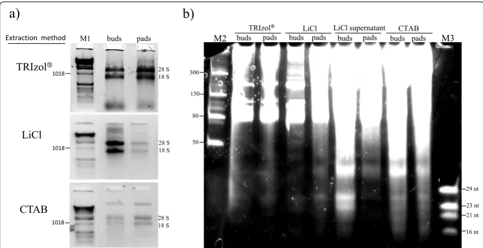
Total RNA was isolated from cactus pads and floral buds as shown in Figure 1a. Differences in yield and quality were observed with respect to the tissue type. Total RNA was best isolated from cactus pads using TRIzol® Reagent, while total RNA from floral buds was best isolated using the LiCl protocol. The 260/280 ratios were similar among the different methods (1.76-2.19), as presented in Table 1. However, 260/230 ratios showed large variations (0.21-2.03), being especially low when TRIzol®Reagent was used. Possibly, guanidine thyocia-nate or other contaminants could be producing this effect. Electrophoretic analysis showed that the LiCl method produced the best RNA quality (Figure 1a). Moreover, this method was easier in comparison with the CTAB and TRIzol® methods, as the mucilage, pre-sent in the cactus tissue, greatly difficulted handling and separation of the upper phases, requiring considerably long times for these steps.
Small RNA enrichment
RNAs, especially when isolated from floral buds (as shown in Figure 1b). Both the tissue type and the isola-tion method contributed to the yield. From the isolaisola-tion method comparison, it was concluded that LMW RNA from the LiCl supernatants, and total RNA obtained with the LiCl method showed higher yields and quality.
As sRNAs were obtained both from the supernatant and the total RNA pellet (Figure 1b), it was tested whether the extraction could be streamlined avoiding
the total RNA separation step. Different strategies were tested to optimize the extraction protocol in a way that sRNAs could be directly obtained by omitting the LiCl total RNA precipitation step. For this, the original LiCl protocol [18] was modified in four different ways by consequentially removing steps (Figure 2). As a control, the modifications were also tested with the model plant Arabidopsis. In the first modification, the enrichment step was performed using the supernatant directly after
buds pads buds pads buds pads buds pads
TRIzol LiCl LiCl supernatant CTAB
M2
29 nt
23 nt 21 nt
16 nt
a)
b)
50 80 150 300
TRIzol
CTAB
LiCl
buds pads
M1
Extraction method
1018
1018
28 S 18 S
28 S 18 S
28 S 18 S 1018
M3
Figure 1Comparison of total RNA and low-molecular-weight (LMW) RNA samples isolated from cactus (Opuntia robusta) with different extraction methods. a) Total RNA was extracted (in triplicate) with three different RNA extraction methods. 0.1 g of tissue was used for each sample and 1/10 volume of each sample was loaded on a 1% agarose gel (gel stained with ethidium bromide). b) LMW RNA extracted from total RNA samples obtained with different extraction methods, 1/10 volume (1-3 ug) of each sample was loaded on a 12.5%
polyacrylamide gel (gel stained with SYBR gold). M1, 1 kb DNA ladder (Invitrogen); M2, Low Range ssRNA Ladder (NEB); and M3, oligonucleotide ladder.
Table 1 Concentration* of total RNA and LMW RNA samples obtained using different extraction methods
Method Tissue Average 260/280 Average 260/230 total RNA yield (μg) 260/280 260/230 LWM RNA yield (μg)
TRIzol® buds 1.84 0.21 76.3 1.96 1.73 56.34
pads 1.83 0.25 50.28 1.96 1.59 38.4
LiCl buds 2.19 2.03 34.18 1.85 2.25 16.33
pads 1.76 1.72 4.20 1.85 2.24 1.57
LiCl supernant buds — — — 2.03 1.94 29.88
pads — — — 1.87 1.61 24.42
CTAB buds 2.19 2.01 24.65 2.12 2.22 16.81
pads 1.86 1.29 17.60 2.05 2.15 10.26
total RNA precipitation (without using the pellet con-taining HMW RNA), which produced positive results (Figure 3a and 3b). In the second modification, LMW enrichment was performed directly after the overnight LiCl precipitation. In the third modification, enrichment was done directly after the addition of LiCl. Finally, and since good LMW RNA yields were still obtained by removing the various steps, the complete LiCl precipita-tion step was omitted in the fourth modificaprecipita-tion. LMW RNA enrichment was directly performed using the upper phase after the chloroform extraction (modifica-tion 4 in Figure 2), which, strikingly, worked very well for both plant species (Figure 3a and 3b).
The extractions were performed in triplicate and the LMW RNA extracts were electrophoresed using dena-turing polyacrylamide gels (Figure 3). The presence of sRNAs obtained with the different strategies was observed. The final strategy (where the enrichment step was performed right after the chloroform extraction) showed a less intense band corresponding to 40 nt RNAs and produced a cleaner extract compared to the other methods (Figure 3 and Table 2). Furthermore, spectrophotometric analysis showed that all the 260/280 ratios were high when using the different methods to extract LMW RNA from cactus tissue, indicating a good purity of the samples (Table 2). However, the 260/230 ratios were very low in all cases, both for cactus as for Arabidopsis. Nevertheless, LMW RNA analyzed in poly-acrylamide gels showed well defined 5.8S, 5S, and tRNA bands, which suggests a good recovery (Figure 3). More-over, a well defined 24 nt band (sRNAs) could be directly observed in the polyacrylamide gel. This band
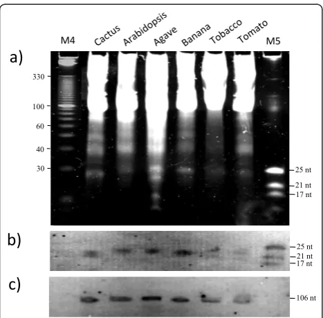
was observed and well defined when using the different methods. Interestingly, the sRNAs isolated using modifi-cation 4 (where the LiCl precipitation step was omitted) showed less background in the gel and a higher concen-tration of LMW RNA, showing that this strategy is effi-cient for LMW RNA isolation from plant species with high polysaccharide content, leading to increased yield in a reduced number of steps using conventional lab chemicals. This method does not only allow efficient LMW RNA recovery from cactus or Arabidopsis, but it also works very well for other plant species, such as agave, banana, tomato, and tobacco (Figure 4a). Further-more, when necessary, HMW RNA may still be recov-ered from the pellet after the polyethylene glycol precipitation (step 9 of theSmall RNA extraction proto-col; data not shown).
Chloroform extraction
Centrifuge
LiCl 8M addition
Precipitation (overnight)
Isolation and enrichment of LMW RNA
3 4
Total RNA Recovered supernatant
2 1
Centrifuge
LiCl/phenol extraction
Centrifuge
Figure 2Overview of the LiCl extraction method modifications. 1) LMW RNA enrichment from the supernatants obtained after precipitation of total RNA, 2) LiCl overnight precipitation, followed by LMW RNA enrichment, 3) LiCl addition, immediately followed by enrichment of LMW RNA, and 4) Enrichment of LMW RNA directly after the chloroform extraction, omitting the LiCl precipitation step.
M4 3 4 2 1 M5
a)
b)
100
60
40
30 330
M4 3 4 2 1 M5
25 nt
21 nt 17 nt 25 nt
21 nt 17 nt
100
60
40
30 330
Northern blot analysis
As described above, positive results were obtained for the isolation of LMW RNAs using the fourth modifica-tion and well defined sRNA bands could be visualized in a gel for 6 different plant species (Figure 4a). To test
whether the LMW RNA observed in the gel represented intact molecules and not degradation products, a north-ern blot assay was performed. Figure 4b and 4c shows that indeed, LMW RNA from cactus and Arabidopsis isolated with the shortest protocol version can be successfully detected in northern blot hybridization experiments using the miRNA159a probe, detecting 21 nt sRNA molecules, as well as using the small nucleolar RNA U6 probe, which detects 106 nt LMW RNA species (Figure 4b and 4c, respectively).
Conclusion
Here we provide a simple and effective method suitable for sRNA extraction from polysaccharide-rich material such as cactus, agave, banana, and tomato tissues, which also works well for less complex plant tissues form e.g., Arabidopsis and tobacco. This modified extraction method gives good yield and quality of LMW RNA spe-cies. Moreover, the LMW RNA obtained from the differ-ent plant species was successfully used for northern blot assays. Well defined bands were detected when using the miRNA159a and the small nucleolar RNA U6 probes. Therefore, the sRNA molecules that can be obtained with this low-cost short method are suitable for down-stream assays like northern blot hybridization, and most likely also for cloning and sequencing of sRNAs.
Acknowledgements
We thank Dr. Candelario Mondragón-Jacobo at INIFAP Norte de Guanajuato for providing cactus material, Alba Ramos-Olmos for technical advice with northern blot assays, and two anonymous reviewers for their valuable comments. We also thank the Mexican Science Council (CONACyT) for PhD fellowships to FFRC (199450) and to NDF (159754). The work in de Folter laboratory is financed by CONACyT 82826.
Author details 1
Laboratorio Nacional de Genómica para la Biodiversidad (LANGEBIO), CINVESTAV-IPN, Km. 9.6 Libramiento Norte, Carretera Irapuato-León, CP Table 2 Concentrations* of LWM RNA samples obtained using the LiCl method with different modifications
Tissue Modification ng/μl Average 260/280 Average 260/230 Yield (μg)
Cactus 1) LMW RNA enrichment from the supernatants obtained after precipitation of total RNA
382.30 1.82 1.81 7.65
2) LiCl precipitating (overnight), followed by LMW RNA enrichment 527.75 1.81 1.90 10.56 3) Addition of LiCl, directly followed by LMW RNA enrichment 291.42 1.66 1.35 5.83 4) Enrichment of LMW RNA directly after the chloroform extraction
without addition of LiCl
1008.42 2.02 1.04 20.17
Arabidopsis 1) LMW RNA enrichment from the supernatants obtained after precipitation of total RNA
582.52 1.68 1.23 11.65
2) LiCl precipitating (overnight), followed by LMW RNA enrichment 333.68 1.66 1.21 6.67 3) Addition of LiCl, directly followed by LMW RNA enrichment 609.38 1.53 1.13 12.19 4) Enrichment of LMW RNA directly after the chloroform extraction
without addition of LiCl
1418.77 1.53 0.96 28.38
*Yields and spectrophotometric A260/A230 and A260/A280 ratios of LMW RNA isolated with LiCl extraction method modifications from Prickly pear cactus and Arabidopsis. 0.1 g of tissue was used for the extraction and quantified using a Nanodrop spectrophotometer. LMW RNA was extracted two times in triplicate (6 independent samples).
a)
b)
c)
100 60
40 30 330
25 nt 21 nt 17 nt
M4 M5
25 nt 21 nt 17 nt
106 nt
36821 Irapuato, Guanajuato, México.2Facultad de Ciencias Naturales-Biología, Universidad Autónoma de Querétaro, CP 76360 Juriquilla, Querétaro, México.
Authors’contributions
FFRC did the major experimental work. NDF and JPVC contributed to the northern blot assays and with technical advice. FFRC, ACH, NMM, and SDF conceived the project and designed the experiments. FFRC, NMM, and SDF drafted the manuscript. All authors read and approved the final manuscript.
Competing interests
The authors declare that they have no competing interests.
Received: 28 October 2010 Accepted: 24 February 2011 Published: 24 February 2011
References
1. Carthew RW, Sontheimer EJ:Origins and Mechanisms of miRNAs and siRNAs.Cell2009,136:642-655.
2. Chen X:Small RNAs and their roles in plant development.Annu Rev Cell Dev Biol2009,25:21-44.
3. Chen X:Small RNAs - secrets and surprises of the genome.Plant J2010, 61:941-958.
4. Voinnet O:Origin, biogenesis, and activity of plant microRNAs.Cell2009, 136:669-687.
5. Chen D, Meng Y, Ma X, Mao C, Bai Y, Cao J, Gu H, Wu P, Chen M:Small RNAs in angiosperms: sequence characteristics, distribution and generation.Bioinformatics2010,26:1391-1394.
6. Lu C, Tej SS, Luo S, Haudenschild CD, Meyers BC, Green PJ:Elucidation of the Small RNA Component of the Transcriptome.Science2005, 309:1567-1569.
7. Carra A, Gambino G, Schubert A:A cetyltrimethylammonium bromide-based method to extract low-molecular-weight RNA from
polysaccharide-rich plant tissues.Anal Biochem2007,360:318-320. 8. Carra A, Mica E, Gambino G, Pindo M, Moser C, Pe ME, Schubert A:Cloning
and characterization of small non-coding RNAs from grape.Plant J2009, 59:750-763.
9. Pilcher RL, Moxon S, Pakseresht N, Moulton V, Manning K, Seymour G, Dalmay T:Identification of novel small RNAs in tomato (Solanum lycopersicum).Planta2007,226:709-717.
10. Chappell L, Baulcombe D, Molnár A:Isolation and Cloning of Small RNAs from Virus-Infected Plants.Current Protocols in Microbiology2005, UNIT 16H.2.
11. Meyers BC, Green PJ:Plant MicroRNAs - Methods and Protocols.Humana Press; 2009.
12. Moxon S, Jing R, Szittya G, Schwach F, Rusholme Pilcher RL, Moulton V, Dalmay T:Deep sequencing of tomato short RNAs identifies microRNAs targeting genes involved in fruit ripening.Genome Res2008,
18:1602-1609.
13. Itaya A, Bundschuh R, Archual AJ, Joung JG, Fei Z, Dai X, Zhao PX, Tang Y, Nelson RS, Ding B:Small RNAs in tomato fruit and leaf development. Biochim Biophys Acta2008,1779:99-107.
14. Hutvagner G, Mlynarova L, Nap JP:Detailed characterization of the posttranscriptional silencing-related small RNA in a GUS gene-silenced tobacco.RNA2000,6:1445-1454.
15. Chomczynski P, Sacchi N:Single-step method of RNA isolation by acid guanidinium thiocyanate-phenol-chloroform extraction.Anal Biochem 1987,162:156-159.
16. Chomczynski P, Sacchi N:The single-step method of RNA isolation by acid guanidinium thiocyanate-phenol-chloroform extraction: twenty-something years on.Nat Protoc2006,1:581-585.
17. Pall GS, Hamilton AJ:Improved northern blot method for enhanced detection of small RNA.Nat Protoc2008,3:1077-1084.
18. Verwoerd TC, Dekker BM, Hoekema A:A small-scale procedure for the rapid isolation of plant RNAs.Nucleic Acids Res1989,17:2362.
doi:10.1186/1746-4811-7-4
Cite this article as:Rosas-Cárdenaset al.:A simple and efficient method for isolating small RNAs from different plant species.Plant Methods2011 7:4.
Submit your next manuscript to BioMed Central and take full advantage of:
• Convenient online submission
• Thorough peer review
• No space constraints or color figure charges
• Immediate publication on acceptance
• Inclusion in PubMed, CAS, Scopus and Google Scholar
• Research which is freely available for redistribution