Xing-Hua Wang
1, A–F, Ying Shi
1, B, Huan-Hu Zhang
2, B, Xia Ma
1, B–D,
Characteristics of Focal Liver Lesions in Arterial Phase
on Contrast-enhanced Ultrasound and Contrast-
-enhanced Computed Tomography – Comparative Study*
1 Department of Ultrasound, the 2nd Clinical College of Shanxi Medical University, Taiyuan, China2 Department of Hepatology, the 2nd Clinical College of Shanxi Medical University, Taiyuan, China
A – research concept and design; B – collection and/or assembly of data; C – data analysis and interpretation;
D – writing the article; E – critical revision of the article; F – final approval of article; G – other
Abstract
Background. To compare the features of focal liver lesions in the arterial phase on contrast-enhanced ultrasonog-raphy (CEUS) and contrast-enhanced computed tomogultrasonog-raphy (CECT).
Material and Methods. A total of 38 lesions in 29 patients with focal liver lesions (FLL) were examined with CEUS and CECT. The characteristics of the enhancement were determined, especially in the early arterial phase (0~25 s). The enhancement of FLL in CEUS and CECT graded as follows: grade 0 – no enhancement; grade I – periph-eral enhancement or spotty enhancement at the center; grade II – spoke-like, honeycomb-like or heterogeneous enhancement; grade II – entire enhancement.
Results. On CEUS, the arriving time within 25 s was found in 36 out of 38 lesions (94.73%) and the peak time within 25 s in 29 lesions (76.32%). The number of grade II–III FLL was 25 (65.79%) on CEUS, and 13 (34.21%) on CECT showing a significant difference (p < 0.05).
Conclusions. Compared with CECT, CEUS plays an important role in the diagnosis of FLL with enhancement in the early arterial phase (< 25 s) (Adv Clin Exp Med 2014, 23, 1, 85–89).
Key words: ultrasonography, helical computed tomography, contrast, focal liver lesions.
Adv Clin Exp Med 2014, 23, 1, 85–89 ISSN 1899–5276
ORIGINAL PAPERS
© Copyright by Wroclaw Medical University
The conventional ultrasonography is a pre-ferred method for the diagnosis of focal liver le-sion (FLL) and has been widely used in screen-ing liver lesions. However, due to the lack of data on enhanced images, ultrasonography is found to be less accurate than contrast enhanced comput-ed tomography (CECT) in identifying liver neo-plasms [1–3]. With its great advantages of imaging techniques, the contrast enhanced ultrasonogra-phy (CEUS) has made a breakthrough and plays crucial roles in the diagnosis of FLL [4–9]. CEUS allows a whole-course observation of contrast en-hancement, whereas CECT is performed at pre-de-signed time points (generally in arterial, portal and equilibrium phases). On CECT, the arterial phase usually occurs from 25 s to 30 s and the presen-tations before the arterial phase (0–25 s) is often overlooked, although it may provide important
diagnostic information. Therefore, in the pres-ent study, the characteristics of FLL on CEUS and CEUT were compared in an effort to improve di-agnostic accuracy.
Material and Methods
Subjects
A total of 29 patients with FLL (38 lesions) were recruited from our hospital from August 2009 to April 2011. There were 17 males and 12 fe-males with a mean age of 50.3 ± 6.9 years (range: 24~76 years). The mean diameter of lesions was 4.7 ± 0.8 cm (range: 1.4~14.2 cm). For patients with multi-lesions, 2~3 lesions which were clearly
displayed on CEUS were selected for further analy-sis. Procedures were explained to each subject and informed consent was obtained before the study. The whole study was approved by the Ethics Com-mittee of the Shanxi Medical University.
The 38 lesions were confirmed by pathologi-cal examination, angiography or follow-up and included hepatic cell carcinoma (HCC) (n = 9), metastatic liver cancer (n = 10), hepatic hemangi-omas (n = 9), focal nodular hyperplasia of the liver (n = 4), hepatic abscess (n = 1), liver tuberculosis (n = 1), regenetrative nodules in the liver (n = 3) and heterogeneous fatty liver (n = 1).
Material and Methods
CEUS was performed with a Philips iU22 Ul-trasound System along with a 2~5-MHz probe at a low mechanical index (0.06–0.08) using a con-trast pulse sequence. The concon-trast agent was Sono-Vue. Before CEUS, the contrast was added to 5 mL of normal saline for preparation of contrast sus-pension. Firstly, the entire liver was detected in the conventional grey-scale ultrasound mode to search FLL. The location, size and number of FLLs were observed and the appearance, boundary and inner echo were recorded. Then, the liver was observed in a color ultrasound mode to observe the blood flow inside and surrounding the FLL. After the op-timal section was identified, the probe was fixed and the contrast enhanced mode was switched. Then, 2.4 mL of SonoVue suspension was injected intravenously as a bolus through the elbow vein, followed by a 5 mL injection of normal saline. The time and patterns of enhancement and the changes in enhancement over time were observed. Especial-ly, the arrival time, peak time, and the patterns of enhancement in early arterial phase (0–25s) were observed, and were carried out for at least 4 min. The characteristics of enhancement in the arterial phase were compared on CEUS and CECT.
CECT was conducted with a GE 16-slice he-lical CT scanner. The plain scanning and subse-quent contrast-enhanced scanning were applied. The contrast agent was 90–100 mL of Iohexol, which was injected with a high-pressure injector at a rate of 3 mL/s through the elbow vein. The en-tire liver was scanned in 3 phases, which included arterial phase (postinjection 25–30 s), portal phase (60–70 s) and equilibrium phase (120–180 s). The patterns of FLL enhancement were recorded. All the data was entered into a computer.
The enhancement varies among FLLs with dif-ferent blood supply. The contrast filled area exhib-ited enhancement and the contrast free area was slightly or not enhanced. According to the patterns
of enhancement in the arterial phase, these lesions were graded: grade 0 – no enhancement; grade I – peripheral enhancement or spotty enhancement at the center; grade II – spoke-like, honeycomb-like or heterogeneous enhancement; grade III – entire enhancement.
Statistical Analysis
SPSS13.0 (SPSS, USA) was used for statistical analysis. Qualitative data was compared using chi square test. A value of p < 0.05 was considered sta-tistically significant.
Results
Time of Enhancement
CEUS showed that 36 of 38 lesions (94.73%) had the enhancement time of < 25 s and 29 (76.32%) had the peak time of < 25 s. The informa-tion on blood perfusion before 25 s (arterial phase) was absent on CECT and thus not provided in the present study.
Patterns of Enhancement
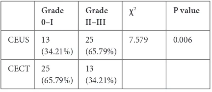
The patterns of enhancement of 38 FLLs are shown in Table 1 and the results of comparison of enhancement patterns between CEUS and CE-CT in Table 2. On CEUS, the proportion of grade 0–I lesions was significantly lower and that of grade II–III markedly higher than those on CECT (P< 0.05) (Table 2).
Table 1. Patterns of enhancement of 38 FLLs in arterial phase
Grade
0 Grade I Grade II Grade III
CEUS 1 12 9 16
CECT 2 23 7 6
Table 2. Enhancement patterns between CEUS and CECT
Grade
0–I Grade II–III χ
2 P value
CEUS 13
(34.21%) 25 (65.79%) 7.579 0.006 CECT 25
Misdiagnosis
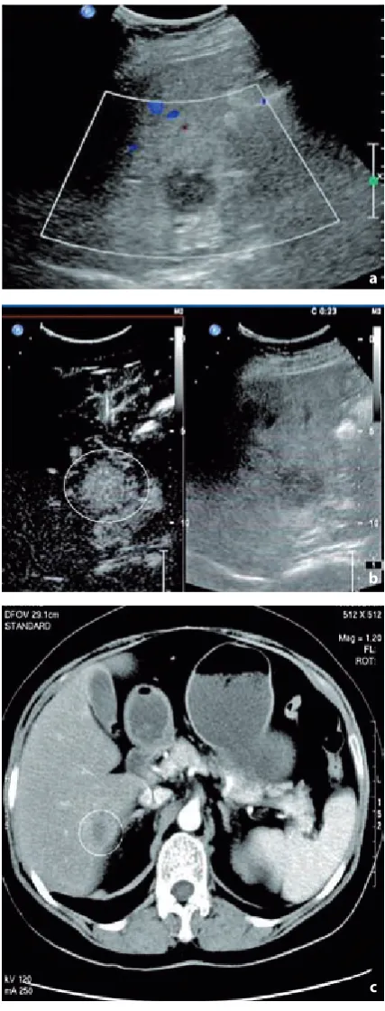
Of 38 lesions, 6 were identified by CEUS but misdiagnosed by CECT. All 6 lesions had the ar-rival time of < 25 s, of which 5 had the peak time of < 25 s. The ultrasound images in the first 25 s provided important information for an accu-rate diagnosis. These 6 lesions included blood sup-ply-rich small liver cancer (n = 1, Fig. 1), blood
supply-rich metastatic liver cancer (n = 1, Fig. 2) and focal nodular hyperplasia of the liver (n = 3).
Of 38 lesions, 3 were identified by CECT but misdiagnosed or not identified by CEUS, including
Fig. 1. Small liver cancer: A – conventional ultrasound showed a 1.0 0.9 cm isoecho in the right lobe of the liver, unclear boundary (white arrow) and no blood flow signal; B – CEUS showed diffuse enhancement within 18 s (white circle); C – CECT displayed non-enhancement in arterial phase (white circle) and the liver abscess was considered
Fig. 2. Metastatic liver cancer with rich blood supply: A – conventional ultrasound showed hypoecho in the right lobe of the liver and no evident blood flow signals (white circle); B – CEUS showed diffuse enhancement within 23 s (white circle); C – CECT displayed periph-eral enhancement in arterial phase (white circle) and the hemangioma was considered
c c
b
b a
2 HCCs secondary to hepatic cirrhosis and 1 right hepatic hemangioma close to the diagram. All 3 le-sions did not present evident space-occupying fea-tures on CEUS and had hypointense on the plain CT.
Discussion
CEUS is a novel imaging technique and CE-CT is one of the major diagnostic modalities of FLL in clinical practice. CEUS can obtain the real- -time enhancement of lesions and does not require pre-designing time points for scanning. Howev-er, the CECT images are collected intermittently at different time points starting from 25~30 s (ar-terial phase), to 60~70 s (portal phase) and to 120~180 s following contrast injection. CEUS al-lows continuous assessment of the overall en-hancement course – starting from the injection of contrast to the excretion of contrast from the body, especially at the early stage of enhancement (first 25 s following the injection). The enhancement at the early stage may provide important information on lesions, particularly about severe arteriosclero-sis and lesions occurring in the early arterial phase such as blood supply-rich small liver cancer and fo-cal nodular hyperplasia of the liver. However, these cannot be presented on CECT. Hemodynamic fea-tures are the important basis of imaging exami-nation to identify FLL. The characteristics of en-hancement in the arterial phase are crucial for the differential diagnosis. CEUS and CECT have their own advantages in the early stage (first 25 s follow-ing contrast agent injection), resultfollow-ing in differenc-es in the characteristics of enhancement which is beneficial for the final diagnosis.
Some studies have been conducted to compare the ability of CECT and CEUS to identify FLL and predominantly focus on the specificity, sensitivity and accuracy of both techniques [10–14]. Howev-er, the characteristics of enhancement in the arteri-al phase between 2 techniques are seldom reported. Most studies [9–14] demonstrate that the patterns of enhancement in the same phase are compara-ble between 2 techniques and the ability of both techniques to identify FLL is also similar. In addi-tion, other studies [15] suggest that CEUS is more sensitive, specific and accurate in displaying FLLs and more likely to display the small liver cancer as compared to CECT.
In this study, 36 of 38 lesions (94.73%) had the arrival time of < 25 s and 29 (76.32%) had the peak time of < 25 s. In the arterial phase, the proportion of Grade II~III lesions on CECT was significantly lower than that on CEUS (25 vs.13) (P < 0.05). We further analyzed 6 lesions which were identified
by CEUS but misdiagnosed by CECT and the re-sults showed that all lesions were severe arterial diseases, including blood supply-rich small HCC (n = 2), blood supply-rich metastatic liver cancer (n = 1) and focal nodular hyperplasia of the liver (n = 3). The 6 lesions had the arrival time of < 25 s, of which 5 had the peak time of < 25 s. The accura-cy of CEUS in the diagnosis of 6 lesions is attribut-ed to the enhancement in the first 25 s, which pro-vides the important information for diagnosis. The scanning in CECT is intermittently done and in-formation within the first 25 s cannot be obtained, which may cause a misdiagnosis of lesions with en-hancement in the early arterial phase.
In this study, three lesions were misdiagnosed or not identified by CEUS. Two were HCC in the same patient. They were identified as hepatic cir-rhosis companied by multiple nodules in the right lobe of the liver on CECT; 2 selected nodules had no rapid enhancement and were identified as cir-rhotic nodules by CEUS. However, these 2 nod-ules were identified as huge lesions occupying the whole right lobe of the liver on plain CT, which displayed an irregular, slight enhancement in the arterial phase but no rapid extinction in the portal and delayed phases. These lesions were identified as a giant hepatic carcinoma by CECT. The 2 nod-ules were confirmed as a giant hepatic carcinoma in the follow-up period. CECT is superior to CEUS in displaying the entire liver and the giant hepat-ic carcinoma. The 2 nodules located in the giant space-occupying lesions in the right lobe of the liv-er. The CEUS cannot display the outline of space-occupying lesions, and the liver parenchyma sur-rounding the nodules is virtually the tumor tissue. Another lesion was the hepatic hemangioma in the context of severe cirrhosis. It is located in the right lobe of the liver close to the diaphragm. The con-ventional ultrasonography cannot display this re-gion and thus fails to find this lesion, thus causing a misdiagnosis.
targeted lesions in different sections. In addition, CEUS is based on the two-dimensional ultrasound and generally visualize the lesions which are found
in the two-dimensional ultrasound whereas CECT can diagnose independently and display lesions which are undetectable in the plain CT.
References
[1] Oudkerk M, Van Ooijen B, Mali SP, Tjiam SL, Schmitz PI, Wigers T: Liver metastases from colorectal carci-noma: detection with continuous CT angiography. Radiology 1992, 185, 157–161.
[2] Takayasu K, Muramatsu Y, Furukawa H, Wakao F, Moriyama N, Takayama T, Yamasaki S, Sakamoto M, Hirohashi S: Early hepatocellular carcinoma: appearance at CT during arterial portography and CT arteriography with pathologic correlation. Radiology 1995, 194, 101–105.
[3] Seltzer SE, Getty DJ, Pickett RM, Swets JA, Sica G, Brown J, Saini S, Mattrey RF, Harmon B, Francis IR, Chezmar J, Schnall MO, Siegelman ES, Ballerini R, Bhat S: Multimodality diagnosis of liver tumors: feature anal-ysis with CT, liver-specific and contrast-enhanced MR, and a computer model. Acad Radiol 2002, 9, 256–269.
[4] Lencioni R: European federation of societies for ultrasound in medicine and biology (EFSUMB) guidelines for the use of contrast agents in liver ultrasound: what is the impact in clinical practice? Eur Radiol 2005, Suppl 5, E98–103.
[5] Salvatore V, Borghi A, Piscaglia F: Contrast-enhanced ultrasound for liver imaging: recent advances. Curr Pharm Des 2012, 18, 2236–2252.
[6] Quaia E: Solid focal liver lesions indeterminate by contrast-enhanced CT or MR imaging: the added diagnostic value of contrast-enhanced ultrasound. Abdom Imaging 2012, 37, 580–590.
[7] Martie A, Sporea I, Popescu A, Sirli R, Dănilă M, Serban C, Ardelean M, Bota S, Sendroiu M, Chisevescu D: Contrast enhanced ultrasound for the characterization of hepatocellular carcinoma. Med Ultrason 2011, 13, 108– 113.
[8] Sirli R, Sporea I, Popescu A, Dănilă M, Martie A, Bota S, Jurchis A, Sendroiu M: Contrast enhanced ultrasound for the diagnosis of liver hemangiomas in clinical practice. Med Ultrason 2011, 13, 95–101.
[9] Quaia E: The real capabilities of contrast-enhanced ultrasound in the characterization of solid focal liver lesions. Eur Radiol 2011, 21, 457–462.
[10] Dietrich CF, Kratzer W, Strobe D, Danse E, Fessl R, Bunk A, Vossas U, Hauenstein K, Koch W, Blank W, Oudkerk M, Hahn D, Greis C: Assessment of metastatic liver disease in patients with primary extrahepatic tumors by contrast-enhanced sonography versus CT and MRI. World J Gastroenterol 2006, 12, 1699–1705.
[11] Gaiani S, Celli N, Piscaglia F, Cecilioni L, Losinno F, Giangregorio F, Mancini M, Pini P, Fornari F, Bolondi L:
Usefulness of contrast-enhanced perfusional sonography in the assessment of hepatocellular carcinoma hypervas-cular at spiral computed tomography. J Hepatol 2004, 41, 421–426.
[12] Burns PN, Wilson SR: Focal liver masses: enhancement patterns on contrast-enhanced images-concordance of US scans with CT scans and MR images. Radiology 2007, 242, 162–174.
[13] Trillaud H, Bruel JM, Valette PJ, Vilgrain V, Schmutz G, Oyen R, Jakubowski W, Danes J, Valek V, Greis C: Characterization of focal liver lesions with SonoVue-enhanced sonography: international nulticenter-study in comparison to CT and MRI. World J Gastroenterol 2009, 15, 3748–3756.
[14] Giorgio A, Ferraioli G, Tarantino L, de Stefano G, Scala V, Scarano F, Coppola C, Del Viscovo L: Contrast- -enhanced sonographic appearance of hepatocellular carcinoma in patients with cirrhosis: comparison with con-trast-enhanced helical CT appearance. AJR Am J Roentgenol 2004, 183, 1319–1326.
[15] Maruyama H, Takahashi M, Ishibashi H, Yoshikawa M, Yokosuka O: Contrast-enhanced ultrasound for char-acterisation of hepatic lesions appearing non- hypervascular on CT in chronic liver diseases. Br J Radiol 2012, 85, 351–357.
Address for correspondence:
Xinghua Wang
Department of Ultrasound
The 2nd Hospital of Shanxi Medical University Taiyuan, 030001
China
Tel.: + 86 158 340 32 169 E-mail: [email protected] Conflict of interest: None declared Received: 19.02.2013