Włodzimierz Więckiewicz
1, A, B, E, F, Andrzej Bieniek
2, A, B, E, F,
Mieszko Więckiewicz
3, C, D, F, Łukasz Sroczyk
4, B–DInterdisciplinary Treatment of Basal Cell Carcinoma
Located on the Nose – Review of Literature
Wielospecjalistyczne leczenie raka podstawnokomórkowego nosa
– przegląd piśmiennictwa
1 Department of Prosthetic Dentistry, Wroclaw Medical University, Wroclaw, Poland
2 Department of Plastic Surgery, Clinic of Dermatology, Venereology and Allergology, Wroclaw Medical
University, Wroclaw, Poland
3 Division of Dental Materials, Wroclaw Medical University, Wroclaw, Poland
4Department of Maxillofacial Orthopaedics and Orthodontics, Wroclaw Medical University, Wroclaw, Poland
A – research concept and design; B – collection and/or assembly of data; C – data analysis and interpretation;
D – writing the article; E – critical revision of the article; F – final approval of article; G – other
Abstract
Skin cancers are a large group of all diagnosed face cancers. Surgical treatment of tumours with a margin of healthy tissue around the face is a challenge for dermatology, oral and maxillofacial surgery, plastic surgery and reconstruc-tive prosthetics. The location of cancer in an exposed location has significant effects on the psyche of the patients. Removal of a part or all of the nose and multi-stage reconstruction requires the cooperation of many professionals throughout the treatment period. This paper describes the procedure of treatment from diagnosis to definitive surgery. The aim of this article is intended to present the method of interdisciplinary cure of basal cell carcinoma located on the wing of the nose (Adv Clin Exp Med 2013, 22, 2, 289–293).
Key words: nose basal cell carcinoma, nose epithesis, plastic surgery.
Streszczenie
Raki skóry są dużą grupą wszystkich rozpoznawanych nowotworów złośliwych twarzy. Chirurgiczne leczenie nowotworów z marginesem zdrowych tkanek okolicy twarzy jest wyzwaniem dla dermatologii, chirurgii szczęko-wo-twarzowej, chirurgii plastycznej oraz protetyki rekonstrukcyjnej. Umiejscowienie nowotworów w miejscach eksponowanych wpływa znacząco na psychikę pacjentów. Usunięcie części skrzydła nosa oraz wieloetapowa rekon-strukcja wymaga współpracy wielu specjalistów przez cały okres leczenia. W pracy opisano procedurę leczenia od rozpoznania do ostatecznego zabiegu chirurgicznego. Celem pracy jest przedstawienie metody wielospecjalistycz-nego leczenia raka podstawnokomórkowego umiejscowiowielospecjalistycz-nego na skrzydle nosa (Adv Clin Exp Med 2013, 22, 2, 289–293).
Słowa kluczowe: rak podstawnokomórkowy nosa, epitaza nosa, chirurgia plastyczna.
Adv Clin Exp Med 2013, 22, 2, 289–293 ISSN 1899–5276
REVIEWS
© Copyright by Wroclaw Medical University
In recent years there has been an increase in the incidence of face-skin cancer. At the core of this phenomenon are genetic factors, excessive exposure to radiation – especially UVB, lack of exercise, pollution (effects of preservatives in food, artificial and natural radiation and cigarette smoke), and the extension of human life
group of malignant neoplasms that are located in the skin of the face.
ICD – International Classification of Diseases divides skin cancers into melanoma (ICD C43) and non-melanoma cancers (ICD C44). One of the non-melanoma cancers is the basal cell carcinoma
(BCC), this type constitutes 65–70% of all
occur-ring skin cancers. This cancer is characterized by the slow growth and almost total absence of me-tastases (less than 0.01% of cases) and local viru-lence [2]. In addition, it should be noted that parts of the face such as the nose, lips and cheeks play an important role in the act of speech, breathing, eating food, and therefore in basic activities of life. In the case when it comes to removing the tumour of different face parts, there is a need to perform a face epitheses (facial prosthesis, extraoral pros-thesis) [3–7]. An important element of a properly done epithesis is to restore these functions, which directly translates into the restoration of patient’s mental balance [8–10]. In addition, it should be noted that plastic surgery allows for reconstruc-tion of such loss [11–14].
The paper presents the procedure of interdis-ciplinary treatment of basal cell carcinoma located on the wing of the nose.
Procedure
After a physical and subjective examination (Fig. 1) histopathological examination should be performed in order to unambiguously confirm the diagnosis of BCC. The procedure to remove the tumour from the wing and the side wall of the nose, should be carried out from a vertical cut with lateral margin – 5 mm and a deep margin covering the full thickness of the walls of the nose at this spot (Figs. 2, 3).
After the surgery, the patient should be advised of the necessary follow-up visits and the need for referral to a specialist to perform a temporary ep-itheses of the nose wing. Prosthetic procedure can begin after the healing of tissues (Fig. 4). Before embarking on the prosthesis procedure, a thor-ough control of teeth is recommended, and in the case extensive tooth loss is found, their immediate reconstruction is recommended using appropri-ate dentures to ensure proper functioning of the masticatory organ and facial soft tissue support. Various stages of implementation of an epitheses should be spread over time, which will affect the normal healing of tissue around the operated area and will improve the quality of use of the nose pros-thesis (Fig. 5). After about 36 months after surgery and significant minimization of the risk of recur-rence of the disease, subsequent surgery should
be performed. This concerns the reconstructive procedure of the right wing of the nose with an Indian flap from the patient’s forehead (Figs. 6, 7). Reconstruction using a vertical flap containing the supratrochlear artery was used. Transposition
Fig. 1. Picture of the nose before surgery
Ryc. 1. Zdjęcie nosa przed zabiegiem operacyjnym
Fig. 2. Plan of tumor resection in accordance with rules of the micrographic surgery
Ryc. 2. Plan wycięcia guza zgodnie z zasadami chirur-gii mikrograficznej
Fig. 3. Nose after surgery
Fig. 5. Nose complemented by partial epitheses
Ryc. 5. Uzupełniony nos częściową epitezą
Fig. 4. Nose after healing
Ryc. 4. Nos po wygojeniu
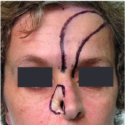
Fig. 6. Preparing for surgical reconstruction of the nose
Ryc. 6. Przygotowanie do zabiegu plastyki nosa
Fig. 7. Lateral part of nose was prepared for the graft and prepared the Indian flap from forehead
Ryc. 7. Wypreparowana do przeszczepu boczna część nosa oraz przygotowany płat indyjski z czoła
Fig. 8. Two weeks after final surgery
Ryc. 8. Dwa tygodnie po zabiegu
Fig. 9. Fourteen weeks after final surgery
flap has been moved above nasal dorsum to cover of nose tissue lack. Inner layer of the nostril was created by folding the most distal part of the flap. The secondary defect was closed immediately by layered suture (Fig. 8). After 3 weeks, when the flap cicatrizes initially to adjacent tissue, the flap pedicle was excised. After treatment, follow-up visits are necessary (Fig. 9).
In case of recurrence of the disease, the au-thors recommend performing a criodestruction of the tumour and radical tumour resection surgery with a margin of 5 mm and microscopic control of post-surgical wound edges. The resulting loss of tissue should be covered with a skin graft of inter-mediate thickness with the following recommen-dations: washing the graft with soap and water and oiling it with Vaseline, use of the No-Scar product, periodic checks every three months.
During the treatment the patient should be under constant supervision of the surgeon, pros-thetist and a psychologist.
Discussion
According to the authors, the interdisciplinary treatment of craniofacial cancer procedures should be recommended in cases where this type of thera-py may be used. The use of epitheses as temporary prosthesis, allows for the proper development of the periphery of tissue after surgery and ensures the patient’s psychological comfort during the pe-riod between procedures. An important issue is keeping the prosthesis on the prosthetic field. For small and medium-sized nasal tissue losses, the authors recommend using anatomical and arising after surgery spaces in order to hold the epithesis in place. According to Konstantinović et al. for large losses of nose tissue, one possible solution is to use implants to improve prosthesis retention [6]. However, Ciocca et al., for large losses,
pro-pose increasing of retention of the nose epithesis by its connection with glasses, using CAD / CAM technology [15]. At the final stage of the plastic surgery of tissue loss, one should apply pedicle flaps providing proper nourishment of the tissues. However, the use of pedicle flaps in the craniofa-cial area has its limitations. According to Anghel et al. this technique is suitable for covering small and medium-sized tissue losses in the wings and the nose wall [12]. In the case of large losses, Grajek et al. propose the use of free flaps taken from the ear and forearm [16]. When there is a need for resec-tion of the nasal cartilage, Son et al. use of grafts from a part of the ear cartilage [17]. There are also single-step nasal-tissue reconstruction techniques. Kannan et al. propose the use of a single-step re-construction of the wings and the nose wall in basalioma using the biloped flap [18]. However, the one-step procedure for malignant tumours strongly infiltrating the surrounding tissue does not seem to be the optimal solution. Despite this, many authors recommend this technique [19–21]. The multi-stage procedure allows observation of the tissue edge of the operated site and does not burden the patient with another surgery, the effect of which can be undone in the case of the disease recurring.
The authors should stress the need for inter-disciplinary treatment of the patient with facial tis-sue loss. Treatment of patients after radical surger-ies in the craniofacial area constitutes a complex problem of surgical-prosthetic therapy [22]. The patient should be placed under the special care of various specialists: a maxillo-facial surgeon, a plas-tic surgeon, an oncologist, ENT specialist, oph-thalmologist, dermatologist, prosthetitian, perio-dontologist, psychiatrist, psychologist, phoniatrist and dental technician. The number and high qual-ifications of the specialists who may be involved in the treatment of cancer patients emphasize the complexity of the problem and its great difficulty.
References
[1] Cottel WI: Skin tumors I: basal celland squamous cell carcinoma. Sel Readings Plast Surg 1992, 7, 6, 1–34.
[2] Brana I, Siu LL: Locally advanced head and neck squamous cell cancer: treatment choice based on risk factors and optimizing drug prescription. Ann Oncol 2012, 23, 10, 178–185.
[3] Conley J: Cancer of the skin of the nose. Ann Otol Rhinol Laryngol 1974, 83, 1, 2–8.
[4] Chambers MS, Lemon JC, Martin JW: Anterior key method for indexing orbital prosteses. J Prosth Dent 2002, 87, 1, 102–105.
[5] Selçuk CT, Sahin Ü, Çelebioglu S, Erbas O, Aydin C, Yüce S: Complex craniofacial reconstruction with prosthe-ses as an alternative method to autogenous reconstruction. J Craniofac Surg 2011, 22, 6, 2090–2093.
[6] Konstantinović VS, Lazić VM, Stefan I: Nasal epithesis retained by basal (disk) implants. J Craniofac Surg 2010, 21, 1, 33–36.
[7] Disant F: Role of epitheses in the rehabilitation of substance losses of the face. Rev Laryngol Otol Rhinol 1997, 118, 2, 109–112.
[9] Pigmo M, Funk J: Augmentation of obturator retention by extension into the nasal aperture: a clinical report. J Prosth Dent 2001, 85, 4, 349–351.
[10] Sailer HP, Haers PE, Zollikofer CP, Warnke T, Carls FR, Stucki P: The value of Stereolightographic Models for Preoperative Diagnosis of Craniofacial Deformities and Planning of Surgical Corrections. Int J Oral Maxillofac Surg 1998, 27, 5, 327–333.
[11] Tolman DE, Taylor PF: Bone achored craniofacial prosthesis study: Irradiated patients. Int J Oral Maxillofac Implants 1996, 11, 5, 612–619.
[12] Anghel I, Anghel AG: Reconstructive rhinoplasty in cases with basal cell carcinoma of the nose. Chirurgia 2012, 107, 3, 373–378.
[13] Lunatschek C, Schwipper V, Scheithauer M: Soft tissue reconstruction of the nose. Facial Plast Surg 2011, 27, 3, 249–257.
[14] Anghel I: Using of facial skin flaps in surgery of nose carcinomas. Otorinolaringologia 2000, 23, 1–2, 80–83.
[15] Ciocca L, Fantini M, De Crescenzio F, Persiani F, Scotti R: New protocol for construction of eyeglasses-support-ed provisional nasal prosthesis using CAD/CAM techniques. J Rehabil Res Dev 2010, 47, 7, 595–604.
[16] Grajek M, Maciejewski A, Szumniak R, Krakowczyk Ł: The use of three free flaps in the simultaneus reconstruc-tion of the nose after extensive resecreconstruc-tion due to malignant cancer. Pol Przegl Chir 2012, 84, 2, 99–101.
[17] Son D, Kwak M, Yun S, Yeo H, Kim J, Han K: Large auricular chondrocutaneous composite graft for nasal alar and columellar reconstruction. Arch Plast Surg 2012, 39, 4, 323–328.
[18] Kannan R, Reena J: Reconstruction of ala of nose with biloped flap. J Maxillofac Oral Surg 2011, 10, 1, 57–59.
[19] Teltzrow T, Arens A, Schwipper V: One-stage reconstruction of nasal defects: evaluation of the use of modified auricular composite grafts. Facial Plast Surg 2011, 27, 3, 243–248.
[20] Mahlberg MJ, Leach BC, Cook J: The spiral flap for nasal alar reconstruction: our experience with 63 patients. Dermatol Surg 2012, 38, 3, 373–380.
[21] Hassanpour SE, Shariati SM: One stage reconstruction of nasal defect by reverse flow retroauricular island flap – case series and discussion. J Plast Reconstr Aesthet Surg 2008, 61, 8, 949–952.
[22] Baker SR, Swanson NA: Management of nasal cutaneous malignant neoplasms. An interdisciplinary approach. Arch Otolaryngol 1983, 109, 7, 473–479.
Address for correspondence:
Mieszko Więckiewicz Division of Dental Materials Wroclaw Medical University Krakowska 26
50-425 Wrocław Poland
Tel.: +48 660 47 87 59
E-mail: [email protected]
Conflict of interest: None declared