Relaxation of the Supporting Structures of
the Female Pelvis
DAVID H. NICHOLS, M.D.
Professor of Obstetrics and Gynecology, School of Medicine, State University of New Yori< at Buffalo, Buffalo, New Yori<, and Head of the Department of Obstetrics and Gynecology, Buffalo General Hospital, Buffalo, New Yori<
The purpose of this discussion is to share with you some thoughts about pelvic relaxation, its mysteries, some technical minutiae helpful in identifying them and some of the surgical prob lems involved. Thus, by looking at a number of these diagnostic challenges, you may be stimu lated to some diagnostic thinking in an office setting.
Let me start with a few of the more decep tively simple challenges in pelvic relaxation, the goals we seek to achieve, and how to accom plish them.
We have seen within my generation a re examination of sexual thinking whereby aging persons presume to continue a reasonably comfortable and satisfactory coital relationship well into their senior years. Solving a problem of procidentia or vaginal vault inversion by closing the vagina by LeFort colpocleisis or removing it by colpectomy is no longer a solution equally acceptable to all concerned. If we are going to consider a surgical approach to reconstruction and rebuild for someone a vagina that is both physiologically and sexually useful, we must have some idea of the purpose and benefits we hope to achieve by reconstruction.
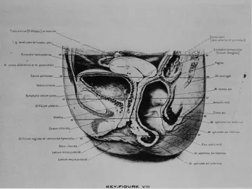
Consider, for example, the old illustration from Cross-sectional Anatomy by Eycleshymer
The following is an edited transcript of remarks by Dr. Nichols at the 51 st Annual McGuire Lecture Series, October 19, 1979, Medical College of Virginia, Richmond, Virginia.
Correspondence and reprint requests to Dr. David H. Nichols, Chairman, Section of Obstetrics and Gynecology, Brown University Program in Medicine, Providence. Rhode Island 0291 2.
and Shoemaker (Fig 1). and notice that the va gina in this instance has an almost vertical axis. Is that then the goal we seek when we reposi tion a misplaced vagina? Or is the vagina being displaced anteriorly by a full rectum, which was full at the time of death, as this illustration obvi ously was from a cadaver dissection?
To resolve the issue, the vaginas of nulli parous young women can be lightly painted with a barium paste. This would not distort the vagina but would render it radio-opaque. Lateral colpograms taken of these women demonstrate that the vagina does have an S-shaped curve with a horizontal inclination to the axis of the up per vagina (Fig 2) If we were to ask these nul liparous patients to bear down, as by a Valsalva maneuver, this upper axis would become even more horizontal (Fig 3).
We can confirm this for ourselves daily in the office during vaginal examination of a nul liparous patient by letting the examining fingers follow the axis of the vagina. Usually the finger tips will end up in the hollow of the sacrum.
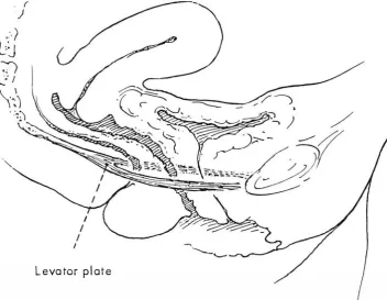
The usual upper horizontal vaginal axis is clinically significant and I will now discuss how this axis can be destroyed or changed and some of the surgery that can be done to restore it. The vagina in this situation rests on the gen erally empty rectum. The only time the rectum is filled is in the patient with a large rectocele, or in someone during the act of defecation, or in someone recently deceased. The empty rectum in turn "sits" on the levator ani. The portion of the levator ani behind the rectum upon which it rests is called the levator plate It is formed from
T11�uttrina [Fa!loppi,) ftlon.n"m ••. ··· ... Lii le1e1 uttfi •t fundus uttri
-
...
···-. •. _M ,c.-cti.,s ,1bdom1n11 et m. pyra,mdatis_ .•...
_.E.o.uvatiorecto11:,1<na (C,1v11m Ooi,51u,J
·•• .• M.sphint1or 1n, 1nt�rn11s
'
KEY·�IOUFUC VIII
Fig 1-0rawing of sagittal section through the embalmed cadaver showing the axis of the vagina to be in an almost vertical position. It is displaced anteriorly by the dilated rectum, a relationship not often found in the living.
the fusion of the two halves of the pubococ cygeal muscle posterior to the rectum (Fig 4).
How many different anatomic entities or "systems" are there concerned with keeping the vagina inside the body? It is really an in vagination, and it is unlikely that without help it would remain one. lnvagination has many con ceptual similarities to the finger of an in-turned rubber glove; by putting air into the glove and squeezing an in-turned finger promptly pops out. Why then does the vagina not "pop out" or evert more frequently than it does? Wh.at are the systems that affected, damaged, or missing parts can influence? Why is the birth canal where it is?
There are actually at least six responsible but independent systems that influence in vagination. The first is the bony pelvis to which most of the soft tissues of the pelvis are ulti mately attached. If there is a defect in the bony pelvis, for example, a congenital defect coinci dent with extrophy of the bladder, the mid-por tion of the pubis may be missing in which case
the rectus muscles and pubococcygeal muscles have a defective attachment influencing greatly the architecture of the anchoring supports of the vagina.
Second among the six systems is the round and broad ligament complex; third, the cardinal-uterosacral ligament complex; fourth, the pelvic diaphragm; fifth, the urogenital dia phragm; and sixth, the perineum and the peri nea! body. Let us examine these in some detail to see why they influence pelvic support.
We know, for example, that the round and broad ligament complex often influences pelvic support in a negative way. If the broad liga ments have been involved with either endome triosis or ligneous fibrosis as a result of previous infection, or sometimes by cancer, what would appear to be an easy vaginal hysterectomy isn't easy at all. The uterus is arrested in its descent by pathologic fixation from the broad ligament. Certainly the role of the round ligaments as at tributed to retro displacement of the uterus is al ways up for constant reexamination, particularly
Fig 2-A normal vaginal depth and axis. The vaginal walls of a 25-year-old, healthy nulligravida have been painted with bar ium. The perinea! curve of the lower vagina 1s shown along with a more horizontal axis of the upper vagina.
Fig 3-The same patient is straining as by a Valsalva maneuver which accentuates the horizontal axis of the upper vagina.
Levator plate
Fig 4-0rawing of normal vaginal axis of the l,v,ng showing almost horizontal upper vagina and rectum lying on and parallel to the levator plate.
in view of Blue Shield's national decision to ex clude suspension operations from those autho rized for payment. I do not believe that retro version causes uterine prolapse, but that in many instances the same qualities that lead to retroversion may also lead to the development of genital prolapse. These conditions may be separate results of common etiologic circum stances. The significance of this observation is that suspension of a retroverted uterus does not of itself prevent subsequent prolapse.
We consider the cardinal ligamehts to gether with the uterosacral ligaments as a single anatomicosurgical suspensory unit: the car dinal-uterosacral ligament complex.
The pelvic diaphragm consists of the leva tor ani and its fascia! covering, the medial por tion of the levator ani. The pubococcygeal mus cles fuse behind the rectum, constituting the levator plate which is so important in pelvic sup port. The normally horizontal axis of this plate will sag if the diaphram has lost its integrity. Not
only will the hiatus or the distance between the anterior margin of the levator plate and the pubis increase, but the greater the sagging the greater the tendency for anything that rests on top of this levator plate to slide over and down, accentuated by the pull of gravity (Fig 5).
The urogenital diaphragm is a sort of sandwich between the two pubic rami. It is pen etrated by the urethra and vagina, and also helps to support the urethra. The pubourethral ligamentous support of the urethra is, in fact, continuous with and part of the urogenital dia phragm, and has much clinical significance. Obstetrically, the posterior portion of this liga ment is the one most likely to be damaged. Pathologic stretching may cause rotational de scent of the bladder neck predisposing to stress incontinence.
There is a big difference in the perinea! body between persons Defect of the perinea! body may be inherited, and occasionally a peri nea! body will be missing even in nulliparous
women. A defective perinea! body is sometimes confused with a rectocele, and if congenital in origin, is usually asymptomatic unless there is a total absence of the perineum, in which case the patient is said to have a so-called "double barrelled shotgun" type of vagina, and the vagi nal canal is often contaminated by rectal soiling; such a patient has a virtual cloaca. The urethra essentially lies upon the perinea! body; in in stances in which a urethral repair has been done and a perinea! defect is evident, correc tion of the perinea! defect is complimentary to the support of the urethra and anterior vaginal wall.
The combinations of damage to the six different anatomic systems responsible for pel vic support that can be produced indicate why there is no one standard surgical procedure, for example, vaginal hysterectomy and repair or the Manchester procedure, that will solve all combinations of problems equally well.
There can be permanent elongation and stretching of the cardinal and uterosacral liga ment complex permitting descent of the upper vagina but without any particular cystocele or rectocele. This may be acquired and is often quite significant. The process may be set in mo tion in someone who has had the misfortune of
being told to bear down during labor prior to full dilation of the cervix, and in so doing has pushed the cervix in front of the presenting part of the fetus. Many times that advice to bear down prior to full dilation has been accepted by the patient as a hopeful, though false and un knowingly dangerous means of shortening la bor. It has often done intractable and per manent damage to the supports of the vagina.
In another situation, failure to perform an episiotomy in someone in whom vaginal elastic ity is defective may result in a cystocele and rectocele, even in a patient who did not bear down prior to full dilation of the cervix. We are in an era where there are some young women who don't want an episiotomy and who are questioning circumcision, as well as the need for hospital delivery even if operative inter vention of any kind in the conduct of their labor is indicated. We can rest assured that some can get by without episiotomy, but many can not without sustaining serious damage. How can one tell the difference? Probably the most im portant single point is separating the care of those who have elasticity of the vagina and peri neum from those who do not.
The so-called older primapara, the patient over 35, generally has reduced elasticity of the vagina and particularly the lower portion and the perineum One can also distinguish the patient with poor elastic tissue by the presence of striae on the sides of the abdomen. The patient with many abdominal or breast striae is usually an obvious candidate for episiotomy if excessive vaginal damage is to be prevented Episiotomy must be performed at a proper time; not just to prevent a tear, but before irreparable damage has been done to the soft tissues. Lastly, an episiotomy must be properly repaired with the goal of reuniting structures which were tran sected by episiotomy, and not simply just stop ping the bleeding and putting the skin together.
Anterior colporrhaphy is not so simple as it would appear from the three or four pages given in the average surgical text, so let us look at some of the reasons why.
A typical conception of cystocele shows some pathologic stretching of the bladder that displaces the vagina downward. Are the symp toms that may be produced so predictable that reconstruction is always simple? The relation ship between the bladder, the urethra and the
posterior surface of the pubis has much clinical significance with or without a coincident cys tocele
Consider some of the various alterations that an anterior vaginal wall can undergo. Many years ago a characteristic descent of the base of the bladder during the act of voiding was emphasized so that the vesicourethral junction may be represented as a straight line. While this is physiologic during the act of void ing, it is not to a patient at rest who is not void ing; thus, when this flattening is apparent in someone who is not voiding, it indicates rota tional descent or "wheeling" of the vesicoureth ral junction with elongation of the supporting tis ues concerned with holding the urethra in its normal position. Such an altered relationship may be a significant factor in the production of stress incontinence as the vesicourethrai junction may now be the lowest portion of the hydrostatic column of water. We know that in continence may result from an abnormal rela tionship between intraurethral pressure and in travesical pressure as a function of urethral tone and not solely from positioning of the vesicou rethral junction. A cystocele may or may not coexist. The anterior vaginal wall may thus bring down with it the base and neck of the bladder, but not the bladder proper; this is not a cys tocele in the usual sense. Sometimes rotational descent of the bladder neck has been called "urethrocele," and one will find many refer ences to this fallacy in the literature, but a true urethrocele is a pathologic dilatation of the ure thra and is very rare. I think in my whole oper ative experience I've seen but three or four true urethroceles. Some of these people with a true urethrocele are perfectly continent. Rotational descent of the bladder neck is also sometimes called · · pseudocystocele. · ·
In addition, there may be funneling of the urethra. If the urethra is funneled in addition to the rotational descent of the bladder neck, a yet different set of statistics prevails, whereby the bottom of the hydrostatic column of urine nor mally located at the base of the bladder is now located at the base of the funnel, thus reducing intraurethral tone and pressure and increasing the susceptibility to stress urinary incontinence. If a patient has a cystocele serious enough to require anterior colporrhaphy, the op erator should probably repair the full length of
the vagina and make sure that the vesical neck is adequately supported and that the cystocele is not repaired with more tissue than required, thus risking the development of iatrogenic stress incontinence from unwitting flattening of the posterior urethrovesical angle. Neither should a cystocele be repaired with less tissue than required. The vagina should be precisely trimmed to a size suitable to the particular needs of the patient.
A perinea! defect with a rectocele is a fac tor in the defective support of the urethra. If the urethra and anterior vagina rest on nothing of substance, any operation that has been done to them will tend to have less mechanical support than if the anterior vaginal wall rests on some thing of strength. Rectoceles come in various sizes and shapes within the pelvis; some pro duce symptoms and some do not. It is difficult to improve a patient's comfort by surgical cor rection of something that is asymptomatic and a surgeon would not normally operate on a recto cele or cystocele if it were producing no symp toms distressing to the patient and if the repair were not part of additional pelvic surgery. But if a vaginal hysterectomy were performed be cause of menorrhagia, prolapse, fibroid or something of that nature on a woman who has a coincident cystocele and rectocele, certainly the cystocele and rectocele ought to be re paired at the same time.
Let us now briefly consider enterocele. It is sometimes described simply as a peritoneal hernia. Sometimes symptoms are produced, sometimes not. Symptoms associated with an enterocele are caused by gravity-induced trac tion upon the contents of the sac: small bowel or omentum. If the sac is in fact empty, the pa tient will be relatively asymptomatic. But the sac is a potential site for further distress and should be removed if surgery is being performed on other parts of the pelvis.
Consider the difference in the relationship between the cul-de-sac and the vagina in the congenital type of enterocele versus pseudo enterocele. The latter may be seen in a person in whom a high rectocele was treated by peri neorrhaphy alone. Perhaps the doctor was not aware how high this rectocele extended be cause with the patient asleep and in the lith otomy position it is sometimes difficult to judge how it looks when the patient is standing.
Fail-ure to repair the full length of the rectocele gives rise to this condition which resembles an en terocele but isn't and is usually symptomatic, the symptom being inability to completely empty the bowel. The sac, which is unrepaired rectocele, fills with fecal material, and the pa tient cannot evacuate it except by digital pres sure in the vagina. A simple maneuver distin guishes enterocele from high or midvaginal rectocele or dropped cul-de-sac which is a function of support of the vault of the vagina. Because enterocele is most evident when the patient is standing, she should be examined in that position: index finger in the rectum, thumb in the apex of the fully replaced vagina and the patient asked to bear down. If enterocele is present, the physician can feel it fill with either small intestine or omen tum. When gently squeezed between the thumb and forefinger, discomfort is evident. In that way the physician can distinguish preoperatively between entero cele, which is a sliding hernia, and descent of the cul-de-sac in someone in whom the vagina has dropped.
Failure to recognize and treat variances by appropriate surgery usually leads to post operative disability.
One of the different approaches to the problem of massive eversion of the vagina has been ventral suspension or fixation of the ever ted vagina. Sometimes it is successful, but the 90 degree change in the usual axis of the va gina imposes a risk that would otherwise not be present. Pulling the vagina forward without oblit erating the cul-de-sac has made the latter vul nerable to enterocele; a future enterocele that might not have been there had the cul-de-sac been obliterated. This risk is also present follow ing some other surgical procedures that may change the axis of the vagina, if no attempt has been made to obliterate the cul-de-sac. For ex ample, unless the peritoneum is opened and the cul-de-sac deliberately obliterated, the Marshall Marchetti retropubic pin-up type of operation will be followed by a significant incidence of subsequent enterocele requiring a second oper ation
A transabdominal approach to eversion of the vagina with the aim of restoring a sexually useful vagina is sacropexy Affixing the vault of the vagina to the hollow of the sacrum has an advantage over ventral suspension in that the
normal vaginal axis is restored and the cul-de sac is no longer vulnerable.
We have considered a variety of different kinds of damage to the supports of the pelvis. Let us consider the causes of damage.
1 . Congenital defect.· not only a missing or underdeveloped tissue or organ but a defective innervation. Striated muscle deprived of an ade quate nerve supply may be hypotonic.
2. Increased intra-abdominal pressure: An
obese woman may wear a girdle that is too tight; maybe she is doing heavy work; or there is the older woman in a situation where a loved one, sometimes a parent, has become disabled by a stroke. That disabled patient must be man ually lifted in and out of the bed, in and out of the bathtub, on and off the commode, produc ing a massive increase in the intra-abdominal pressure of the person doing the lifting. Often there is no one else to do the lifting, but we see the consequences of that damage.
3. Obstetric damage can be caused in a
number of different ways In the 1940s we par ticipated in problems of home delivery, usually among the poor in the large cities, as part of the home delivery service. Delivery was conducted reasonably comfortably, but the consequences of soft tissue damage during labor were
some-times greater than would have been seen in the.
hospital. Now there is some revival of interest in
nonsupervised obstetric performance. Women.
are delivering one another, and there may be a significant return of some of these problems of genital prolapse that have not been so common recently. Obstetric damage, even if in a hospi tal, is often related to the conduct of labor. We are considering the supports of tr,e urethra. Re member the damage they can sustain. There are at least six different combinations of dam age to the anterior vaginal wall. If the baby's head is very closely applied to the undersurface of the pubic arch during labor, it may damage these tissues in contrast to circumstances in which, because of either a larger fetal head or a narrow arch, the baby's head is pushed away from these tissues beneath the urethra (Fig 6). Two more types of damage to the vagina, and secondary damage to the bladder, have to do with the conduct of labor. In the first instance, the vagina has been damaged from within out. An "explosion" type of damage has occurred in which the vaginal wall has been stretched beyond its limits of elasticity-much like a piece of crepe paper that has been stretched and at at a certain point loses its elasticity and is permanently stretched out of shape. In contrast
Fig 6--0bstetric damag_e to the anterior vaginal wall may be greater when the baby's head has come in close contact with the tissues beneath the pubic arch (left). Posterior displacement of the baby's head during labor (right), is more likely to concen trate damage on the attachments of the posterior vaginal wall, sparing some anterior damage.
is a circumstance in which the labor patient has been told or has found herself bearing down prior to full dilatation of the cervix and pushes the cervix in front of the baby, overstretching the cardinal and uterosacral connective tissue supports of the cervix. Even when the cervix is fully dilated, the vagina may fail to dilate and the patient may push the vagina in front of the baby, almost like a doughnut, stretching the va gina itself in addition to stretching the supports, accounting for some of the combination dam age. So our old friend the cystocele is not quite so simple as it appears on the surface. If one adds the distention cystocele, the one produced by overstretching, and the one from dis placement to those six kinds of damage pre viously alluded to, there are now eight types.
With displacement cystocele, the vaginal wall may not have been stretched out of shape, but it is in the wrong place. There are reason ably good rugal folds in each vaginal wall. This patient requires major attention to restoration of the supports of the cervix and the vagina and less attention to reconstruction of the anterior vaginal wall itself. With displacement cystocele, the bladder has been pulled down from its nor mal position by the prolapsed uterus and by its attachment to the cervix.
The goal of most surgery for urinary stress incontinence includes elevation of the vesico urethral junction to a point where it is once again within the abdomen and, therefore, pre sumably responsive along with the bladder to changes in intra-abdominal pressure.
The symptoms of genital prolapse are those of pelvic heaviness, backache, vaginal mass, dyspareunia and disorders of function primarily related to coitus or inability to empty the bladder or rectum. The patient may have to manually elevate the bladder in order to empty it or make digital pressure within the vagina to overcome inability to empty the rectum. When these symptoms are sufficiently disabling, or if they are progressive, the patient should indeed be treated.
The treatment includes different kinds of prophylaxis. If the patient is a heavy smoker with emphysema, asthma, or chronic bron chitis, successfully getting her to stop smoking will lessen the insults placed on the supporting
tissues by coughing. If the patient is too fat, she should lose some weight. If she's wearing a girdle that is too tight, she should stop wearing it and either lose the weight or buy a larger gar ment. If she is working at an occupation that re quires heavy lifting, for example, a dipper in a bumper replating factory, or someone in nursing who is required to lift patients and is developing a genital prolapse, she should try another occu pation. The pessary isn't used very much any more, although it is frequently used temporarily to replace the dropped tissues in someone awaiting surgery. Inserted and left as a definitive treatment, a patient will be temporarily comfort able enough that she may postpone surgery un til a time when she is much older and unable to respond to the stress of surgery as smoothly as when younger. Pubococcygeal perinea! resis tive excercises and voluntary contraction of the pubococcygeal muscles, the Kegel exercises, are certainly helpful in relieving many of the symptoms They must be done often enough: 20 contractions in a row, three seconds each, six times a day, for at least three months. Ad vise the patient that she can do them while in public, as the contractions can't be seen. The physician can usually perceive the patient's pubococcygeal strength by putting one finger in her vagina and asking her to squeeze her va gina shut. One can feel whether her pub ococcygeal muscles are strong, and if they are not, estrogen replacement supplemental to sur gery will improve healing qualities and tissue strength.
We have considered many aspects of genital prolapse including the clinical anatomy, sites of damage, causes and prevention of gen ital prolapse and a variety of treatments. Most can be readily recognized if we but take the time to look for them.
Figure 1 is reproduced with permission from Carter, et
al, Cross-Sectional Anatomy: Computed Tomography and
Ultrasound Correlation, New York, Appleton-Century-Crofts, 1977.
Figures 2, 3 and 4 are reproduced with permission
from Obstetrics and Gynecology (36:251-256, 1970).
Figures 5 and 6 are reproduced with permission from Nichots, et al, Vaginal Surgery, Baltimore. Williams & Wil kins Company, 1 9 77.