Design and development of an in-house multiplex RT-PCR assay for
simultaneous detection of HIV-1 and HCV in plasma samples
Paryan M1, Forouzandeh Moghadam M2*, Kia V2, Mohammadi-Yeganeh S1, Abbasali Raz A1, Mirab Samiee S3
1Biotechnology Research Center, Pasteur Institute of Iran, Tehran, Iran. 2Department of Medical Biotechnology,
Tarbiat Modares University, Tehran, Iran. 3Food and Drug Laboratory Research Center, Ministry of Health
and Medical Education, Tehran, Iran.
Received: December 2011, Accepted: February 2012.
ABSTRACT
Background and Objectives: HIV-1 and HCV infections are life threatening problems in patients who receive blood products. Serological methods have proven useful in detecting these infections, but there are setbacks that make it challenging to detect these infectious agents. By the advent of Nucleic Acid Testing (NAT) methods, especially in multiplex format, more precise detection is possible.
Materials and Methods: We have developed a multiplex RT-PCR assay for simultaneous detection of HIV-1 and HCV. Primers were designed for highly conserved region of genome of each virus. Using these primers and standard plasmids, we determined the limit of detection, clinical and analytical specificity and sensitivity of the assay. Monoplex and multiplex RT-PCR were performed.
Results: Analytical sensitivity was considered to be 100 and 200 copies/ml for HIV-1 and HCV, respectively. High concentration of one virus had no significant effect on the detection of the other one with low concentration. By analysis of 40 samples, clinical sensitivity of the assay was determined to be 97.5%. Using different viral and human genome samples, the specificity of the assay was evaluated to be 100%.
Conclusions: The aim of this study was to develop a reliable, rapid and cost effective method to detect HIV-1 and HCV simultaneously. Results showed that this simple and rapid method is perfectly capable of detecting two viruses in clinical samples.
Keywords: HIV-1, HCV, Multiplex RT-PCR, Co-infection
INTRODUCTION
Infection with HIV-1 and HCV, especially co-infection, is a life threatening problem in patients who receive blood products. Therefore, it is absolutely necessary to be certain about the safety of donated bloods and
blood products. In the United States and Europe, co-infection with these two viruses is relatively prevalent and the incidence rate is 10% and 25%, respectively (1, 2).
HCV infection in HIV-1 infected patients is usually caused by drug injection (90%) and blood or blood products transfusion (70%) (3, 4). The major cause of death in HIV-1 infected patients who receive HAART which prevents the progress of HIV-1 infection is the HCV mediated hepatotoxicity (5, 6). In addition, simultaneous HIV-1 infection and HCV-related hepatic diseases, like cirrhosis and hepatocarcinoma, rapidly increases in these patients (7, 8).
In addition to co-infection, these viruses are most
* Corresponding author: Mahdi Forouzandeh Moghadam Ph.D Address: School of Medical Sciences, Department of Medical Biotechnology, Tarbiat Modares University, Jalal Ale Ahmad Highway, Tehran, Iran.
noticeable infectious agents transmitted by blood. One of the most considerable solutions to overcome the problem is serological tests for screening the antibodies produced against HIV-1 and HCV (9, 10). Serological methods have been used for years to detect viral infections. Although culturing of viruses and direct examination are emphasized, serological methods have proven to be useful in detecting these infections (11).
HIV-1 and HCV could be transmitted in window period in which the infection is not detectable with serological tests (12). Other reasons that disqualify serological tests are antigenic variations of viruses, infections with different serotypes of a virus, presence of silent carriers and lack of antibody in the beginning of the infection. Another disadvantage of these methods is the presence of maternal antibodies which make it impossible to detect infection in new borns from mothers who are infected with HIV-1 or HCV. Furthermore, by means of serological methods, distinguishing patients with active infection from those who completely cured (and they have anti-HCV IgG ) is not possible (13-15).
To overcome these setbacks, nucleic acid based test-ing (NAT) methods for detecttest-ing the viral genome have been developed. One of the Advantages of NAT methods in comparison with serological methods includes direct examination of the infectious agent’s genome with high specificity and sensitivity. Although the possibility and functionality of NAT have been proven, large scale exploitation of it is limited because of high cost (16, 17). Moreover, pooling of samples employed by several molecular approaches determines a relatively high rate of false-negative results and loss of sensitivity when a low-positive sample is diluted into the pool. To conquer these problems, Multiplex NAT has been developed to make it possible to simultaneously analyze a single sample for multiple infections. So the time and cost needed for NAT assays are significantly reduced (18-21). By the advent of Multiplex RT-PCR, it is now possible to detect simultaneous viral infections with high precision and accuracy. Recently this simple, rapid and cost-effective method has been used to detect bacterial, protozoan and other infectious agents (21-27).
MATeRIAlS AND MeTHODS
Clinical samples. Eighty different plasma samples were entered in this study. These included 30 samples co-infected with HIV and HCV, 10 HIV positive
and HCV negative, 20 HCV positive and HIV-1 negative and 20 HIV-1 and HCV negative samples from healthy individuals. The co-infected and HIV positive samples were obtained from patients of Imam Khomeyni hospital, whose infections had been confirmed by ELISA, Western blot and RT-PCR. HCV positive and healthy samples were obtained from Day hospital, and were confirmed by ElectroChemiLuminescense (ECL) and RT-PCR. All of the samples were transferred to our laboratory considering the biosafety regulations.
Primer design. Primers were designed based on conserved regions of genome for each virus so they were able to recognize all the genotypes of each virus. Totally, 471 sequences of HCV viruses and 1242 sequences of HIV-1 viruses were retrieved from GenBank, NCBI (http://www.ncbi.nlm.nih.gov) and were aligned using MEGA-4 software to identify the conserved regions. pol gene of HIV-1 and 5’NCR of HCV genome were selected (Table 1).The length of amplicons for HIV-1 and HCV were 179 bp (nucleotides 4354 to4533) and 241 bp (nucleotide 84 to325), respectively. Moreover, Oligo 6 and AlleleID 6 softwares were used to inspect the compatibility of primer pairs.
Viral RNA extraction. Blood samples were collect-ed in tubes containing EDTA and were centrifugcollect-ed at 2500 RPM for 20 minutes. The plasmas were stored at -80oC until use.
To extract viral genomes from 200 µl of plasma samples, High Pure Viral RNA Kit (Roche Diagnostics, Germany) were used according to manufacturer’s instructions.
Reverse Transcription. Viral RNA were reverse transcribed using M-MuLV enzyme (Fermentas, Germany). 1 µl of HIV-R primer (2 µM) and 1 µl of HCV-R primer (2 µM) were added to 5 µl of extracted RNA samples of HIV-1 and HCV in two different 0.2 ml tubes, respectively. RNase and DNase free distilled water were added to a final volume of 12 µl. Then tubes were incubated for 10 minutes in 70oC.
Finally, 5 µl enzyme buffer, 2 µl dNTP (10 mM), 0.5 µl RNase Inhibitor (20 U/ µl) and 1 µl M-MuLV RT (200U/ µl) were added and then tubes were incubated at 42oC for 60 minutes. To stop the reaction, tubes
were incubated at 70oC for 10 minutes.
To eliminate residual RNA, reverse transcribed
cDNAs were treated with RNase H for 20 minutes at 37oC. cDNAs were store at -80oC until use.
Cloning of cDNAs in order to construct standard Plasmid. Cloning was carried out using Fermentas T/A cloning kit K1213. The cDNAs from genotype 3a of HCV and genotype M of HIV-1 were cloned in PTZ 57R/T according to manufacturer’s instructions. Then Escherichia coli DH5α strain was transformed
chemically according to the instructions mentioned in Sambrook molecular cloning. Plasmids were extracted from bacteria using Qiaprep® Spin Miniprep Kit (Qiagen, Germany). Then plasmids were treated by
EcoRI restriction enzyme to obtain linear plasmid to use as standards. By running the plasmid on 0.8% agarose gel the quality of plasmids were assessed. Then the concentrations of plasmids were determined using a Nanodrop, and standards were made based on copy number/ml. 10-fold serial dilutions were constructed from 106 to 102 copies/ml.
Performing RT-PCR using designed primer. In order to amplify the target sequence of each virus, RT-PCR was carried out for each virus cDNA, separately and products were run on 2% agarose gel. 179 bp and
241 bp bands indicated the amplification of HIV-1 and HCV target sequences, respectively.
Detection of HIV-1 and HCV genomes using multiplex RT-PCR. To assess the assay, multiplex PCR was performed in 25 µl PCR reaction using Multiplex PCR kit (Qiagen, Germany). 12.5 µl of 2× Multiplex PCR Master Mix (Qiagen, Germany), 0.4 µl of 10 mM dNTPs mix, 0.4 µl of Taq DNA polymerase (5 U/ µl), 1 µl of 25 mM MgCl2, 0.5 µM of HIV-1 and HCV oligonucleotide primers and 5 µl cDNA were added to the master mix according to manufacturer’s instruction. Multiplex PCR were carried out according to the following temperature profile: 95°C for 5 minutes, “40 cycles of 95°C for 20 seconds, 58°C for 30 seconds and 72°C for 35 seconds” and a final extension for 5 minutes. The PCR reactions were performed using ABI 2720 thermocycler. The PCR products were run on 3% agarose gel.
Sensitivity and specificity. To determine the diagnostic specificity, ten samples of human genome and some blood transmitted viruses were analyzed, as well as performing BLAST using NCBI BLAST program. To determine the clinical specificity, 20
location Sequence
Name
4354 5’ GTA CAG TGC AGG GGA AAG 3’
HIV-F
4533 5’ AAC CAG AGI AG[C/T] TTT GCT G 3’
HIV-R
84 5’ CAT GGC GTT AGT ATG AGT G 3’
HCV-F
325 5’ AA AAC TAT CAG GCA GTA CCA CAA G 3’
HCV-R
Sequences of each virus were retrieved from GenBank, NCBI and were aligned to locate highly conserved regions. pol regions of HIV-1 and 5’NCR of HCV were chosen for primer designing. HIV-F: HIV-1 forward primer, HIV-R: HIV-1 reverse prime HCV-F: HCV forward primer, HCV-R: HCV reverse primer.
Table 1. Primer sequences for HIV-1 and HCV.
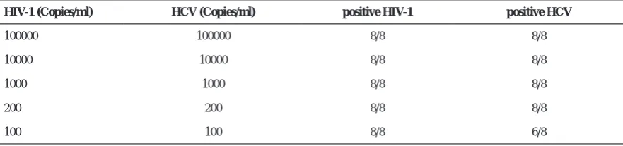
HIV-1 (Copies/ml) HCV (Copies/ml) positive HIV-1 positive HCV
100000 100000 8/8 8/8
10000 10000 8/8 8/8
1000 1000 8/8 8/8
200 200 8/8 8/8
100 100 8/8 6/8
* Eight multiplex reactions in which the concentration of each viral plasmid standard was equal. All eight reactions in each
group of standards showed positive results for HIV-1. For HCV, 6 out of 8 reactions were positive for 102 standard. In
other groups of standards all eight reactions were positive for HCV. Therefore the analytical sensitivity of the assay considered 100 and 200 copies/ml for HIV-1 and HCV, respectively.
negative samples, were used. These samples were tested previously using serological and molecular methods and confirmed that they were HIV-1 and HCV negative. To determine the analytical sensitivity, the plasmids mentioned above were used. A serial dilution of each standard plasmid from 106-102 copies/
ml was prepared. Multiplex-PCR was performed on each standard in a quadruplicate mode and repeated the next day to inspect the limit of detection (LOD) of the assay.
Finally to assess the clinical sensitivity, 40 positive samples including 30 samples from co-infected HIV-1 and HCV patients and ten manually spiked samples, prepared from mixing plasma of patients with HIV-1 or HCV infection with plasma of healthy individuals, were tested.
ReSUlTS
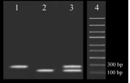
Performing multiplex RT-PCR and running 3% agarose gel electrophoresis. Fig. 1 Shows the gel electrophoresis related to Multiplex RT-PCR in which both specific targets have been amplified and separated based on their size.
Multiplex RT-PCR sensitivity. In order to determine the analytical sensitivity of the multiplex assay, eight multiplex RT-PCR of each standard plasmid were performed (105 copies/ml, 104 copies/ml, 103 copies/
ml, 200 copies/ml, 100 copies/ml (Table 2). In each group of reactions, the load of each standard was equal. The analytical sensitivity of the assay was 100
copies/ml and 200 copies/ml for HIV-1 and HCV, respectively.
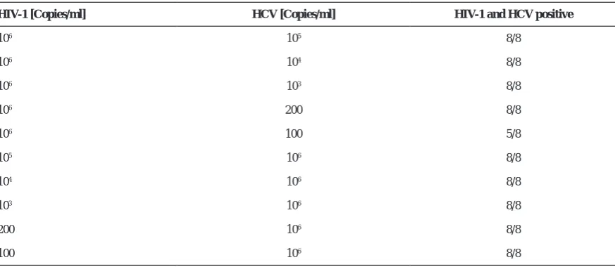
Since the analytical sensitivity of multiplex PCR could be affected by high concentration of each target sequence, a serial dilution of each viral standard were tested against a high copy number/ml [106 ] standard
of the other one. As shown in the Table 3 high viral load of one target sequence does not influence the analytical sensitivity of the other virus with low concentration.
To inspect the clinical sensitivity, 40 co-infected samples were analyzed. Ten samples were manually prepared by adding HIV-1 and HCV infected plasma within healthy plasma. It should be noted that in those ten manually prepared samples, the load of each infected sample was one log higher than the analytical sensitivity of the assay, in order to assess the capability of the assay when the viral load is low.
Using the designed assay, 39 out of 40 samples were positive for both viruses and one sample was positive only for HCV (Fig. 2). Therefore the clinical sensitivity of this assay was 97.5%.
Determining the specificity. In order to determine the analytical specificity, first, a BLAST was carried out using NCBI Nucleotide BLAST software. It was proved that the primers do not attach to any other sequences except HIV-1 or HCV. Then, ten different samples including human genome and some blood transmitted viruses, like HBV, HTLV-1, TTV, B19, HSV-1, HSV-2, HHV-6, HHV-7, HCMV and EBV were examined. Only HIV-1 and HCV and no other
HIV-1 [Copies/ml] HCV [Copies/ml] HIV-1 and HCV positive
106 105 8/8
106 104 8/8
106 103 8/8
106 200 8/8
106 100 5/8
105 106 8/8
104 106 8/8
103 106 8/8
200 106 8/8
100 106 8/8
* This experiment was performed to inspect the effect of high concentration of one virus on the amplification of the other one.
High concentration of one target has no significant effect on the amplification of the other target with low concentration.
Table 3*. Analytical sensitivity of the Multiplex assay with different standard concentration.
irrelevant genomes were detected (data not shown). Clinical specificity was assessed by using 25 negative samples which were proved to be negative by standard serological and molecular methods previously mentioned. No positive results were observed, thus the clinical specificity of the assay considered 100%.
DSICUSSION
The objective of developing this multiplex RT-PCR was the simultaneous detection of HIV-1 and HCV in patients’ plasma. To do so, conserved regions of genomes of each virus were amplified using specific primer pairs. The concentration of MgCl2 and primers were optimized, as these are fundamental factors affecting specificity and sensitivity. Analysis of the multiplex RT-PCR products is based on 3% agarose gel electrophoresis which shows the amplification of the templates. Gel electrophoresis showed two different bands of 179 and 241 bp indicating the amplification of HIV-1 and HCV, respectively (Fig. 1). The first multiplex assay for the detection of HIV-1 and HCV was based on monoplex and simultaneously flowcytometric detection which was cumbersome (28). Other multiplex methods like AMPLINAT PMX test (16) and TMA-based assay (29) and multiplex real-time PCR (18) are sensitive methods for the detection of HIV-1 and HCV, but these methods are expensive and could not be used as a screening method. In contrast, multiplex RT-PCR has considerable advantages, such as being less time consuming because of multiplexing, high sensitivity, reproducibility and straightforwardness.
Candiotti et al. (2004) developed a multiplex real-time PCR for the simultaneous detection of HBV, HCV and HIV-1 based on Hydrolysis Probes (19). Although this method has high sensitivity, it is much more expensive than Multiplex RT-PCR which makes it less optimal as a simple screening method.
In another experiment, De Cringes et al. (2010) used a SYBR Green I Multiplex Real-Time PCR which was able to differentiate HIV-1 and HCV in DBS1 samples based on amplicon melting temperature (30). The analytical sensitivity of the assay was 400 copies/ ml for HIV-1 and 2500 copies/ml for HCV which was not highly sensitive. In an article published by Giblini in 2006, it was shown that by using SYBR Green I multiplex real-Time PCR, it is possible to detect the co-infection of HIV-1 and HCV in plasma samples with the sensitivity of 500 copies/ml. The primers were designed for gag region of HIV-1 and 5’UTR of HCV (3). Tang et al. developed a new detection tool-visual DNA microarray for simultaneous and specific detection of human immunodeficiency virus type-1 and hepatitis C virus. The sensitivity of visual DNA microarray was (103copies/ml) (31).
In this article we describe the development of a Multiplex RT-PCR assay for simultaneous detection of HIV-1 and HCV viruses in plasma samples. This assay is able to detect and differentiate these viruses based on the difference in amplicon size on 3% agarose gel with a high sensitivity. In order to design primers, a multiple alignment was performed and the most conserved regions were selected. Based on the BLAST results and inspection of other interfering factors, like DNA extracted from human samples and viruses transmitted by blood, it was shown that the designed primers, and in other words the assay is capable of detecting HIV-1 and HCV and there is
20
Fig. 1.
Agarose gel electrophoresis analysis of HIV-1 and HCV monoplex and multiplex
amplification. Lane 1 and 2 are the monoplex reactions and lane 3 is the multiplex
reaction. HIV-1-specific amplicon is 197bp and HCV-specific amplicon is 241bp. Lane 4
is 100 Kb DNA marker.
Fig. 2.
Determining the clinical sensitivity using co-infected samples . The agarose gel
above shows part of the results. Lanes 1 through 6 are manually prepared samples by
adding HIV-1 and HCV infected plasma together with healthy plasma. M: 100 bp DNA
marker, NTC: No Template Control
Fig. 1. Agarose gel electrophoresis analysis of HIV-1 and HCV monoplex and multiplex amplification. Lane 1 and 2 are the monoplex reactions and lane 3 is the multiplex reaction. HIV-1-specific amplicon is 197bp and HCV-specific amplicon is 241bp. Lane 4 is 100 Kb DNA marker.
Fig. 2. Determining the clinical sensitivity using co-infected samples. The agarose gel above shows part of the results. Lanes 1 through 6 are manually prepared samples by adding HIV-1 and HCV infected plasma together with healthy plasma. M: 100 bp DNA marker, NTC: No Template Control.
no cross reaction with other viruses. The analytical sensitivity of the assay for HIV-1 and HCV was 100 and 200 copies/ml, respectively. As shown in (Table 3) although the presence of high concentration of either virus slightly lowers the sensitivity of the multiplex assay (especially in low concentrations of HCV), it has no effect on analytical sensitivity of the assay. In order to determine the analytical sensitivity, samples from HIV-1 and HCV infected patients with known copy number were used. In order to reduce the error in determining the initial copy number of samples, a mean of three quantifications in three different days using standard commercial kits were calculated and used. It should be noted that in this process, the coefficient of variation was lower than 5% by analyzing 40 HIV-1 and HCV positive samples and 20 samples from healthy individuals. The clinical sensitivity and specificity of the assay were determined to be 97.5% and 100%, respectively. A considerable matter in calculating the clinical sensitivity is that at least, 20 samples out of 40 positive co-infected ones, have low copy numbers (maximally one log more than the analytical sensitivity) of either virus. And this shows the capability of the developed assay to detect low copy number of virus in patients’ plasma; which is crucial in HIV-1 HCV patients.
In addition to high sensitivity and specificity, low cost and simplicity are the major advantages of this assay. Furthermore, the assay is rapid and practical for screening of HIV-1 and HCV in patient plasma and could be exploited in blood screening at blood transfusion organizations.
ACKNOWOleDGeMeNT
This work was supported by a grant from Tarbiat Modares University of Medical Sciences.
ReFeReNCeS
Alter MJ, Margolis HS, Krawczynski K, Judson FN, 1.
Mares A, Alexander WJ, et al. The natural history of community-acquired hepatitis C in the United States. The Sentinel Counties Chronic non-A, non-B Hepatitis Study Team. N Engl J Med 1992; 327: 1899-1905. Sherman KE, Rouster SD, Chung RT, Rajicic N. 2.
Hepatitis C Virus prevalence among patients infected with Human Immunodeficiency Virus: a cross-sectional analysis of the US adult AIDS Clinical Trials Group.
Clin Infect Dis 2002; 34: 831-837.
Gibellini D, Gardini F, Vitone F, Schiavone P, Furlini 3.
G, Re MC. Simultaneous detection of HCV and HIV-1
by SYBR Green real time multiplex RT-PCR technique in plasma samples. Mol Cell Probes 2006; 20: 223-229. Thimme R, Spangenberg HC, Blum HE. Hepatitis 4.
B or hepatitis C and human immunodeficiency virus infection. J Hepatol 2005; 42 Suppl (1): S37-44. Gibb DM, Goodall RL, Dunn DT, Healy M, Neave P, 5.
Cafferkey M, et al. Mother-to-child transmission of hepatitis C virus: evidence for preventable peripartum transmission. Lancet 2000; 356(9233): 904-907. Michaels SH, Clark R, Kissinger P. Declining morbidity 6.
and mortality among patients with advanced human immunodeficiency virus infection. N Engl J Med 1998; 339: 405-406.
Sulkowski MS, Mast EE, Seeff LB, Thomas DL. 7.
Hepatitis C virus infection as an opportunistic disease in persons infected with human immunodeficiency virus.
Clin Infect Dis 2000 Suppl 1(1): S77-84.
Sulkowski MS, Moore RD, Mehta SH, Chaisson RE, 8.
Thomas DL. Hepatitis C and progression of HIV disease. JAMA 2002; 288: 199-206.
Greub G, Ledergerber B, Battegay M, Grob P, Perrin 9.
L, Furrer H, et al. Clinical progression, survival, and immune recovery during antiretroviral therapy in patients with HIV-1 and hepatitis C virus coinfection: the Swiss HIV Cohort Study. Lancet. 2000 Nov 25; 356(9244): 1800-5.
Monga HK, Rodriguez-Barradas MC, Breaux K, 10.
Khattak K, Troisi CL, Velez M, et al. Hepatitis C virus infection-related morbidity and mortality among patients with human immunodeficiency virus infection.
Clin Infect Dis 2001; 33: 240-247.
Busch MP, Kleinman SH, Jackson B, Stramer SL, 11.
Hewlett I, Preston S. Committee report. Nucleic acid amplification testing of blood donors for transfusion-transmitted infectious diseases: Report of the Interorganizational Task Force on Nucleic Acid Amplification Testing of Blood Donors. Transfusion
2000 ; 40: 143-159.
Pawlotsky JM. Use and interpretation of virological 12.
tests for hepatitis C. Hepatology 2002; 36 (5 Suppl 1): S65-73.
Allain JP. Will genome detection replace serology in 13.
blood screening for microbial agents? Baillieres Best Pract Res Clin Haematol 2000; 13: 615-629.
Reichelderfer PS, Coombs RW. Virologic parameters as 14.
surrogate markers for clinical outcome in HIV-1 disease: verification, variation, and validation. J Acquir Immune Defic Syndr Hum Retrovirol 1995;10 Suppl 2: S19-24. Stramer SL, Caglioti S, Strong DM. NAT of the United 15.
States and Canadian blood supply. Transfusion 2000; 40: 1165-1168.
Meng Q, Wong C, Rangachari A, Tamatsukuri S, Sasaki 16.
M, Fiss E, et al. Automated multiplex assay system for simultaneous detection of hepatitis B virus DNA, hepatitis C virus RNA, and human immunodeficiency virus type 1 RNA. J Clin Microbiol 2001; 39: 2937-2945. Mine H, Emura H, Miyamoto M, Tomono T, Minegishi 17.
K, Murokawa H, et al. High throughput screening of 16 million serologically negative blood donors for hepatitis
B virus, hepatitis C virus and human immunodeficiency virus type-1 by nucleic acid amplification testing with specific and sensitive multiplex reagent in Japan. J Virol Methods 2003; 112(1-2): 145-151.
Adami V, Falasca E, Dorotea L, Malangone W, Astori 18.
G, Marini L, et al. Qualitative multiplex RT-PCR for simultaneous detection of hepatitis C virus and human immunodeficiency virus in plasma samples. Clin Microbiol Infect 2004; 102: 1075-1080.
Candotti D, Temple J, Owusu-Ofori S, Allain JP. 19.
Multiplex real-time quantitative RT-PCR assay for hepatitis B virus, hepatitis C virus, and human immunodeficiency virus type 1. J Virol Methods 2004; 118: 39-47.
Giachetti C, Linnen JM, Kolk DP, Dockter J, Gillotte-20.
Taylor K, Park M, et al. Highly sensitive multiplex assay for detection of human immunodeficiency virus type 1 and hepatitis C virus RNA. J Clin Microbiol 2002; 40: 2408-2419.
Shrestha NK, Tuohy MJ, Hall GS, Reischl U, Gordon 21.
SM, Procop GW. Detection and differentiation of Mycobacterium tuberculosis and nontuberculous mycobacterial isolates by real-time PCR. J Clin Microbiol 2003; 41: 5121-5126.
Berry N, Herrera C, Cranage M. Detection, quantification, 22.
and characterisation of HIV/SIV. Methods Mol Biol. 2011; 665: 133-60.
Beuret C. Simultaneous detection of enteric viruses by 23.
multiplex real-time RT-PCR. J Virol Methods 2004; 115: 1-8.
Goire N, Freeman K, Tapsall JW, Lambert SB, 24.
Nissen MD, Sloots TP, et al. Enhancing gonococcal antimicrobial resistance surveillance: a real-time PCR assay for detection of penicillinase-producing Neisseria gonorrhoeae using non-cultured clinical samples. J Clin
Microbiol 15; 2010: 2015.
Richards GP, Watson MA, Kingsley DH. A SYBR 25.
green, real-time RT-PCR method to detect and quantitate Norwalk virus in stools. J Virol Methods 2004 1; 116: 63-70.
Wei X, Youngpairoj AS, Garrido C, Zahonero N, 26.
Corral A, de Mendoza C, et al. Minority HIV mutation detection in dried blood spots indicates high specimen integrity and reveals hidden archived drug resistance. J Clin Virol 2011; 50: 148-152.
Weidner J, Cassens U, Gohde W, Sibrowski W, Odaibo 27.
G, Olaleye D, et al. An improved PCR method for detection of HIV-1 proviral DNA of a wide range of subtypes and recombinant forms circulating globally. J Virol Methods 2011; 172: 22-26.
Defoort JP, Martin M, Casano B, Prato S, Camilla C, 28.
Fert V. Simultaneous detection of multiplex-amplified human immunodeficiency virus type 1 RNA, hepatitis C virus RNA, and hepatitis B virus DNA using a flow cytometer microsphere-based hybridization assay. J Clin Microbiol 2000; 38: 1066-1071.
Vargo J, Smith K, Knott C, Wang S, Fang C, McDonough 29.
S, et al. Clinical specificity and sensitivity of a blood screening assay for detection of HIV-1 and HCV RNA.
Transfusion. 2002; 42: 876-885.
De Crignis E, Re MC, Cimatti L, Zecchi L, Gibellini 30.
D. HIV-1 and HCV detection in dried blood spots by SYBR Green multiplex real-time RT-PCR. J Virol Methods 2010; 165: 51-56.
Tang J, Zhou L, Duan LL, Feng Y, Cao X, Wang Y. 31.