Research Article
Serum Level of Vascular Endothelial Growth Factor (VEGF) can be used
to Assess Response of Radiation Therapy in Cervical Cancer
Kadar Serum Vascular Endothelial Growth Factor (VEGF) dapat Digunakan
untuk Menilai Respons Terapi Radiasi pada Kanker Serviks
Ferry Armanza1, Andrijono1, Bambang Sutrisna2 1Division of Oncology Gynecology,
Department Of obstetrics and Gynecology Faculty of Medicine University of Indonesia/ 2Faculty of Public Health University of Indonesia
Dr. Cipto Mangunkusumo General Hospital Jakarta
INTRODUCTION
Cervical cancer is the primary cancer of the cer-vix (cervical canal and/or portio). Cervical cancer
ranks first in overall cancer incidence in Indone-sian women.1 Approximately 70% of cervical can-cer patients came at advanced stage (> II-B).
Re-Abstract
Objective: To compare the sensitivity and specificity of Squamous Cell Carcinoma (SCC) and Vascular Endothelial Growth Factor (VEGF) levels to assess the response of radiation therapy.
Method: The study was conducted by the method of analytic obser-vational cohort study in 24 patients with cervical cancer stage II-B and III-B in RSCM that met inclusion criteria. Examination of VEGF and SCC in serum samples was performed in the Prodia Laboratory Jakarta. The examination was conducted twice before and after ra-diation therapy. The subjects were treated by rara-diation therapy/ chemoradiation according to standard procedures. After the com-pletion of radiation was declared, the response of radiation therapy was conducted by clinical assessment.
Result: Of the 24 subjects, we obtained a mean level of SCC pre-ra-diation was 23.43 ± 5.84 ng/ml and post-radiation was 2.19 ± 0.68 ng/ml. The mean VEGF pre-radiation was 790.41 ± 111.06 pg/ml and post-radiation was 497.47 ± 79.26 pg/ml. ROC curves of each tu-mor marker obtained SCC pre-radiation AUC 40%, p 0.53 (CI 0.18-0.68) and SCC post-radiation AUC 48.1%, p 0.91 (CI 0.21-0,75) can not be used as a diagnostic and prognostic factors of response to ra-diation therapy. VEGF pre-rara-diation produced an AUC of 17.5%, p 0.04 (CI 0.00-0.36), thus cannot be used as a prognostic factor for re-sponse to radiation therapy. VEGF after radiation produced an AUC of 92.5%, p 0.01 (CI 0.81-1.00), thus can be a diagnostic factor for response to radiation therapy. VEGF post-radiation with cut-off point 614.75 pg/ml had a sensitivity 80%, specificity 75%, NDP 94.12%, NDN 42.86%; RKP 3.2; RKN 0.26 and accuracy 79.16%. There is a significant correlation between the decrease of serum VEGF level post-radiation and a positive response of radiation the-rapy (p 0.01, CI 1.00-3.23).
Conclusion: Examination of VEGF levels can be used to assess the response of radiation therapy with a sensitivity of 80% and specifi-city of 75%.
[Indones J Obstet Gynecol 2014; 2-1: 40-45]
Keywords: cervical cancer, SCC, therapeutic response, VEGF
Abstrak
Tujuan: Penelitian ini bertujuan membandingkan sensitivitas dan spesifisitas kadar SCC dengan VEGF untuk menilai respons terapi ra-diasi.
Metode: Penelitian dengan metode studi kohort analitik observasional terhadap 24 subjek penelitian yaitu penderita kanker serviks stadium II-B dan III-B di RSCM yang memenuhi kriteria inklusi. Pemeriksaan VEGF dan SCC pada sampel serum dilakukan di Lab. Prodia Pusat Ja-karta, sebanyak dua kali pemeriksaan yaitu sebelum dan sesudah te-rapi radiasi. Subjek ditatalaksana dengan tete-rapi radiasi/kemoradiasi sesuai prosedur standar. Setelah terapi dinyatakan selesai, kemudian dilakukan penilaian respons terapi secara klinis.
Hasil: Dari 24 subjek penelitian didapatkan rerata kadar petanda tu-mor SCC praradiasi 23,43 ± 5,84 ng/ml dan pascaradiasi 2,19 ± 0,68 ng/ml. Rerata VEGF praradiasi 790,41 ± 111,06 pg/ml dan pascara-diasi 497,47 ± 79,26 pg/ml. Dari kurva ROC masing-masing petanda tumor didapatkan SCC praradiasi AUC 40%; p 0,53 (IK 0,18-0,68) dan SCC pascaradiasi AUC 48,1%; p 0,91 (IK 0,21-0,75) tidak dapat dipakai sebagai faktor prognostik maupun diagnostik terhadap respons terapi radiasi pada kanker serviks. VEGF praradiasi AUC 17,5%; p 0,04 (IK 0,00-0,36) tidak dapat dipakai sebagai faktor prognostik terhadap respons terapi radiasi pada kanker serviks. VEGF pascaradiasi AUC 92,5%; p 0,01 (IK 0,81-1,00) dapat menjadi faktor diagnostik terhadap respons terapi radiasi pada kanker serviks. VEGF pascaradiasi dengan titik potong (cut-off point) 614,75 pg/ml memiliki sensitivitas 80%; spesifisitas 75%; NDP 94,12%; NDN 42,86%; RKP 3,2; RKN 0,26 dan akurasi 79,16%. Terdapat korelasi bermakna antara penurunan ka-dar serum VEGF pascaterapi dengan respons radiasi positif (p 0,01; IK 1,00-3,23).
Kesimpulan: Pemeriksaan kadar VEGF dapat digunakan untuk meni-lai respons terapi radiasi dengan sensitivitas 80% dan spesifisitas 75%.
[Maj Obstet Ginekol Indones 2014; 2-1: 40-45] Kata kunci: kanker serviks, respons terapi, SCC, VEGF
search by Sarika (2006) found from the 465 new cases of cervical cancer, most cases were diagnosed as stage III-B with the most common histopathol-ogy was squamous cell carcinoma.2
Primary therapy of advanced cervical cancer is radiation. But radiation therapy is known to be able to cause complications, such as fibrosis that causes stiffness in the radiated tissue. Advanced cervical cancer generally have a large tumor size and extension. Radiation therapy may not kill can-cer cells in the central region. Assessment of clini-cal response is based solely on the findings of the response at the surface, so it will face obstacles in the face of treatment response. One method to as-sess treatment response is the examination of tu-mor markers.
SCC antigen is a tumor marker for the presence of squamous cells that produce serine protease in-hibitor. Some studies found that serum SCC can be used to monitor the onset of cervical squamous cell carcinoma after primary therapy. SCC serum levels were still high and an increase after treatment showed a tumor or progressive disease. Using a cut-off value of SCC 3.0 ng/ml Strauss HG (2002) et al detect levels of serum SCC antigen pre-opera-tive as an independent prognostic factor in squa-mous cell cervical cancer, both of reccurence-free and overall survival. From the analysis concluded that SCC antigen as tumor markers correlate with prognosis in operability of cervical cancer, tumor size, status of pelvic nodes, cervical stromal infil-tration, and the degree of spread of the tumor into the parametrium.3
Hong JH (1998) et al who examined the levels of squamous cell carcinoma (SCC) found that levels of SCC antigen > 10 ng/ml is an independent pre-dictor of poor prognosis in cervical cancer and can be used as a prognostic factor in selecting patients for intensive therapy. SCC levels were still high af-ter radiation therapy is a strong predictor of treat-ment failure.4
Vascular endothelial growth factor (VEGF) plays an important role in tumor angiogenesis by in-creasing vascular permeability, endothelial cell growth, proliferation, migration and differentia-tion. VEGF can also facilitate the extravasation of tumor cells and then metastasize by way of de-stroying the tumor extracellular matrix wall by ac-tivating the proteolytic enzymes.5
Although a lot of evidence to suggest that VEGF
plays a role in microinvasion at an early stage, there has been no extensive studies to assess VEGF expression as a prognostic factor in locally ad-vanced cervical cancer, including radiation therapy outcomes.6,7
Evaluation of the expression levels of VEGF are also useful in assessing response to therapy, such as post-radical hysterectomy, chemoradiation, and even monitor the results of treatment with anti-angiogenesis drugs. This research was undertaken to assess whether serum VEGF levels have prog-nostic value and can be used in the treatment of cervical cancer patient.8,9
As tumor markers both VEGF and SCC need to be explored to determine which one is more sen-sitive and specific for assessing response to radia-tion therapy in cervical cancer.
METHODS
This study was a cohort study, which examined the serum levels of VEGF and SCC antigen in patients with cervical cancer stage IIB - IIIB before and after radiation therapy. The objective was to determine the relationship between the serum level of VEGF and SCC with radiation therapy response and then compare the sensitivity and specificity of VEGF and SCC to the response of radiation therapy.
The study population was all cervical cancer pa-tients who came to RSUPN Dr. Cipto Mangun-kusumo Jakarta with the following inclusion crite-ria, clinical stage IIB-IIIB (according to FIGO) with histopathology results squamous cell carcinoma; underwent chemoradiation/radiation; does not have any other systemic diseases that affect the levels of serum SCC antigen and VEGF and has not received treatment for cancer.
Patients were managed by chemoradiation or radiation in accordance with the study protocol. Af-ter chemoradiation or radiation has completed, an-other venous blood sampling was taken again about 2 (two) weeks after radiation therapy com-pleted, to examine VEGF and SCC antigen serum levels and do clinical examination to assess the re-sponse of chemoradiation therapy.
progressive. In this study, interpretation was di-vided into two groups of responses, positive res-ponse = complete resres-ponse and negative resres-ponse = response beside complete response.
RESULTS
The study subjects was 24 patients with advanced cervical cancer, which comprised of 9 patients stage II-B and 15 patients stage III-B. The tumor markers, Squamous Cell Carcinoma Antigen (SCC antigen) and Vascular Endothelial Growth Factor (VEGF), was measured twice, before and after ra-diation. The mean level of SCC pre-radiation was 23.43 ± 5.84 ng/ml and post-radiation was 2.19 ± 0.68 ng/ml, and the mean level of VEGF pre-radia-tion was 790.41 ± 111.06 pg/ml and post-radiation was 497.47 ± 79.26 pg/ml.
Measurement of tumor markers pre-radiation was performed to assess whether the SCC and VEGF could be used as a prognostic factor of res-ponse to radiation therapy. While the post-radia-tion was examined to assess whether the SCC and VEGF have diagnostic value to replace the clinical judgement that had been done before. Based on measurements using ROC curves, we obtained re-spectively SCC tumor markers and VEGF for thera-peutic response. (Table 1)
Table 1. Results of ROC Curves of Tumor Markers and Response of Radiation Therapy
Tumor markers AUC (%) p CI 95%
SCC:
Pre-radiation 40.0 0.53 0.18 - 0.68 Post-radiation 48.1 0.91 0.21 - 0.75
VEGF:
Pre-radiation 17.5 0.04 0.00 - 0.36 Post-radiation 92.5 0.01 0.81 - 1.00 AUC: area under curve
VEGF post-radiation with AUC 92.5% results, show that VEGF levels are excellent (AUC> 90%) to be used as a diagnostic factor in diagnosing re-sponse of radiation therapy in cervical cancer with p: 0.01 (CI: 0.81 - 1.00). (Fig. I)
To get the cut-off point of serum VEGF levels af-ter radiation, we made a graphof VEGF sensitivity and specificity values. (Fig. II)
Post radiation serum VEGF levels at 614.75 pg/ml had a highest sensitivity and specificity val-ues, with sensitivity 80% and specificity 75%. Ac-cording to the 2x2 table with a value of 614.75 pg/ml as the cut-off point on the response of ra-diation therapy, we obtained the following results. (Table 2)
Figure 1. ROC Curve of VEGF Serum Levels in Post-radiation Patients.
Table 2. Diagnostic Value of VEGF (pg/ml) Post-Radia-tion and Response of RadiaPost-Radia-tion Therapy.
VEGF value
Response of therapy
Total Positive (%) Negative (%)
≤614.75 16 94.1 1 5.9 17
>614.75 4 57.1 3 42.9 7
Total 20 83.3 4 16.7 24
From the table above, the value obtained sensi-tivity 80%, specificity 75%, positive predictive va-lue (PPV) 94.12%, negative predictive vava-lue (NPV) 42.86%, positive likelihood ratio (PLR) 3.2; nega-tive likelihood ratio (NLR) 0.26 and accuracy 79.16%.
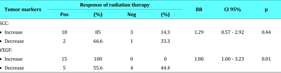
Table 3 below shows the trend of up and down inboth serum tumor markers SCC and VEGF for therapeutic response.
It appears that the decline in serum levels of SCC from pre to post-radiation did not correlate with a positive response (p 0.44; CI 0.57 to 2.92). While the decline in serum levels of VEGF in pre and post-radiation therapy correlated with a positive res-ponse (p 0.01, CI 1.00 to 3.23).
DISCUSSION
Based on the pathology report in 2002, cervical cancer ranks first out of 10 cases of most cancers in both men and women and cancer in women only, with a total of 2532 cases.10 From the results of tumor markers measurements, it appears that the SCC tumor markers can not be used to assess response of radiation therapy. SCC pre-radiation levels reached only 40% AUC with p: 0.53 (CI: 0.18 to 0.68). These results indicate that the SCC
pre-radiation can not be used to assess the response to radiation therapy in cervical cancer. The SCC le-vels reached 48.1% AUC after radiation with p: 0.91 (CI: 0.21 to 0.75) indicating that the SCC also can not substitute the clinical assessment of thera-peutic response.
VEGF as tumor markers was also measured be-fore and after radiation. VEGF results obtained pre-radiation covers only AUC 17.5%, although the p: 0.04 (CI: 0.00 - 0.36), as same as SCC, can not be used for assessing response to radiation therapy in cervical cancer. But VEGF post-radiation with AUC 92.5% results, show that VEGF levels are excellent (AUC> 90%) to be used as a diagnostic factor in diagnosing response of radiation therapy in cervi-cal cancer with p: 0.01 (CI: 0.81 - 1.00).
Hong JH (1998) et al examined the levels of squamous cell carcinoma (SCC) and found that
le-vels of SCC antigen > 10 ng/ml was an independent predictor of poor prognosis in cervical cancer, and could be used as a prognostic factor in selecting patients for intensive therapy. High levels of SCC antigen after radiation therapy was a strong pre-dictor of treatment failure.4
Some studies find that serum SCC antigen can be used to monitor the incidence of cervical squa-mous cell carcinoma after primary therapy. SCC an-tigen serum levels which remains high or even in-crease after treatment showed a tumor or progres-sive disease.3,11,12
Radiation therapy is the primary treatment mo-dality in invasive cervical cancer and can achieve satisfactory results in patients with early stage. While in patients with advanced stage therapeutic modalities are still many failures. Preliminary re-Table 3. Classification of Tumor Markers pre and Post Radiation and Therapeutic Response.
Tumor markers Response of radiation therapy RR CI 95% p
Pos (%) Neg (%)
SCC:
• Increase 18 85 3 14.3 1.29 0.57 - 2.92 0.44
• Decrease 2 66.6 1 33.3
VEGF:
• Increase 15 100 0 0 1.80 1.00 - 3.23 0.01
search conducted by Hong JH et al (1998) showed that there is a failure rate of 30% in patients with squamous cell carcinoma of the cervix with large lesions (bulky) stage IB-IIA and IIB after definitive radiation therapy, and will increase to 50% in pa-tients with stage III.3,4,7
Evaluation of the expression levels of VEGF are also useful in assessing therapeutic response, such as post-radical hysterectomy, chemoradiation, and even monitor the results of treatment with anti-angiogenesis drugs.7,8
Serum levels of VEGF post-radiation with AUC 92.5% results, show that VEGF levels are excellent (AUC > 90%) to be used as a diagnostic factor in diagnosing response of radiation therapy in cervi-cal cancer with p: 0.01 (CI: 0.81 - 1.00).
Cheng WF get a mean (median) intra-tumoral VEGF protein in patients with cervical cancer was 180 pg/mg, whereas in normal tissue 0 pg/mg. Large lesions > 4 cm than < 4cm (1030 compared to 118). Limfo-vascular invasion than not (568 compared to 118) and patients with lymph-nodes metastasis than without metastasis (795.5 com-pared to 121 pg/mg). Over-expression of VEGF ob-tained from immuno-histochemical examination of 10/20 (50%) in cases with metastatic nodes, while the nodes without metastasis at 16/84 with p = 0.002.13
In this study, analysis of ROC curve and sensi-tivity and specificity graphs showed that VEGF lev-els of 614.75 pg/ml had a highest sensitivity (80%) and specificity (75%). The results suggest that se-rum VEGF levels after radiation with a cut-off 614.75 pg/ml can be used to diagnose the response of radiation therapy in cervical cancer, as mention from the values of sensitivity, specificity, PPV, NPV and sufficient accuracy, although the value of PLR 3.2 (<10) and NLR 0.26 (> 0.1)is still inadequate.
Hansgen et al which examined serum VEGF in 42 patients with locally advanced (FIGO II-IV) squamous cell cervical cancer who were treated with radiation, found that the concentration of VEGF did not correlate with tumor stage. Compari-son of VEGF levels with clinical outcome after 6 months of therapy with patients obtained a com-plete response occurred significantly decreased levels of VEGF (304 pg/ml ± 188) compared to pa-tients with symptoms of tumor (892 pg/ml ± 756, p <0.0005). Thus concluded that high serum VEGF levels before therapy associated with poor
res-ponse to radiation therapy in locally advanced cer-vical cancer.14-16
This study also found that the reduction in VEGF serum levels pre and post-radiation were associ-ated with a positive treatment response (p 0.01, CI 1.00 to 3.23). Thus if serum VEGF decrease after radiation therapy, the therapeutic response will be positive.
Loncaster et alexamined 100 patients with lo-cally advanced cervical cancer (IB large lesions up to IIIB), which consisted of 94 squamous cell car-cinomas, 5 adenocarcinomas and 1 adenosqua-mous carcinoma, found that there is no correlation between VEGF expression with disease stage, tu-mor differentiation, patient age and tutu-mor radio-sensitivity. In survival analysis, VEGF expression was an independent prognostic factor of the most significant (p = 0.001).6,13-17
This study suggests that serum VEGF levels post radiation can be used to be a diagnostic factor for response of radiation therapy in advanced cervical cancer with AUC 92.5%, p 0.01 (CI: 0.81 to 1.00). VEGF serum levels with cut-off 614.75 pg/ml had a sensitivity of 80%, specificity 75%, PPV 94.12%, NPV 42.86%, PLR 3.2, NLR 0.26 and accuracy 79.16%. And there is a significant correlation be-tween the decrease in serum VEGF levels after ra-diation therapy with a positive response of radia-tion therapy (p 0.01, CI 1.00 to 3.23).
CONCLUSION
Examination of VEGF levels can be used to assess the response of radiation therapy with a cut off point of 614.75 pg/ml, with high sensitivity (80%) and specificity(75%). There is also a significant correlation between the decrease in serum VEGF levels after radiation therapy with a positive re-sponse of radiation therapy.
REFERENCES
1. Andrijono. Kanker serviks uteri. Sinopsis Kanker Ginekologi. 3 ed. Jakarta: Pustaka Spirit; 2009: 59.
2. Sarika DT. Korelasi stadium dengan usia penderita kanker serviks di Departemen Patologi Anatomik RSCM tahun 2006. http://lontar.ui.ac.id/opac/themes/libri2/detail.jsp? id=123616&lokasi=lokal
The prognostic significance of pre- and posttreatment SCC levels in patients with squamous cell carcinoma of the cer-vix treated by radiotherapy. Int J Radiat Oncol Biol Phys. 1998; 41(4): 823-30.
5. Clauss M. Molecular biology of the VEGF and the VEGF re-ceptor family. Semin Thromb Hemost. 2000; 26: 561-70. 6. Loncaster JA, Cooper RA, Logue JP, Davidson SE, Hunter RD,
West CML. Vascular endothelial growth factor (VEGF) es-pression is a prognostic factor for radiotherapy outcome in advanced carcinoma of the cervix. Bri J Cancer. 2000; 83(5): 620-5.
7. Tandle A, Blazer DG, III, Libutti SK. Antiangiogenic gene therapy of cancer: recent developments. J Transl Med. 2004; 2(1): 22.
8. Libutti SK, Feldman AL. Antiangiogenic gene therapy. In: Lattime EC, Gerson SL, editors. Gene therapy of cancer: translational approaches from preclinical studies to clinical implementation. 2nd ed. San Diego: Academic Press; 2002:
405-15.
9. Brem S. Angiogenesis and Cancer Control: From Concept to Therapeutic Trial. Cancer Control. 1999; 6(5): 436-58. 10. Aziz MF. Gynecological cancer in Indonesia. J Gynecol Oncol.
2009; 20(1): 8-10.
11. Ngan HYS, Cheng GTS, Yeung WSB, Wong LC, Ma HK. The prognostic value of TPA and SCC in squamous cell carci-noma of the cervix. Gynecol Oncol. 1994; 52: 63-8.
12. Duk JM, Groenier KH, De Bruijn HWA, Hollema H, Ten Hoor KA, Van der Zee AGJ, et al. Pretreatment serum squamous cell carcinoma antigen: a newly identified prognostic factor in early-stage cervical carcinoma. J Clin Oncol. 1996; 14: 111-8.
13. Cheng WF, Chen CA, Lee CN, Chen TM, Hsieh FJ, Hsieh CY. Vascular endothelial growth factor in cervical carcinoma. Obstet Gynecol. 1999; 93(5): 761-4.
14. Bachtiary B, Selzer E, Knocke TH, Potter R, Obermair A. Se-rum VEGF levels in patients undergoing primary radiothe-rapy for cervical cancer: impact on progression-free sur-vival. Cancer Letters. 2002; 179: 197-203.
15. Hansgen G, Becker A, Hintner I, Dunst J. Vascular Endothe-lial Growth Factor (VEGF) in sera of patients with cervical cancer and the impact of platelets. Cancer cell biology and angiogenesis. 1999: 107.
16. Choi CH, Song SY, Choi JJ et al. Prognostic significance of VEGF expression in patients with bulky cervical carcinoma undergoing neoadjuvant chemotherapy. BMC Cancer. 2008; 8: 295.