Original Article
Expression of CD34 and CD31 in Central and Peripheral Giant Cell Granulomas
Donia Sadri 1, Fatemeh Shahsavari 1, Maliheh Hezarkhani 1, Maryam Shafizadeh 1
1
Dept. of Oral and Maxillofacial Pathology, Dental Branch of Tehran, Islamic Azad University, Tehran, Iran.
KEY WORDS
Angiogenesis; Pathologic;
Giant cell Granuloma; CD34;
CD31; Jaw;
Received October 2017;
Received in Revised form December 2017; Accepted February 2018;
ABSTRACT
Statement of the Problem: There are some differences between clinical features of central giant cell granulomas (CGCGs) and peripheral giant cell granulomas (CGCGs) despite their same microscopic features. The possible role of angiogene-sis in this issue is still a matter of debate.
Purpose: The aim of the present study was to compare microvessel density (MVD) between CGCGs and PGCGs of the oral cavity using CD31 and CD34.
Materials and Method: Immunohistochemical staining was performed on 18 PGCGs and 19 CGCGs using a monoclonal antibody against CD34 and CD31. MVD was assessed and compared between the lesions using t-test for statistical analysis. p< 0.05 was considered significant.
Results: The expression levels of both CD34 and CD31 were significantly higher in CGCGs compared to PGCGs (p< 0.002 and p< 0.001, respectively). Significant differences in MVD assessed by both markers were observed between males and females in PGCGs (p< 0.05), but not CGCGs (p< 0.2).
Conclusion: The combined evaluation of old- and newly-formed vessels by pan-endothelial cell markers showed differences between CGCGs and PGCGs, sup-porting the possible vascular-proliferative nature of the former. Whether this dif-ference has a part in their diverse biologic behaviors and the role which pre-existent vessels play in comparison to neo-formed vasculature, requires further investigation.
Corresponding Author:Sadri D., Dept. of Oral and Maxillofacial Pathology, Dental Branch of Tehran,
Islamic Azad University, Tehran, Iran. Email: [email protected] Tel: +98-2122763449
Cite this article as: Sadri D., Shahsavari F., Hezarkhani M., Shafizadeh M. Expression of CD34 and CD31 in Central and Peripheral Giant Cell Granulomas. J Dent Shiraz Univ Med Sci., March 2019; 20(1): 10-15.
Introduction
Intra- and extra-osseous lesions occur within the head and neck region, some of which are known counterparts like central and peripheral ameloblastomas and ghost cell odontogenic tumors. [1] However, this relationship is not so clear for giant cell lesions and it is still debatable whether these are separate entities or variants of a single lesion, which can be found at different locations. [2-3]
Peripheral giant cell granulomas (PGCGs) devel-op in response to local irritation or trauma, occasionally erode the underlying bone, and have a low recurrence rate, especially after adequate treatment. On the other hand, the etiology of central giant cell granulomas (CGCG) is controversial and they are known to
demon-strate diverse clinical features and behavior. Some cases demonstrate an indolent behavior and minimal symp-toms, while others develop in a younger age group, be-have aggressively and tend to recur. Despite their clini-cal differences, these intra- and extra-osseous lesions have similar histologic characteristics .They are com-prised of variable amounts of multinucleated giant cells in a background of the oval to spindle-shaped mononu-clear cells. [3] This contradiction has been a major con-cern among researchers leading to studies on various cytomorphometric, immunohistochemical, and ultra-structural aspects of these lesions. [2, 4-6]
has been shown that angiogenesis would affect the bio-logic behavior of various neoplastic and non-neoplastic diseases. This phenomenon is evaluated through as-sessment of MVD using various endothelial cell mark-ers [7] such as CD34 and CD31. CD34 is a 110-kDa cell surface glycoprotein and functions as a cell-cell adhesion factor. It may also mediate the attachment of stem cells to the bone marrow extracellular matrix or directly to stromal cells.Cells expressing CD34 (CD34+ cell) are normally found in the bone marrow as hemato-poietic cells, or in mesenchymal stem cells, endothelial progenitor cells, endothelial cells of blood vessel. [7-8]
CD31 is a 130-kDa glycoprotein that appears on blood endothelial cells, platelets, macrophages and lym-phocytes (T cells, B cells, and NK cells) and osteoclast by immunohistochemistry technique, CD31 is used to demonstrate the presence of endothelial cells in histo-logical tissue sections that helping to evaluate the de-gree of tumor angiogenesis. [7]
Vascular endothelial growth factor (VEGF) has been previously investigated in giant cell lesions and has been suggested that those situated in the jawbones, particularly, lie within the range of primary proliferative vascular lesions. [4] However, this notion was not sup-ported by Kahn et al. [9]
Antigenic factors like VEGF and basic fibroblast growth factor (bFGF) have been reported to have a closer relationship with osteoclast genesis than angio-genesis. [4, 9] Microvessel density (MVD) and mi-crovessel count using endothelial cell markers have been evaluated and compared between CGCG and PGCG with contradictory results. [4, 10-13]
Considering the importance of this process and the fact that endothelial cells not only function in angiogen-esis-related activities but also have a role in various phenomena, we aimed to evaluate angiogenesis in PGCG and CGCG using CD34 and CD31. We were not able to find previous research in this field using the lat-ter pan-endothelial protein.
Materials and Method
After obtaining ethical approval from the ethics com-mittee of our University, patient records were reviewed from 2004 to 2015 and clinical/demographic data for subjects with a diagnosis of giant cell granuloma were extracted. [3, 9] Considering clinical and radiographic
manifestations, all histologic slides were re-evaluated to confirm the diagnosis. [3] Samples with necrotic and/or inadequate tissue, extensive hemorrhage, or incomplete clinical information were excluded. Moreover, other giant-cell-containing lesions like aneurysmal bone cyst, brown tumor of hyperparathyroidism (confirmed by laboratory tests), cherubism, and peripheral ossifying fibroma were excluded from the study.
Paraffin-embedded blocks were retrieved cut into 3µm sections and immunohistochemically stained using the streptavidin-biotin method. All sections were dewaxed, rehydrated, and subjected to endogenous pe-roxidase blocking. This was followed by immersion in a fresh solution of 10mM citrate buffer at pH 6.0 and placing in a microwave for 10 minutes. After cooling at room temperature, they were rinsed in phosphate buffer saline and incubated in monoclonal antibody against CD31 (Dako, ready-to-use monoclonal mouse anti-human, clone JC70A, Denmark) and CD34 (Novocas-traTM ready-to-use mouse monoclonal antibody, product code: RTU-END, Germany) for 50 minutes. The sec-tions were then rinsed in PBS and reacted with biotinyl-ated secondary antibodies for 30 minutes followed by a second rinse in PBS and incubation with streptavidin-peroxidase (30 minutes) and a final rinse in PBS. Color was developed by exposure of the slides to 3-3ʹ dia-minobenzidine after which counterstaining with Harris’ hematoxylin was performed. Positive controls included pyogenic granuloma and solitary fibrous tumor for CD31 and CD34, respectively and endothelial cells in normal tissue vasculature were used as internal controls. Primary antibodies were omitted for both groups as negative controls. [14]
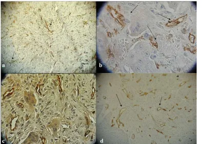
Figure 1a: Blood vessels immunostained by anti-CD34 antigen in CGCG (Nikon light microscope 400×), b: Blood vessels and stromal cells immunostained by anti-CD34 antigen in PGCG (Nikon light microscope 1000×), c: Blood vessels and stromal cells im-munostained by anti-CD31 antigen in CGCG (Nikon light microscope 1000×), d: Blood vessels imim-munostained by anti CD31 antigen in PGCG (Nikon light microscope 400×).
between the observers were resolved by consensus. Statistical analysis was performed using t-test and
p<0.05 was considered significant.
Results
According to the inclusion and exclusion criteria, our study sample consisted of 18 PGCGs of which 6 and 12 occurred in males and females, respectively (p= 0.23). Of these, 10 were found in the mandible and 8 in the maxilla. The youngest patient was a 13-year-old boy with a lesion on the left posterior mandibular gingiva and the oldest subject was 71 with a maxillary PGCG on the gingiva of the canine region. The number of CGCGs was 19 which occurred in 3 men and 16 women (p=0.004). A total of 17 were found in the mandible and 2 in the maxilla. A 17-year-old girl with a right mandib-ular lesion and a 47-year-old woman with a lesion in the left canine-premolar area constituted the youngest and oldest patients with CGCG in the current investigation. The mean and median ages for individuals with PGCG were 43.2 years and 42.5 years and for CGCG were 33.5 and 34 years, respectively. Median ages were used as the cut-off point to divide patients into younger and older groups as proposed previously. [10]
The expression level of CD34 was 17.6±5.7 and 24.5±6.6 in PGCGs and CGCGs, respectively (p< 0.002). (Figure 1a), (Figure 1b) correspondingly, these values were 10.6±2.3 and 19.6±5.3 (p< 0.001) for CD31. (Figure 1c), (Figure 1d)
There was an increased intensity/staining of both markers in the peripheral areas of the PGCG samples (Figure 1d).
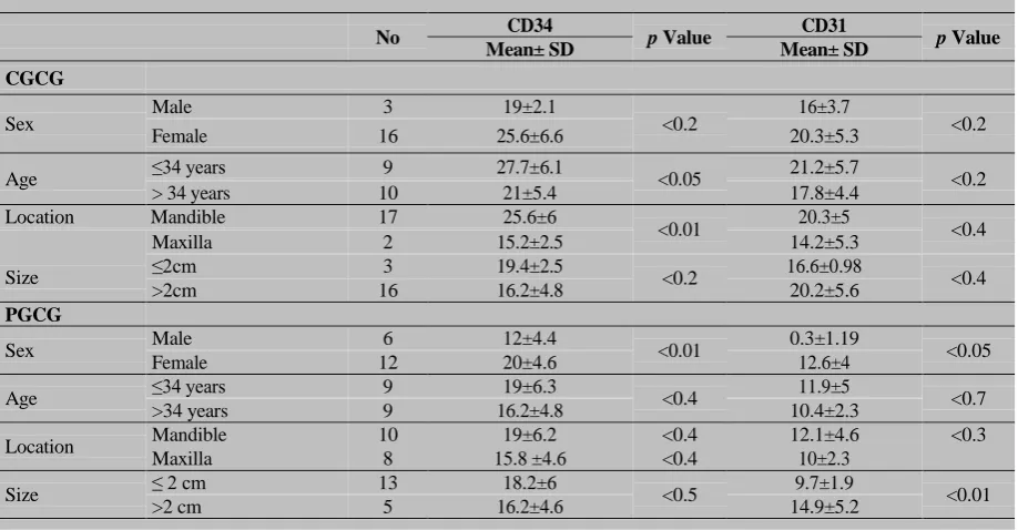
Table 1 shows immunostaining values of CD34 and CD31 according to the demographic features of patients with both lesions. A significant difference in CD34(p< 0.01) and CD31 (p< 0.05) was found between men and women in PGCGs. Expression levels of CD34 were significantly different between the two age groups (p< 0.05) in CGCG, while CD31 showed statistically significant difference in lesion size (p< 0.01) in PGCGs.
Discussion
Table 1: Mean of CD31 and CD34 expression according to clinical and demographic factors
No CD34 p Value CD31 p Value
Mean± SD Mean± SD
CGCG
Sex Male 3 19±2.1 <0.2 16±3.7 <0.2
Female 16 25.6±6.6 20.3±5.3
Age ≤34 years 9 27.7±6.1 <0.05 21.2±5.7 <0.2
> 34 years 10 21±5.4 17.8±4.4
Location Mandible 17 25.6±6
<0.01 20.3±5 <0.4
Size
Maxilla 2 15.2±2.5 14.2±5.3
≤2cm 3 19.4±2.5
<0.2 16.6±0.98 <0.4
>2cm 16 16.2±4.8 20.2±5.6
PGCG
Sex Male 6 12±4.4 <0.01 0.3±1.19 <0.05
Female 12 20±4.6 12.6±4
Age ≤34 years 9 19±6.3 <0.4 11.9±5 <0.7
>34 years 9 16.2±4.8 10.4±2.3
Location Mandible 10 19±6.2 <0.4 12.1±4.6 <0.3
Maxilla 8 15.8 ±4.6 <0.4 10±2.3
Size ≤ 2 cm 13 18.2±6 <0.5 9.7±1.9 <0.01
>2 cm 5 16.2±4.6 14.9±5.2
PGCG: peripheral giant cell granuloma, CGCG: central giant cell granuloma
previous reports. [3, 11-12] The age range of the pa-tients fell within those described formerly; however, the mean age was somewhat higher in the current investiga-tion in comparison to some studies. [3, 13-16]
Histopathologically, similar to other studies, [4, 13] we observed well-formed large vessels in the pe-riphery of the PGCGs as opposed to the microvessels found in the central parts of the lesions.
Based on our results, MVD assessed by both markers was significantly higher in CGCG compared to PGCG. Studies that classified CGCG as a proliferative vascular disease and those compared the angiogenesis between aggressive and non-aggressive forms of this lesion merely confirmed our findings. [4, 13-14] A sig-nificantly higher level of vascularity has been reported in aggressive versus non-aggressive forms of CGCG [17-18] that complies with the higher MVD and more aggressive behavior of CGCG reported in the current investigation. In addition, the larger CD68+ cell popula-tion has been reported in CGCGs compared to PGCGs cell population. [19]
A higher amount of antigenic cytokines like VEGF, TGFβ1, TGFα, TNFα, PDGF and thymidine phosphorylate in CGCGs, leads to increased endothelial cell proliferation and angiogenesis, [2] which supports our results. Hallikeri et al. [14] also observed a signifi-cantly higher MVD in CGCGs, similar to the findings of the current investigation. Likewise, Tobón-Arroyave
et al. [15] reported microvessel counts to be larger in aggressive CGCGs compared to peripheral lesions. In-terestingly, microvessel count was similar in PGCGs and non-aggressive CGCGs, but lower in PGCG com-pared to aggressive CGCG. On the other hand, the re-sults obtained in the current investigation are in contrast to those who have found increased angiogenesis in PGCG compared to CGCG. [4, 11] This could be at-tributed to differences in the antibody used for assess-ment of MVD, its clone, or the methodology of meas-urement.
According to our results, both markers showed significantly higher vasculature in women with PGCG compared to men within the same lesion. By evaluating estrogen and progesterone receptor proteins, Whitaker
et al. [16] suggested PGCGs to be under hormonal in-fluence, which can help explain this finding.
CD34 and CD31 are panendothelial markers that are known to stain both old- and newly-formed vessels. In contrast, CD105 strongly reacts with newly formed vasculature in angiogenic tissues but weakly or not at all with endothelial cells of normal tissues. [17-18]
be hypothesized that other functions of vascular struc-tures and endothelial cells such as inflammation, vascu-lar tone, permeability may be more pronounced in these lesions and might have a role in the differences found between them. [13-14] Furthermore, due to the fact that plasma cells, monocytes, fibroblasts, and some compo-nents of the extracellular matrix may also show reactivi-ty for CD31 and CD34, it may be possible that some of them are counted as positive single endothelial cells during MVD assessment, while possessing an entirely different function. [8, 12, 18, 20]
It is noteworthy that drawing definitive conclu-sions about the pathogenesis of PGCG and CGCG based on the current investigation would not be possi-ble; however, collecting information from various stud-ies may be a basis for future research evaluating the biologic behavior of these lesions.
According to previous studies, it seems that CD105, p53, MDM2, PCNA, AgNOR, [17, 2-4]; MMP-9 [4] and Cathepsin D Expression [21] have little, if any, impact on the biologic behavior of PGCG and CGCG. On the other hand, VEGF expression in mono-nucleated and total cells, [4] morphometric parameters of multinucleated giant cells and CD68 immunoreactivi-ty [2] have been shown to differ between these lesions and according to our findings, MVD assessed by panen-dothelial markers could be added to these factors.
Conclusion
Based on our findings, it seems that combination of old and newly formed vessels are different in PGCGs com-pared to CGCGs, which could be possibly responsible for the variation in their biologic behavior.
Acknowledgement
The authors acknowledge to Dr. Etemad- Moghadam and Dr. Alaeddini for their professional supports.
Conflict of Interest
The authors declare that they have no conflict of inter-est.
References
[1] Etemad-Moghadam S, Baghaee F, Dadafarid Z,
Alaed-dini M. A 44-year analysis of ghost cell odontogenic
tu-mour subtypes in an Iranian population. J Craniomaxillo-
fac Surg. 2014; 42: 1154-1159.
[2] Flórez-Moreno GA, Henao-Ruiz M, Santa-Sáenz DM,
Castañeda-Peláez DA, Tobón-Arroyave SI.
Cytomor-phometric and immunohistochemical comparison
be-tween central and peripheral giant cell lesions of the jaws.
Oral Surg Oral Med Oral Pathol Oral Radiol Endod.
2008; 105: 625-632.
[3] Sadri D, Hejazi M, Jahanbani J, Forouzandeh A.
Quanti-tative analysis of argyrophilic nuclear organizer regions
in giant cell lesions of jaws. J Oral Pathol Med. 2010; 39:
431-434.
[4] Matos FR, Nonaka CF, Miguel MC, Galvão HC, de
Sou-za LB, Freitas Rde A. Immunoexpression of MMP-9,
VEGF, and vWF in central and peripheral giant cell
le-sions of the jaws. J Oral Pathol Med. 2011; 40: 338-344.
[5] Souza PE, Mesquita RA, Gomez RS. Evaluation of p53,
PCNA, Ki-67, MDM2 and AgNOR in oralperipheral and
central giant cell lesions. Oral Dis. 2000; 6: 35-39.
[6] Shahsavari F, Sadri D, Miri H. Does Peripheral and
Cen-tral Giant Cell Granuloma Express Different Profile of
Ki67 and P27? Adv Biores. 2016; 7: 180-185.
[7] Sidney LE, Branch MJ, Dunphy SE, Dua HS, Hopkinson
A. Concise review: evidence for CD34 as a common
marker for diverse progenitors. Stem Cells. 2014; 32:
1380-1389.
[8] Kahn A, Chaushu G, Ginene L, Vered M. Age and
Ex-pression of CD163 and Colony-Stimulating Factor
1Receptor (CD115) Are Associated with the Biological
Behavior of Central Giant Cell Granuloma. J Oral
Maxil-lofac Surg. 2017; 75: 1414-1424.
[9] Takeshita N, Hoshino I, Mori M, Akutsu Y, Hanari N,
Yoneyama Y, et al. Serum microRNA expression profile:
miR-1246 as a noveldiagnostic and prognostic biomarker
for oesophageal squamouscell carcinoma. Br J Cancer.
2013; 108: 644-652.
[10]Chaparro-Avendaño AV, Berini-Aytés L, Gay-Escoda C.
Peripheral giant cell granuloma. A report of five cases
and review of the literature. Med Oral Patol Oral Cir
Bu-cal. 2005; 10: 48-57.
[11]Motamedi MH, Eshghyar N, Jafari SM, Lassemi E, Navi
F, Abbas FM, et al. Peripheral and central giant cell
granulomas of the jaws: a demographic study. Oral Surg
Oral Med Oral Pathol Oral Radiol Endod. 2007; 103:
e39-e43.
[12]Peacock ZS, Jordan RC, Schmidt BL. Giant cell lesions
angiogene-sis correlate with behavior? J Oral Maxillofac Surg.
2012; 70: 1860-1866.
[13]Dewsnup NC, Susarla SM, Abulikemu M, Faquin WC,
Kaban LB, August M. Immunohisto-chemical evaluation
of giant cell tumors of the jawsusing CD34 density
analy-sis. J Oral Maxillofac Surg. 2008; 66: 928-933.
[14]Hallikeri K, Acharya S, Koneru A, Trivedi DJ.
Evalua-tion of microvessel density in central and peripheral giant
cell granulomas. J Adv Clin Res Insights. 2015; 2: 20-25.
[15]Tobón-Arroyave SI, Hurtado-García P, García-Quintero
OD, Isaza-Guzmán DM, Flórez-Moreno GA.
Immuno-expression of NF-ĸB and their inhibitory subunits IĸBα and IĸBβ in giant cell lesions of the jaws: implications for
their clinical behavior. J Oral Pathol Med. 2015; 44:
752-760.
[16]Whitaker SB, Bouquot JE. Identification and
semi-quantification of estrogen and progesterone receptors in
peripheral giant cell lesions of the jaws. J Periodontol.
1994; 65: 280-283.
[17]Nair S, Nayak R, Bhat K, Kotrashetti VS, Babji D.
Im-munohistochemical Expression of CD105 and TGF-β1 in
OralSquamous Cell Carcinoma and Adjacent Apparently
Normal OralMucosa and its Correlation with
Clinico-pathologic Features. Appl Immunohistochem Mol
Mor-phol. 2016; 24: 35-41.
[18]Pusztaszeri MP, Seelentag W, Bosman FT.
Immuno-histochemical expression of endothelial markers CD31,
CD34, von Willebrand factor, and Fli-1 in normal human
tissues. J Histochem Cytochem. 2006; 54: 385-395.
[19]Torabinia N, Razavi SM, Shokrolahi Z. A comparative
immunohistochemical evaluation of CD68 and TRAP
protein expression in central and peripheral giant cell
granulomas of the jaws. J Oral Pathol Med. 2011; 40:
334-337.
[20]Khiavi MM, Aghbali AA, Halimi M, Kouhsoltani M,
Hamishehkar H. Immunohistochemical expression of Src
protein in peripheral and central giant cell granulomas of
the jaws. J Oral Maxillofac Pathol. 2013; 17: 358-362.
[21]Zargaran M, Moghimbeigi A, Afsharmoghadam N, Nasr Isfahani M, Hashemi A. A Comparative Study of
Ca-thepsin D Expression in Peripheraland Central Giant Cell
Granuloma of the Jaws by Immunohistochemistry