Original Research
Medical Journal of Islamic Republic of Iran, Vol. 25, No. 2, Aug 2011, pp. 87-93
___________________________________________________________________________________________________ 1 Orthopedic Surgeon, (Hand Microsurgen), Assistant Professor of Kermanshah university of Medical Sciences, Molecular Pathology Research Center, Kermanshah, Iran. Email: kmardanpour@yahoo.com
2. (Correspondence author), Clinical and Surgical Pathologist, Assistant Professor of Kermanshah University of Medical Sciences, Molecular Pathology Research Center, Kermanshah, Iran. Tel: +98831 7259481 Email: mahtabrahbar@yahoo.com
The functional outcome of surgically treated unstable pelvic ring
fractures by open reduction, internal fixation
Kaykhosro Mardanpour1, Mahtab Rahbar2
Molecular Pathology Research Center, Kermanshah University of Medical Sciences, Kermanshah, Iran.
Received: 15 Nov 2010 Revised: 15 Jan 2011 Accepted: 6 Feb 2011 __________________________________________________________________________________________
Abstract
Background: Unstable Pelvic fracture, a result of high energy antero-posterior compression injury, has been managed based on internal fixation and open reduction. The mode of fixation in Unstable Pelvic fracture has, however, been a subject of controversy and some authors have proposed a need to address the issue of partial breach of the pelvic ring elements in these injuries. This study was performed to evaluate the functional and radiological results of treatment of pelvic ring fractures by open reduction, internal fixation.
Methods: Thirty eight patients with unstable pelvic fractures, treated from 2002 to2008 were retrospectively reviewed. The mean age of patients’ was 37 years old (range 20 to 67). Twenty six patients were men and 12 women. The most common cause was a road traffic accident (N=37, 97%). There were 11 type-C and 27 type-B fractures according to Tile’s classification. Thirty six patients sustained additional injuries. The most prevalent additional injuries were lower extremity fractures. Open reduction, internal fixation as a definite management was applied for all patients. Quality of reduction was graded according to the grades proposed by Matta and Majeed’s score was used to assess the clinical outcome. The mean period of follow-up was 25 months (ranged from 6 to 109 months). About 81.6% of patients had either good or excellent radiological reduction.
Results: The functional outcome was excellent in 66%, good in 15%, fair in 11% and poor in 7% of the pa-tients. There were 4 postoperative infections. No sexual function problem was reported. Nerve deficits recov-ered completely in 2 and partially in 3 of 11 patients with preoperative neurologic deficiency. There was no sig-nificant relation between functional outcome and the site of fracture
Conclusion: Unstable pelvic ring fracture injuries should be managed surgically by rigid stabilization that must be carried out as soon as the general condition of the patient permits, and even up to two weeks.
Keywords: unstable pelvic fractures, clinical outcome, internal fixation, open reduction.
_______________________________________________________________________________________
Introduction
Pelvic fractures account for 1-3% of all skeletal fractures and 2% of orthopedic hos-pital admissions. The frequency of pelvic fractures occurs in a bimodal pattern, with peaks observed in persons aged 20-40 years and later in individuals older than 65 years [1] their incidence in trauma patients is quoted to range between 3 % and 8.2% and instability occurs in 13% to 17% of cases [2].
Most of pelvic fractures are stable and oc-cur with a low-energy mechanism of injury. This article focuses on unstable pelvic frac-tures, which are usually caused by high-energy injuries. Therefore, they are often combined with other injuries [2]. The most common high-energy injury is motor vehicle accident. Patients who sustain these injuries not only have the osseous injury but they also often have concomitant life-threatening injuries. Younger people are more likely to be involved in these accidents. Early death
Surgical treatment of unstable pelvic ring fractures
88 MJIRI, Vol. 25, No. 2, August 2011, pp. 87-93 Table 1. Associated injuries with pelvic fracture Associated
injury type prevalence Percentage of subjects Lower
extremi-ty fractures
17 44.7
Upper extremity
fractures 4 10.5
Acetabulom fracture
8 21
Vertebral
frac-ture 2 5.2
Neural injury 11 29
Urologic injury 12 31.5
Intraabdominal
hemorrhage 6 15.7
Gynecologic injury
1 2.6
Head trauma 2 5.2
Chest wall
trauma 1 2.6
after these injuries is usually due to hemorr-hage, multiple organ system failure, or sep-sis (as high as 40 to 50 %)[1].
These unstable high-energy pelvic frac-tures require a multidisciplinary approach for treatment [1]. Open reduction and inter-nal fixation of the unstable pelvic ring frac-tures has been suggested to provide the best stability of fixation and the best clinical out-come [3,4], however rehabilitation period is prolonged. The purpose of this study is to challenge both morbidity and functional out-come of surgically treated unstable pelvic fractures in our department. These case se-ries follow the experience of a single sur-geon on the operative management of unsta-ble pelvic ring fractures at educational hos-pital in Kermanshah medical university dur-ing a 6 years period between 2002 to 2008 patient profile, operative techniques together with functional and radiological outcome were reviewed in this study.
Methods
A retrospective study was conducted on 38 patients who underwent surgical interven-tion for unstable fractures of the pelvic ring between 2001 and 2008. Instability was de-fined according to Matta and Tile’s classifi-cation [5].
All the patients were referred to the hos-pital at different intervals after the injury. Sixteen Patients were excluded of this study because of incomplete documents and if they were less than 6 months after surgery. The including criteria were more than 6 months after surgery, existence of complete medical records, regular physical examinations and radiographies.
According to the Tile classification, 27 pa-tients had type-B fracture and11 papa-tients had type-C fracture. Anterior wall injury distri-butions were as follows; 7 cases with unila-teral ramus fracture, 2 with bilaunila-teral ramus fractures and 28 with open fracture of sym-physis pubis.
Posterior wall injury distribution included; 12 cases with both sacroiliac dislocation and fracture, 2 cases with only sacroiliac disloca-tion, 13 cases with iliac fracture, and 4 cases
with sacrum fracture that was associated with sacroiliac joint fracture.
On admission, the patient was stabilized and appropriate therapeutic and diagnostic measures were instituted. The average age of the patients was 37 years (ranged 20-67 years). The cause of injury was a road traffic accident in 37 patients (97%), and only one (3%) injured due to a fall from a height. Every patient was subjected to history taking and clinical examination to detect any nerve injury or associated injuries.
The most common associated injury was long bone fracture in lower extremity hap-pened in 17 patients. Other associated in-jures were urologic injury in 12 cases (31.5%), 8 of 12 had sustained bladder tear, 3of 12 had urethral rupture, and 1 of 12 had testis injury who were referred to urologist. 11 cases had neurologic injuries (29%) that 5of 11 had acetabulum fracture. Neurologi-cal injury of the lumbosacral plexus was identified in five patients at the initial evaluation; 3 of 5 cases were combined mo-tor and sensory deficits affecting the L4-L5, and S1 nerve roots and the remainder showed an isolated sensory deficit of the S1 and S2 nerve roots.
There were 6 patients with intra abdomin-al hemorrhage which was approved by so-nography they underwent of urgent laparot-omy 2 of the 6 patients had splenic rupture,
K. Mardanpour, et. al.
89 MJIRI, Vol. 25, No. 2, August 2011, pp. 87-93
Table 2. Complications after surgery Complication prevalence Percent age of
subjects Malposition 1 2.6 Deep wound
in-fection
4 10.5
Lateral cutaneous nerve of thigh injury
4 10.5
Symphiseal fusion 1 2.6
Pelvic obliquity 1 5.2
Pulmonary
throm-bo- emboli 2 2.6
Device failure 1 2.6
Nonunion 1 2.6 Urinary tract
in-fection 2 5.2
2 colon rupture and 2 spleen and liver inju-ries.
Two patients had head injury and were re-ferred to neurosurgeon. There were three patients had humerus and scapula fracture, two patients had L4 vertebral body fracture and one patient with pneumothrax (Table 1).
All patients were put on straight traction prior to surgery. For 16 patients with ante-rior wall pelvic fracture, open reduction, in-ternal fixation have been performed by Fne-shtil approach. One of the 16 patients with acetabulum fracture after anterior fixation, the patient was either placed in flappy lateral position for trochanter osteotomy at first and then acetabulum fixation have been done by kocher-langenbeek approach. For 6 patients who had antero–lateral pelvic injury, poste-rior fixation have been done by ilioinguinl approach or extended iliofemoral approach.
For the rest of patients (15 patients) with antero-posterior pelvic wall fractures, pa-tients were either placed in the supine posi-tion for anterior fixaposi-tion by Fneshtil ap-proach at first and then placed on flappy lat-eral position for posterior fixation by ilioin-guinl approach.
One patient who had associated acetabu-lum fracture after antero-posterior fixation, acetabulum fixation has been done by ko-cher-langenbeek approach.
All patients received Cefuroxime preop-eratively. Prophylaxis for Deep Venous Thrombosis (DVT) included low molecular weight heparin (Enoxaparin 20mg sc) was received during the time of hospitalisation. The prophylaxis for heterotopic ossification was used by indomethacin within 45 days after surgery. All patients were advised to gain weight on affected pelvic belt gradu-ally.
Radiological outcome of fixation was de-termined through post-operative plain radio-graphs of with three standard views. In addi-tion, for evaluating of discrimination be-tween two legs length, we needed plain radi-ography of pelvis. All follow up data was collected and patients were recalled for as-sessment of their functional outcome was assessed with a hip scoring system devised
by Matta [3]. The clinical outcome at the time of follow up was assessed via the scor-ing system introduced by Majeed [6]; the current job status was also recorded.
Statistical Analysis
The radiological result was graded by the maximum residual displacement in the pos-terior or anpos-terior pelvic ring injuries as; ex-cellent for 0 to 5mm, good for 6 to 10mm, fair for 11 to 15mm and poor for more than 15mm of displacement or established non union [5]. The functional result was meas-ured using the functional grading scale de-scribed by Majeed [6]. This functional scor-ing system consisted of several questions in seven items. These items included pain, work, sitting, sexual intercourse, walking aids, gait and walking distance. Each score represented a number of points, which make up the total score ranging from 0 to 100. All follow up data were collected and patients were recalled for assessment of their radio-logical and functional outcome using scoring system devised by Majeed scoring system and the current job status was also recorded. Data were subjected to one way analysis of variance (ANOVA), where P<0.05 was con-sidered significant.
Results
38 patients were available for follow up.
Complications
Surgical treatment of unstable pelvic ring fractures
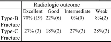
90 MJIRI, Vol. 25, No. 2, August 2011, pp. 87-93 Table 3. Radiological outcomes after surgery.
Radiologic outcome
Excellent Good Intermediate Weak
Type-B Fracture
70%(19) 22%(6) 0%(0) 8%(2)
Type-C Fracture
27% (3) 18%(2) 27%(3) 28%(3)
Table 4. Functional (clinical) outcomes after surgery
Functional outcome
Excellent Good Intermediate Weak
Type-B
Fracture 66%(18) 15%(4) 11%(3) 8%(2)
Type-C
Fracture 46%(5) 27%(5) 27%(3) 0%(0)
Total percent of complication was 34% (13 patients). The most common postopera-tive complication were deep wound infection and heterotopic bone formation, followed by pelvic obliquity and urinary tract infection. One patient developed small pulmonary em-bolus that was treated successfully, one had device failure, one nonunion, one lateral cu-taneous of thigh injury, one symphyseal fu-sion, and one pubis malposition have been reported. There was no evidence of deep vein thrombosis, sexual and urinary dysfunc-tion. All fractures united and there were not implant failures. 4 patients developed super-ficial infection, which were successfully treated by oral antibiotics (Table 2).
Radiological outcome
The radiological result was graded by the maximum residual displacement in the post-erior or antpost-erior pelvic ring injuries as; ex-cellent for 0 to 5mm, good for 6 to 10mm, fair for 11 to 15mm and poor for more than 15mm of displacement or established non union [5]. For 27 cases with type B fracture, radiologic reduction with residual displace-ment of symphysis pubis have been re-ported as, 70% excellent, 22% good, 0% fair and 8% poor. In eleven patients with type-C factures, excellent radiological reduction of symphysis pubis have been reported as, 27% excellent, 18% good, 27% fair and 28% poor (Table 3).
Clinical outcome
According to the Majeed’s score, in type-B pelvic fracture, 66% of patients present excellent, 15% good and 11% fair and 8% weak clinical outcome. In type-C pelvic fracture, 46% of patients present excellent, 27% good, 27% intermediate and 0% weak clinical outcome (Table 4).
Thirteen of 27 patients with B-type frac-tures were pain-free at the time of study as opposed to 4 of 11 patients with C-type frac-tures who were in pain. Three patients suf-fered from severe durable pain who could not go back to their work. Thirty five pa-tients returned to their original job.
Outcome evaluation
Age and sex did not influence on radio-logical and clinical outcome but they were depended on the type of pelvic fractures (P=0.03). Excellent radiological and clinical outcomes in type-B was better than type-C pelvic fractures. However, this could not be statistically proved due to small number of patients that were reviewed. All neurological injuries involving the lumbosacral plexus were fully recovered at the time of follow-up. Duration and severity of pain were more than in type-C pelvic fractures. However the level of pain influenced their performance accordingly.
Discussion
Pelvic fracture injuries are relatively rare injuries; their incidence in trauma patients is quoted to range between 3 % and 8.2 % and instability occurs in 13 % to 17 % of cases [2]. The most common cause of pelvic ring disruption is a Road Traffic Accident [8] which involved 70% of our patients. Be-cause of the large force that is required to disrupt the pelvis, pelvic fractures are indica-tive of a high-energy issue to the patient and therefore, often combined with other injuries [2,7].
Patients with major pelvic injuries need a multidisciplinary approach and should be treated in well-equipped and staffed centres. Stabilisation of vital parameters is the first goal, and a standardised trauma protocol for
K. Mardanpour, et. al.
91 MJIRI, Vol. 25, No. 2, August 2011, pp. 87-93
diagnostic policy as well as for surgical treatment should be followed routinely [7]. Associated intra-abdominal injuries should be evaluated by emergency abdominal ultra-sound to diagnose or exclude intra-abdominal bleeding [17]. Ultrasound was sufficient to diagnose intra-abdominal haemorrhage in both patients with splenic lacerations, however laparotomy was unnec-essary in the third patient.
Haemodynamically unstable patients with pelvic fractures have a mortality of 40 to 50% and this increases markedly if there is an associated head injury [8]. Some authors attribute this high mortality to exsanguinat-ing haemorrhage within the pelvis, others believed that massive bleeding from pelvic fractures is uncommon and that mortality is related to the associated injuries [9]. Exter-nal fixation can be applied in 15 minutes by an orthopaedic surgeon or a physician cre-dentialed in this procedure [10]. This method reduces the relative volume of a fractured pelvis, thereby reducing the potential space for haemorrhage. If the patient remains un-stable despite the resuscitation efforts, should undergo angiographic embolisation or be brought to operating theatre for surgi-cal intervention [10].
None of the 6 haemodynamically unstable patients who were transferred to our A&E department required angiographic embolisa-tion, however all of them underwent an ur-gent laparotomy following initial stabiliza-tion of the pelvic ring by internal fixator. Four were found to have sustained splenic lacerations and retroperitoneal haematoma. The vital signs of the sixth patients were sta-bilised after application of a pelvic internal fixation.
The incidence of an open, unstable pelvic ring disruption with connection occurring between the fracture and skin, rectum or va-gina is 3.5 to 4.5% [2]. Berner et al [14] re-ported that morbidity and mortality of the patients with open pelvic fractures were higher than in patients with closed injuries and Kobak et al [20] reported death due to sepsis in a patient with open fracture. One of our female patients had an open fracture that
was communicated with the vagina; that was treated successfully without complications.
The mortality rate in this small series of unstable pelvic fractures was zero; the fact that two patients with serious brain injury recovered fully and the open fracture was communicating with the rectum rather than vagina, as well as the prompt multidiscipli-nary approach for haemodynamically unsta-ble patients may explain this favouraunsta-ble out-come, which comes in contradiction with the recent literature [7].
Associated injuries occurred in most of our patients (29 out of 38 patients).
Corovessis et al reported that concomitant injuries have negatively affected the clinical outcome of unstable pelvic fractures despite ideal radiographic score of surgical reduc-tion. The presence of associated injuries has certainly increased morbidity in our group of patients and negatively affected the func-tional outcome in urologic injury, a vertebral fracture and vaginal and rectum laceration. Eight out of 38 patients (21%) had an asso-ciated acetabular fracture and no patient progressed hip osteoarthritis within 25 months of injury.
The incidence of neurological injuries in this study was 29% which is comparable with that reported in the literature. Four pa-tients with combined motor and sensory neu-rological deficits affecting the L4, L5, S1 nerve roots were fully recovered at the time of follow-up. One patient with isolated sen-sory deficit of S1 and S2 nerve roots were recovered at the time of follow-up. About 25 months following injury. Low number of patients in this study may be a reason for the most favourable potential for recovery of the neurological injury at the L4, L5, and S1 nerve roots, as compared to the literature [6,11]. Early rigid stabilisation of both ante-rior and posteante-rior pelvic ring injury, which was performed on our patients, has been suggested as a potential reason for favour-able prognosis for these injuries.
Stabilisation of the unstable pelvic ring in-juries can be achieved by external and/or internal fixation [7]. External fixation can be applied fast resulting in reduced
Surgical treatment of unstable pelvic ring fractures
92 MJIRI, Vol. 25, No. 2, August 2011, pp. 87-93 rhage in the intrapelvic space by tamponade
[10]. Open reduction and internal fixation of the unstable pelvic ring fractures provides the best stability for fixation as well as best clinical outcome [3,4]. Unstable type-B1 in-juries should be fixed [7] whereas type-B2 and B3 fractures could be treated non-operatively, because the pelvis has the elas-ticity to restore to a near normal position [12].
Some of the reported long-term morbidity for an unstable pelvic ring fracture include chronic pelvic pain, impaired function of the pelvis for sitting and weight bearing that re-sults from pelvic non-union, pelvic malunion and leg length discrepancy [13].
Non-union or leg length discrepancy of more than 1cm was not occurred in our pa-tients but displacement of symphysis pubis of more than 5mm was associated with re-sidual symptoms such as pain and lateral cu-taneous nerve of thigh.
Our study shows that the antibiotic pro-phylaxis used in the unit is effective as only one patient developed superficial infection. The deep venous thrombosis (DVT) prophy-laxis is also effective as no patient developed clinical deep venous thrombosis.
The review of literature shows several studies have supported this both in biome-chanical studies and clinical trials there is a lot of controversy about the long-term out-come of unstable pelvic injuries. Berner in 1982 reported a rate of 16% unsatisfactory functional, and 17% unsatisfactory radio-logical result in a group of 42 patients treated non-operatively after combined dis-ruptions of the pubic symphyses and the sacro-iliac joint. [14] After treatment of a similar injury with open reduction and inter-nal fixation, the rate of unsatisfactory func-tional rating was 21% and 27% in the radio-logical rating.
Rargnarsson in 1993 reported on 21 pa-tients after SI joint disruption treated with internal fixation using plating [15]. He stated the radiological position of the pelvic ring as “unchanged to the post-operative radio-graphs”, a rate of 14% poor functional re-sults were reported.
One of the reasons for the poor result of previous studies has postulated to a residual displacement of 10mm and more was critical for a significant increase of residual pain [16]. Semba et al also reported a correlation of primary anterior and posterior displace-ment exceeding 10mm being correlated with a markedly higher incidence of severe low back pain [13]. Holdsworth in 1948 reported that 50% of the patients they studied re-turned to their original job [1]. Our study showed that 7 patients returned to their original jobs. In the largest series of patients treated with open reduction and internal fixation of unstable posterior pelvic injuries, 67% returned to their former jobs without restrictions [18]. In other study where all fractures were reduced operatively to less than 10mm of residual displacement; 35% of patients had neurologic injuries, and another 23% had associated injuries inhibiting nor-mal gait. Fenor-males with pelvic fractures tended to have increased urinary complaints and dyspareunia, which were shown to cor-relate with residual displacement of >5mm [19].Our study has shown that anatomical restoration of the pelvic ring correlated with higher probability of a good functional and clinical outcomes.
Kabak S, et al functional outcome of open reduction and internal fixation for complete-ly unstable pelvic ring fractures (type C): a report of 40 cases reported that morbidity and mortality rates were higher in patients with a completely unstable pelvic ring in-jury. Emergency department stabilization and reconstruction of the pelvic ring with optimal operative techniques in these pa-tients can reduce morbidity and mortality rates. Anterior and posterior internal fixation results in satisfactory clinical and radiologic outcomes. The affective status of patients was an important aspect that should be con-sidered during the entire care of the patients [20].
Conclusions
Pelvic fractures by high-energy traumas are severe lesions, with significant mortality rate and a great number of associated
K. Mardanpour, et. al.
93 MJIRI, Vol. 25, No. 2, August 2011, pp. 87-93
sions. In high-energy traumas, pelvic frac-tures should always be suspected and con-ducted together with other lesions. Pelvic fractures are challenging injuries to manage. Stabilisation of vital parameters takes pref-erence and significantly reduces mortality. Associated injuries are common and often have a substantial effect on the patient’s psychological status. Rehabilitation period is prolonged; however proper management yields a satisfactory outcome. Further analy-sis and studies including a larger number of patients are required to identify the prognos-tic factors for the late sequelae. This study should be a valid statistical analysis of out-comes in patients who treated surgically, by internal fixation. Early rigid stabilisation of both anterior and posterior pelvic ring injury with open reduction, internal fixation, which performed in our patients, has been sug-gested as a potential reason for favourable prognosis of these injuries.
References
1. Korovessis P, Baikousis A, Stamatakis M, Ka-tonis P. Medium and long term results of open reduc-tion and internal fixareduc-tion for unstable pelvic ring frac-tures. Orthopedics 2000; 23(11):1165-71.
2. Mucha P, Farnell M. Analysis of pelvic fractures management. J Trauma 1984; 24(5): 379-386.
3. Matta J, Saucedo T. Internal fixation of pelvic ring fractures. Clin Orthop 1989: 242:83-97
4. Tile M. Pelvic ring fractures: should they be fixed? J Bone Joint Surg 1988; 70B:1.
5. Tile M. The management of unstable injuries of the pelvic ring. J Bone Joint Surg 1999; 81-B(6): 941-3.
6. Majeed SA. Grading the outcome of pelvic frac-tures. JBJS Br 1989; 71(2): 304-6.
7. Van Veen IH, van Leeuwen AA, van Popta T, van Luyt, Bode PJ, Vugt AB: Unstable pelvic
frac-tures: a retrospective analysis. Injury 1995; 26(2): 81-5.
8. Moreno C, Moore EE, et al. Haemorrhage asso-ciated with major pelvic fracture: A multispecialty challenge. J Trauma.1986; 26:987-994.
9. Poole Gv, Ward E F, Causes of mortality in pa-tients with pelvic fractures. Orthopaedics 1994; 17: 691-6.
10. Coppola T, Coppola M: Emergency depart-ment evaluation and treatdepart-ment of pelvic fractures. Emerg Med Clin of North Am 2000; 18(1): 1-27.
11. Helfet DL, Koval KJ, Hissa EA et al. Intraop-erative somatosensory evoked potential monitoring during acute pelvic fracture surgery. J Orthop Trauma 1995; 9:28-34
12. Tile M. Pelvic ring fractures: should they be fixed? J Bone Joint Surg 1988; 70 (1): 1-12.
13. Smith J, Western journal of Medicine. San Francisco: Feb 1998; 168:124-6 .
14. Berner W, Oestern HJ, Sorge J. Ligamentäre Beckenringverletzungen: Behandlung und Spätergeb-nisse. Unfallheilkunde 1982; 85:377-387.
15. Rargarsson B, Oleurd C. Anterior square-plate fixation of sacroiliac disruption. Acta Orthop Scand 1993; 64(2):138-142.
16. Semba R, Yasukawa K, Gustillo R Critical analysis of results 53 Malgaigne fractures of pelvis J Trauma 1983; 23(6):535-537.
17. Holdsworth FW: Dislocation and fracture dis-location of pelvis. J Bone Joint Surg Br 1986; 30;461-466.
18. Tornetta P, Dickson K, Matta JM: Outcome of rotationally unstable pelvic ring injuries treated operatively. Clin Orthop 1986; 39:147-151.
19. Copeland CE,Bosse MJ, McCarthy ML: Effect of trauma and pelvic fracture on female genitouri-nary, sexual and reproductive function. J Orthop Trauma 1997; 11:73-81.
20. Kabak S, Halici M, Tuncel M, Avsarogullari L, Baktir A, Basturk M. Functional outcome of open reduction and internal fixation for completely unsta-ble pelvic ring fractures (type C): a report of 40 cases. J Orthop Trauma 2003; 17:555-62.