Introduction
Ulcerative colitis (UC) is a systemic illness with a number of extraintestinal manifestations affecting various organs. The most frequent le-sions are cutaneous, ocular, hepatic and articu-lar. In contrast, pulmonary involvement in UC is thought to be rare [1].
However, pulmonary lesions have been re-ported in inflammatory bowel disease (IBD), including chronic bronchial suppuration in
pa-tients with UC [2], localized obstruction of the upper airways [3], bronchiolitis obliterans, or-ganizing pneumonia [4], diffuse obstructive disease [5], bronchiectasis [6], granulomatous lung disease [6], pulmonary vasculitis [6], and diffuse or localized interstitial lung fibrosis [6]. Some authors described alterations of pul-monary function in asymptomatic patients, most commonly reduced lung diffusion capaci-ty and small airways function and bronchial hy-perreactivity [7-9].
A recent review article showed that respirato-ry symptoms and diagnosed respiratorespirato-ry system
Medical Journal of the Islamic Republic of Iran.Vol. 21, No. 4, February 2008. pp. 196-202
Pulmonary function in ulcerative colitis
A.H. Faghihi-Kashani, MD.1, A. Kabir, MD.2, S.A. Javad-Moosavi, MD.3
Nikan Health Researchers Institute, Gastrointestinal and Liver Disease Research Center & Pulmonary Disease Research Center, Iran University of Medical Sciences, Tehran, Iran.
Abstract
Background: Pulmonary involvement in ulcerative colitis (UC) is thought to be rare. There is not a definite document about the question that "Is the lung a target organ in in-flammatory bowel disease?" The aim of the present study is to compare lung function be-tween cases with UC and healthy controls. This study will also be of interest about searching the outbreak of pulmonary function abnormalities in a sample of Iranian pa-tients with UC and factors associated with severity of UC.
Methods: In an analytic cross sectional study between July 2006 and September 2007, we evaluated 70 patients with histologically confirmed UC and 70 matched healthy people. Our checklist addressed demographic variables, symptoms, smoking be-havior, drugs, laboratory findings and pulmonary function tests.
Results: None of the lung volumes and capacities were significantly different in cases as compared to controls. Severity of UC was mild in 65.7%. It was correlated with smok-ing (P=0.019) and allergy (P=0.017). Patients with moderate UC had lower hemoglobin (P<.001), MCH (P=0.002), MCV (P=0.047), MCHC (P=0.028) and higher REFF (P=0.032) and BF (P=0.01).
Conclusion: The controversies about the relation between UC and lung disease can be due to different sample sizes, activity of UC at the time of measurement of lung vol-umes, methods of measuring lung capacities at the time of PFT and different nationali-ties.
Keywords: ulcerative colitis, inflammatory bowel diseases, respiratory function tests
1. Assistant Professor of Gastroenterology, Gastrointestinal and Liver Disease Research Center, Iran University of Medical Sciences, Tehran, Iran.
2.Corresponding author, Methodology and Statistics Consultant, Nikan Health Researchers Institute, Unit 9, No. 1, 3rd Floor, 3rd Bahar
disorders are more common among patients with IBD than generally appreciated [10]. It has been shown that the incidence of respiratory changes is higher in patients with ulcerative co-litis as opposed to those with Crohn’s disease (CD) [5,6].
It is important to recognize the association between IBD and pulmonary disease so that proper management can be better defined [11].
Is the lung a target organ in inflammatory bowel disease? Despite that it is not a new ques-tion; there are no definite documents yet.
The aim of the present study is to investigate lung function in patients with UC and to com-pare their pulmonary function test results with healthy controls.
Methods
Study population and assessments
In an analytic cross sectional study between July 2006 and September 2007, we evaluated 70 patients with histologically confirmed UC and 70 age-gender matched healthy people (fre-quency matched) as controls.
We completed a checklist for each case and control according to medical history, physical examination and laboratory findings. It ad-dressed demographic variables (age, gender, job, income, area of their home), height, weight, du-ration of the disease, symptoms, smoking be-havior, history of pulmonary diseases, allergy, hypertension, drugs, complete blood count (CBC), platelet, and ESR.
Medical history and pulmonary function tests (PFT) determined the condition of the lung. One expert technician performed all the spirometries by a Fukuda spirometer showing FEV1 (forced expiratory volume in one sec-ond), FVC (forced vital capacity), PEFR (peak expiratory flow rate), VC (vital capacity), FEV1/VC (proportion of FEV1 to VC), MMEF (maximum mid-expiratory flow), MEF25%, MEF50% and MEF75% values. According to predicted values for their age, gender, height and weight, the percentage of obtained values
were recorded and expressed as percent of pa-rameter. Normal range of indices are consid-ered as VC>80%, FEV1>75%, and FEV1/FVC >75% for both men and women [12].
A blood sample determined the value of CBC parameters, platelet, and ESR.
We assessed the severity of ulcerative colitis in all patients according to Truelove Index for UC [13]. This index implements clinical symp-toms (abdominal tenderness and distention), vi-tal signs (temperature and pulse rate), number of defecations per day, blood in stool, laborato-ry findings (Hemoglobin and ESR) and radi-ographic findings (distention, air in colon, ede-matous intestinal wall, and thumb print sign).
Data processing, analysis and ethical con-siderations
Mean±SE (standard error), t-test, chi-square and Pearson’s correlation coefficient were used in description and analysis of data. All tests with P<0.05 were considered statistically sig-nificant. SPSS 13 software (SPSS Inc. Chicago, Illinois, USA) was used in analysis.
The Gastrointestinal and Liver Disease Re-search Center Ethical Committee of Iran Uni-versity reviewed and approved the study proto-col. All patients signed an informed consent.
Results
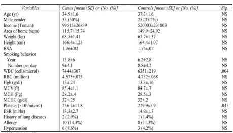
There was no significant difference between cases and controls in demographic variables (age, sex, job, income, area of their home), weight, height, BMI, BSA, smoking behavior, CBC (except WBC and platelet), ESR, history of lung diseases, allergy, and hypertension (Table 1). So, the two groups were matched and compared.
VC less than 80% was found in 4 (5.8%) cas-es and 5 (7%) controls without a statistically significant difference between the two groups.
Severity of UC was mild in 46 cases (65.7%), moderate in 13 cases (18.6%), severe in 1 case (1.4%) and undetermined in 10 cases (14.3%). Severity of disease was correlated with
ing (P=0.019) and allergy (P=0.017). Patients with moderate UC had lower hemoglobin (P<.001), MCH (P=0.002), MCV (P=0.047), MCHC (P=0.028) and higher REFF (P=0.032) and BF (P=0.01).
Patients with UC had dyspnea [16 cases (22.9%)], chest pain [10 cases (14.3%)], cough [8 cases (11.4%)], fever [2 cases (2.9%)], de-fined history of lung disease [2 cases (2.9%)], allergy [10 cases (14.3%)], and hypertension [6 cases (8.6%)]. Abnormal FEV1 was found in only 3 cases (4.3%).
Comparison of cases and controls showed higher WBC (7444 ± 307 vs. 6351 ± 219, P= 0.004) and platelet counts (256734 ± 11826 vs. 229851 ± 5917, P= 0.045) in cases, however both were in normal ranges and were not clini-cally significant.
Chest pain [10 subjects (14.3%) vs. 1 subject (1.4%), P=.004] and dyspnea [16 subjects (22.9%) vs. 6 subjects (8.5%), P=0.018] were more prevalent in cases significantly. However, there was no significant difference in fever and cough between the two groups. None of the
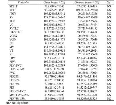
quantitative volumes and lung capacities were significantly different in cases in comparison with controls. Findings of PFT are summarized in Table 2.
Interpretation of PFT showed no significant difference between cases and controls. Mild, moderate and severe expiratory limitation ex-isted in 7 (10.1%), 5 (7.2%) and 1 (1.4%) pa-tient in case and 7 (9.9%) and 1 (1.4%) papa-tient in control groups, respectively. None of the controls demonstrated severe expiratory limita-tion.
Duration of UC was 5.1±.46 years in all cas-es. There was a significant correlation between duration of UC and RV (r=0.26, P=0.04), FEF75% (r=-0.25, P=0.046), history of pul-monary problems (15 ± 5 vs. 4.7 ± .4, P < 0.001), and allergy (7.8 ± 1.36 vs. 4.6 ± .47, P=0.02). Men had UC with more duration (6 ± .7 vs. 4 ± .6, P = 0.03).
Discussion
Little is known about lung function in pa-tients with IBD. Camus et al [6] evaluated pul-NS= Not significant
Table 1. Comparison of cases and controls.
monary function tests in 13 patients with IBD and detected a decline in the FEV1/FVC ratio in bronchiectatic patients. Latent pulmonary in-volvement has also been reported in adult IBD patients recently. Bronchial hyperreactivity (BHR) was seen in 48% of patients with ulcera-tive colitis and CD without any bronchopul-monary symptoms and with normal baseline lung function [14]. These findings may indicate that a latent inflammation exists in the airways of IBD patients which is not detectable by rou-tine pulmonary function tests. In our study, we focused on UC patients because they are clini-cally and histologiclini-cally different from CD. However, the pulmonary evaluation was
as-sessed only by spirometry, a limitation of the present study. Therefore, we did not find signif-icant difference between UC patients and con-trols.
Increased bronchial hyper-responsiveness in patients with IBD with no bronchopulmonary symptoms and with normal baseline lung func-tion has recently been reported [14]. This latter observation may also indicate that an inflam-mation exists in the airways that are not de-tectable by routine pulmonary function tests.
Small peripheral airways (less than 2 mm in diameter) contribute less than 20% of the total airway resistance, and lesions in this region are difficult to detect. However, it is likely that
Table 2. PFT findings.
NS= Not significant FEV1
FEV1/VCMX FEV1/FVC
some of the earliest changes in the airways of patients with IBD would affect primarily the small airways, and thus it is of importance to as-sess the function of the peripheral airways. It can be another cause of not earning significant pulmonary function difference between UC pa-tients and controls in our study despite the pres-ence of some pulmonary problems in IBD pa-tients.
It is also known that high IgE levels are com-mon in IBD despite the absence of atopic symp-toms [15]. These observations suggested the probability of higher prevalence of allergic re-actions and BHR in IBD patients in comparison with normal controls. In our study history of al-lergy was higher in UC patients in comparison with controls. However, it did not reach a sig-nificant difference. Other studies also showed higher levels of allergy in IBD patients [15,16,17,18,19].
In addition, it is noteworthy that there are similarities between colonic and bronchial ep-ithelium: both originate from the primitive gut and both are sensitive to inhaled and ingested irritants.
In our study, there was a positive correlation between duration of UC and RV, history of pul-monary problems, allergy and male gender and a negative correlation with FEF75%. It has been shown that the incidence increases with duration of intestinal disease and is greater in CD than in UC [20].
Pulmonary disease has been described much less frequently than other organ systems associ-ated with IBD. Although the cases with IBD and pulmonary manifestation may be incidental findings, it seems that some are directly associ-ated with CD and UC. However, our methodol-ogy of comparing the UC patients with a matched control group, emphasize on not hav-ing an association between UC and major pul-monary diseases that are found by PFT. In all IBD cases, drug-induced pulmonary disease, a rare side effect of sulfasalazine, mesalamine, and methotrexate, must be ruled out.
Pul-monary disease in IBD ranges from interstitial fibrosis (which can be lethal) to subclinical pul-monary function abnormalities.
Sixteen studies of pulmonary function tests (including> 600 patients with IBD) revealed a spectrum of restrictive disease, small airway disease, bronchial hyper-responsiveness, and hyperinflation during acute exacerbations asso-ciated with IBD [8, 9]. Although some studies are negative ones [21,22], two large trials [7, 23] and another new study [24] showed that more than 50% of patients with UC had abnor-mal pulmonary function tests compared with healthy matched controls. The most commonly described abnormality is a decrease in diffusion capacity of the lungs. In most reports, no rela-tionship was found between these abnormali-ties and 5-ASA usage or disease activity. There was significant correlation between the severity of disease with smoking, allergy, hemoglobin, MCH, MCV, and MCHC in our study. Accord-ing to the authors’ search, there was no congru-ent study about smoking and severity of UC. Our assumption is that according to publication bias, some (or maybe many) congruent studies with our finding (positive association between smoking and severity of UC) are not published. However, we do not have another view about it. Pulmonary function test studies in IBD sug-gest that subclinical pulmonary disease may be present in a large subpopulation of patients [22].
Pulmonary manifestations of IBD may be more common than originally thought. In a re-view article, more than 150 reports of active lung disease, with an even greater number of patients demonstrating subclinical pulmonary findings was found.
Although in most reported cases IBD pre-dates respiratory symptoms, this may represent a reporter bias, because patients who present with preceding pulmonary disease may be overlooked [2].
Many authors who describe pulmonary dis-ease in the setting of IBD stress the relationship
between colectomy and the onset of symptoms. In our review of the literature, we have not found this to be the case. The overall incidence of colectomy in patients with UC is 5% to 10% in patients with a severe first attack and 1% per year in the remainder [25]. The incidence of colectomy among patients with pulmonary manifestations of IBD is similar. Colectomy has never been shown to be a curative treatment of extraintestinal pulmonary disease.
Most of our patients had a mild disease and were inactive at the time of measurement of lung volumes which can be other causes of lack of association between lung abnormalities and UC.
These controversies about the relation be-tween UC and lung disease can be due to differ-ent sample sizes, activity of UC at the time of measurement of lung volumes, methods of measuring lung capacities at the time of PFT and different nationalities. Finally, according to our findings and similar studies, we recom-mend more exhaustive pulmonary evaluation in UC patients in order to find pulmonary ab-normalities.
Simple spirometry is not capable of deter-mining significant pulmonary function differ-ences between UC patients and healthy con-trols. More detailed, basic, structural, mechani-cal and functional evaluation with higher sensi-tivity is needed to rationalize pulmonary abnor-malities in cases with UC.
Acknowledgment
We thank Faezeh Jafarian, Ziba Hosseini-Fard, Safiye Rezazade Lavaf, Seyedeh Khadije Tabatabaee, Mehdi Rezaee and Ala Keikhani for their cooperation in prepar-ing the proposal and gatherprepar-ing data.
References
1. Rankin GB. Extraintestinal and systemic manifes-tations of inflammatory bowel disease. Med Clin North Am 1990; 74:39-50.
2. Higenbottam T, Cochrane GM, Clark TJ, Turner D,
Millis R, Seymour W. Bronchial disease in ulcerative co-litis. Thorax 1980; 35: 581-5.
3. Rickli H, Fretz C, Hoffman M, Walser A, Knoblauch A. Severe inflammatory upper airway steno-sis in ulcerative colitis. Eur Respir J 1994; 7:1899-1902.
4. Wilcox P, Miller R, Miller G, Heath J, Nelems B, Muller N, Ostrow D. Airway involvement in ulcerative colitis. Chest 1987; 92:18-22.
5. Kraft SC, Earle RH, Roesler M, Esterly JR. Unex-plained bronchopulmonary disease with inflammatory bowel disease. Arch Intern Med 1976;136:454-459.
6. Camus PH, Plard F, Aschroft T, Gal AA, Colby TV. The lung in inflammatory bowel disease. Medicine 1993; 72:151–183.
7. Kuzela L, Vavrecka A, Prikazska M, Drugda B, Hronec J, Senkova A, et al. Pulmonary complications in inflammatory bowel disease. Hepatogastroenterology 1999; 46: 1714-1719.
8. Tzanakis N, Bouros D, Samiou M, Panagou P, Mouzas J, Manousos O, et al. Lung function in patients with inflammatory bowel disease. Resp Med 1998; 98: 516-522.
9. Munck A, Murciano D, Pariente R, Cezard J, Navar-ro J. Latent pulmonary abnormalities in children with Crohn’s disease. Eur Respir J 1995; 8: 377-380.
10. Black H, Mendoza M, Murin S. Thoracic manifes-tations of inflammatory bowel disease. Chest 2007; 131(2): 524-32.
11. Storch I, Sachar D, Katz S. Pulmonary Manifesta-tions of Inflammatory Bowel Disease. Inflamm Bowel Dis 2003; 9(2): 104-115.
12. American Thoracic Society: Lung function test-ing: Selection of reference values and interpretative strategies. Am Rev Respir Dis 1991; 144:1202–1218.
13. Hanauer SB. Inflammatory Bowel Disease. N Eng J Med 1996; 334 (13): 841-848.
14. Louis E, Louis R, Drivon V, Bonnet V, Lamproye A, Rodermecker M, et al. Increased frequency of bronchial hyperresponsiveness in patients with inflam-matory bowel disease. Allergy 1995; 50: 729-733.
15. Ceyhan BB, Karakurt S, Cevik H, Sungur M. Bronchial hyperreactivity and allergic status in inflam-matory bowel disease. Respiration 2003; 70(1): 60-6.
16. Louis R, Shute J, Lau L, Franchimont D, Lam-proye A, Radermecker M, et al. Bronchial eosinophilic infiltration in Crohn’s disease in the absence of pul-monary disease. Clin Exp Allergy 1999; 29: 660–666.
17. Fireman Z, Osipov A, Kivity S, Kopelman Y, Sternberg A, Lazarov E, et al. The use of induced sputum in the assessment of pulmonary involvement in Crohn’s disease. Am J Gastroenterol 2000; 95: 730–734.
18. Wallaert B, Colombol JF, Tonnel AB, Bonniere P, Cortot A, Paris JC, et al. Evidence of lymphocyte alveoli-tis in Crohn’s disease. Chest 1985; 87: 363–367.
19. Verleden GM, Koek GH, Evenepoel P, Dupont L, Rutgeerts P. Exhaled nitric oxide correlates with the ac-tivity index of Crohn’s disease and colitis ulcerosa. Am J Respir Crit Care 1999; 159: A862.
20. Veloso F, Carvalho J, Magro F. Immune-related manifestations of inflammatory bowel disease: a prospective study of 792 patients. J Clin Gastroenterol 1996; 23: 29–34.
21. Neilly JB, Main AN, McSharry C, Murray J, Rus-sell RI, Moran F. Pulmonary abnormalities in Crohn’s disease. Respir Med 1989; 83: 487–91.
22. Tunc B, Filik L, Bilgic F, Arda K, Ulker A. Pul-monary function tests, high-resolution computed tomog-raphy findings and inflammatory bowel disease. Acta Gastroenterol Belg 2006; 69 (3): 255-60.
23. Godet PG, Cowie R, Woodman RC, Sutherland LR. Pulmonary function abnormalities in patients with ulcerative colitis. Am J Gastroenterol 1997; 92:1154–56. 24. Mohamed-Hussein AA, Mohamed NA, Ibrahim ME. Changes in pulmonary function in patients with ul-cerative colitis. Respir Med 2007; 101(5):977-82.
25. Feldman M, Scharschmidt B, Sleisenger M. Sleisenger and Fortran’s Gastrointestinal and Liver Dis-ease, 6th ed. Philadelphia: WB Saunders; 1998: 1708-1713.