Original article
1: Ghazvin University of Medical Sciences, Ghazvin, Iran
www.RBMB.net
Comparison of Th1 and Th2 Responses in
Non-Healing and Non-Healing Patients with Cutaneous
Leishmaniasis
Maryam Shahi*
1, Masoud Mohajery
2, Seyyed Ali Akbar Shamsian
2, Hossein
Nahrevanian
3, Seyyed Mohammad Javad Yazdanpanah
2Abstract
Background:Cutaneous leishmaniasis is an endemic disease in many regions of Iran, including the city of Mashhad. In recent years, some cases have not responded to Glucantime, the usual treatment for this disease. The cellular immune response caused by T-helper type 1 (Th1) cells has an important role in protection against leishmaniasis, and activation of the T-helper type 2 (Th2) response causes progression of the disease. By analyzing these responses we hope to find a more effective treatment than that currently in use for leishmaniasis patients.
Methods: The cellular immune responses in 60 cases of non-healing and healing cutaneous leishmaniasis, and individuals in a control group, were analyzed by measuring cytokines released by peripheral blood mononuclear cells (PBMCs) when stimulated with Leishmania major antigens by Enzyme Linked Immuno Sorbent Assay (ELISA).
Results: Subjects from the healing group secreted more interleukin-12 (IL-12) and interferon gamma (IFN-γ) (p<0.05) and less interleukins -4, -5, -10 (IL-4, IL-5, andIL-10) (p<0.005) and -18 (IL-18) (p=0.003) than the non-healing group.
Conclusions: The results demonstrate that secretion of cytokines that activate Th2 response including IL-4, IL-5 and IL-10 in non-healing subjects was higher than healing subjects and secretion of cytokines that activate Th1 response including IL-12 and IFN-γ in healing subjects was higher relative to the non-healing subjects. In this study it has been shown that the level of IL-18 progresses disease in non-healing patients when the level of IL-12 gets decreased.
Keywords: Cutaneous leishmaniasis, Cytokines, Glucantime
Introduction
Leishmaniasis represents a multifarious of diseases with a clinical and epidemiological diversity (1). An estimated 12 million humans are infected, with an incidence of approximately 1.5 million cases of the cutaneous type of the disease (2). Cutaneous leishmaniasis (CL) is an endemic disease in many locations of Iran, including the city of Mashhad. The clinical ending of Leishmania
infection in humans, ranging from relatively mild to severely life-threatening disease, depends on host- and parasite-associated factors (3). The pentavalent antimonials have been used for over 60 years to treat leishmaniasis, but in the recent years, not all cases have responded to these drugs. One drug in this class with reduced efficacy is Glucantime. Successful treatment of
Rep. Biochem. Mol. Biol, Vol. 1, No. 2, Apr 2013 44
leishmaniasis requires not only a decrease in parasite number but also the increase of an effective cell-mediated immune response (2). Th1-type cellular immune responses play an important role in defense against infection with Leishmania parasites, whereas activation of Th2-type cells results in progressive disease (4, 5). In the present study, we investigated cell-mediated immunity in non-healing CL subjects by measuring production of the cytokines interleukins -4, -5, -10, -12, and -18 (IL-4, IL-5, IL-10, IL-12, and IL-18) and interferon gamma (IFN-γ). The responses were compared with those of non-healing CL subjects and healthy controls.
Materials and Methods
Study groups
In this study 60 subjects were selected from number 1 (Abobargh, Vila) clinicand Ghaem Hospital (Mashhad, Iran) between December 2006 and March 2007. The study groups were as follows: the first group consisted of 20 subjects who had recovered after one period of treatment from CL (healing group); the second group consisted of 20 subjects with chronic CL lesions (non-healing group) who had not recovered after three periods of treatment (group 2); the third group consisted of 20 subjects with no history of leishmaniasis who were leishmanin skin test negative (control group). All subjects signed a written consent and the ethical guidelines. All subjects were confirmed negative for human immunodeficiency virus (HIV), human T-cell lymphtropic virus (HTLV-1), and intestinal helminthes that promote Th2-type cell production, and fasting blood sugars (FBS) were determined.
Isolation of mononuclear cells
Blood was collected from healing CL, non-healing CL, and control subjects and treated with ethylenediaminetetraacetic acid (EDTA). PBMCs were isolated by Ficoll (Biogene) density centrifugation at 3000 rpm. The cells were washed twice with phosphate-buffered saline (PBS). Cell viability was ascertained by trypan blue exclusion.
Antigen and mitogen
A clone of Leishmania major (L. major) (MRHO/IR/75/ER, a gift from Dr. H. Nahrevanian, Department of Parasitology, Pasteur Institute, Iran) was used for this study. The parasites were cultured to stationary phase in RPMI 1640 (Gibco) with 17% heat-inactivated fetal calf serum (FCS) (Gibco). The promastigotes were harvested and adjusted to 4×10⁷ parasites/ml followed by rapidly freezing and thawing the parasites 5 times and a final ultrasonication for 5 minutes. This method prepared crude antigen (Ag) of L. major to stimulate PBMCs. The samples were stored at -70 ºC until use. Phytohemagglutinin (PHA) (Gibco) was used at a concentration of 10 μg/ml.
PBMC culture
PBMCs were cultured in RPMI 1640 (Uroclone) enriched with 10% heat-inactivated FCS and 1% penicillin-streptomycin solution (Biosera). The cells were incubated with crude L. major Ag (41.5μ ml) and mitogen (PHA) (10 μg/ml) at g/ 1×10⁶ cells/ml in volumes of 1 ml in 12-well round-bottom microculture plates (Nunc, Denmark). The cultures were incubated for two days at 37 °C under 5% CO2. The supernatants were stored at -70ºC until use.
Cytokine measurements
Cytokines in supernatants were measured by an enzyme-linked immunosorbent assay and the biotin-avidin system according to the manufacturer's guidelines. The assays were calibrated to detect IL-4, IL-5, IL-10, IL-12 and IFN-γ (Biosource Kit) and IL-18 (Bender MedSystems Kit) within the ranges considered in the protocol.
Statistical analysis
Cytokine levels of subjects in each group were analyzed by the analysis of variance (ANOVA) and Kruskal-Wallis in the SPSS software. P<0.05 was considered significant.
Results
Of the 60 subjects evaluated in this study, 20 were identified as non-healing, 20 as healing, and 20 as controls. Clinical findings for the study groups are summarized in Table 1.
Table 1. Major characteristics of healing, non-healing, and control groups.
Characteristic
Study group
Healing Non-healing Control
No. 20 20 20
Mean Age year (range) 23 (15-35) 27 (15-50) 39 (20-54)
M/F ratio 8/12 8/12 4/16
Mean no. of lesions
(range) 2.35 (1-9) 3.45 (1-5) _ Treatment type
(systemic/local) 10/10 10/10 -
Cytokine production
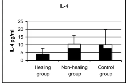
PBMCs from subjects were incubated with Ag and mitogen and the levels of each cytokine were determined in the supernatants. IFN-γ was significantly higher in the healing (P=0.033) and control (P<0.0001) groups than in the non-healing group, while no significant difference was observed between the healing and control groups (Fig. 1). IL-4 was significantly higher in the healing than in the non-healing group (P<0.0001), and higher in the control than in the healing group (P=0.033), while no significant difference in was observed between the non-healing and control groups (Fig. 2). IL-5 was significantly higher in the non-healing than in the healing group (P<0.001) and significantly higher in the control than in the healing group (P=0.029), while no significant difference was observed between the non-healing and control groups (Fig. 3).
IL-10 was significantly lower in the healing than in the non-healing group (P<0.0001) and significantly higher in the control than in the healing group (P=0.001), while no significant difference was observed between the non-healing and control groups (Fig. 4).
IL-12 was significantly higher in the healing (P=0.031) and control (P=0.04) groups than in the non-healing group, while no significant difference was observed between the healing and control groups (Fig. 5).
IL-18 was significantly higher in the non-healing than in the non-healing (P=0.003) and control
(P=0.004) groups, while no significant difference was observed between the control and healing groups (Fig. 6).
Fig. 1. Concentrations of IFN-γ in culture supernatants of PBMCs from healing, non-healing, and control groups stimulated with Ag and mitogen.
Fig. 2. Concentrations of IL-4 in culture supernatants of PBMCs from healing, non-healing, and control groups stimulated with Ag and mitogen.
Discussion
In this study we measured cytokines produced from PBMCs ex vivo after stimulation with
Leishmania Ag and PHA in supernatants. Chemotherapeutic cure of leishmaniasis is largely dependent upon the development of an effective immune response that activates macrophages to produce toxic nitrogen and oxygen intermediates to exterminate the amastigotes. CD4+ T cell populations were found to be an essential issue either in disease progression associated with IL-4 or in disease progression associated with IL-4 or in disease prevention related to IFN-γ (6).
IFN-γ
0 200 400 600 800 1000 1200
Healing group
Non-healing group
Control group
IF
N
-Y
IL-4
0 5 10 15 20 25
Healing group
Non-healing group
Control group
IL
-4
p
g
/m
l
Rep. Biochem. Mol. Biol, Vol. 1, No. 2, Apr 2013 46
Fig. 3. Concentrations of IL-5 in culture supernatants of PBMCs from healing, non-healing, and control groups stimulated with Ag and mitogen.
Fig. 5. Concentrations of IL-12 in culture supernatants of PBMCs from healing, non-healing, and control groups stimulated with Ag and mitogen.
Fig. 4. Concentrations of IL-10 in culture supernatants of PBMCs from healing, non-healing, and control groups stimulated with Ag and mitogen.
Fig. 6. Concentrations of IL-18 in culture supernatants of PBMCs from healing, non-healing, and control groups stimulated with Ag and mitogen.
Th1-type cellular immune responses within a suitable cytokine (IFN-γ, IL-12) play a significant role in protection against infection with Leishmania
parasites, whereas activation of Th2-type cells results in progressive disease. CL is often a self-healing disease; however, persistence of lesions lasting for several years is known to occur (non-healing form) (5, 12). There are few reports on the immune status of non-healing patients, particularly those infected with L. major. In this study PBMCs from the healing and control groups showed high levels of IL-12. In contrast, a low level of IL-12 was produced in the non-healing group. Similar data were previously reported by Habibi et al. IL-12 is essential for the stimulation of Th1 phenotype-dependent protection (8, 9).
It has been reported that IFN-γ improved the efficacy of antimonials in the treatment of visceral leishmaniasis (VL) and CL (6). IFN-γ is crucial, but not sufficient, to control leishmaniasis. It is known that IFN-γ is one of the major
macrophage-activating cytokines, and activated macrophages are a major source of IL-12, which induces autocrine macrophage activation (10). Our data reveals that higher levels of IFN-γ were secreted by PBMCs from the healing and control groups than the non-healing group. Leishmania-specific central memory CD4+ T cells require IL-12 to produce IFN-γ, demonstrating that this population needs additional signals to develop into Th1 cells (11).
Ajdary et al. showed strong IFN-γ production from PBMCs from healing cases in response to soluble Leishmania antigen (SLA) (5).
Our results show that IL-4 was secreted at a high level in the non-healing and control groups compared to the healing group. It has been shown that IL-4 is associated with the down-modulation of IFN-γ-mediated macrophage activation and the development of vulnerability in leishmaniasis (8, 12). Habibi et al. reported the expression of IFN-γ and IL-12 gene in PBMCs from non-healing cases infected with L. major in response to in vitro stimulation IL-5
0 20 40 60 80 100 120 140
Healing group
Non-healing group
Control group
IL
-5
p
g
/m
l
IL-12
0 100 200 300 400
Healing group
Non-healing group
Control group
IL
-1
2
p
g
/m
l
IL-10
0 10 20 30 40 50 60
Healing group
Non-healing group
Control group
IL
-1
0
p
g
/m
l
IL-18
0 200 400 600 800
Healing group
Non-healing group
Control group
IL
-1
8
p
g
/m
l
with recombinant gp63 (rgp63) was decreased, but the expression of IL-4 was increased, in these cases (8).
The findings of the present investigation demonstrated high levels of IL-10 in non-healing and control groups and a low level of IL-10 in the healing group. Non-healing forms of leishmaniasis in humans are commonly associated with elevated levels of the deactivating cytokine IL-10, and in the mouse, normally chronic infections can be cleared in the absence of IL-10. These findings are similar to those reported by Anderson et al., which demonstrated IL-10-producing T cells, activated early in a strong inflammatory setting as a mechanism of feedback control, are the major mediators of T cell-derived IL-10-dependent immune suppression in a chronic intracellular infection (13). In the present study the level of IL-5 was low in the healing group relative to the non-healing and control groups. A Th2 response, with production of interleukin IL-5, can aggravate the disease (14).
Interleukin-18 (IL-18), originally recognized as the IFN-γ-inducing factor, plays an important role in both innate and adaptive immune responses against intracellular pathogens (15). Unexpectedly, the present investigation showed strong IL-18 production from PBMCs from non-healing cases in response to Leishmania Ag relative to the healing and control groups. No reports have compared secretion of IL-18 from PBMCs in healing and non-healing CL cases in response to in vitro stimulation with Leishmania Ag in human. Despite its importance in inducing and regulating immune responses, relatively little is known about its production in Leishmania infections. Wei et al. have shown that BALB/c IL-18-/- mice were more resistant to L. major infection than WT BALB/c mice, whereas DBA/1 IL-18-/- mice were markedly more susceptible than their WT littermates. BALB/c IL-18-/- mice produced less IFN-γ and IL-4, whereas DBA/1 IL-18ko mice produced more IFN-γ and IL-4 than their respective WT controls. These results clearly demonstrate that the role of IL-18 in resistance or susceptibility to L. major is determined by host genetic background (16). Nakanishi et al. demonstrated IL-18 alone or without help of IL-12 could stimulate Th2 cytokine production as well as allergic inflammation. Therefore, the functions of IL-18 in vivo are very heterogeneous and complicated. In principle, IL-18 enhances IL-12-driven Th1 immune responses, but it can also stimulate Th2 immune
responses in the absence of 12 (17). However IL-12 synergizes with IL-18 in exciting IFN-γ production by NK cells (18). IL-18 regulates either Th1 or Th2 responses depending on the cytokine microenvironment. One study in BALB/c mice has shown administration of recombinant IL-18 (rIL-18) alone does not advance Th1 response, but rather induces Th2 response and exacerbates L major
infection in susceptible BALB/c mice (19). The findings of the present investigation demonstrate low levels of IL-12 secreted in the non-healing group. In addition, the level of IL-18 that was previously thought to enhance secretion of IL-12 and activate Th1 responses, in this study showed higher secretion in non-healing patients than healing patients and induced Th2 responses with disease progression. In addition to the above mentioned factors, this unexpected finding maybe depend on decreasing IL-12 secretion from PBMC in non-healing patients.
In the present investigation the level of secreted cytokine was determined after incubating PBMC with Ag and mitogen for two days. The most highly-secreted cytokine was IFN-γ, and Th2 cytokines were secreted at lower levels than Th1 cytokines.
IFN-γ was the most highly-secreted cytokine in PBMCs from the control group following exposure to Leishmania Ag. In this study all the evaluated cytokines in the three groups were secreted in measurable amounts, whereas Ajdary et al. reported PBMCs from control and non-healing donors produced low or negligible levels of IFN- γ (5).
In conclusion, the response of PBMCs from subjects with healing cutaneous leishmaniasis to the stimulus of
Leishmania Ag and mitogen contrasts with that of non-healing subjects. PBMCs from the non-healing group produced significantly more IFN-γ and IL-12 than the non-healing group. However, non-healing group PBMCs secreted higher levels of IL-4, IL-5, andIL-10, which activate the Th2 response along with IL-18 in non-healing CL subjects with lack of IL-12, activates the Th2 response and causes progression of the disease.
It is tempting to evaluate these cytokines and other immune cells in these study groups with other techniques such as PCR, especially IL-18 to speculate an association between the increased secretion of IL-18 from PBMCs from the non-healing group and their lack of IFN-γ and IL-12 in company with progression of their disease. Finally, we propose it is possible to find an effective treatment policy for these patients.
Rep. Biochem. Mol. Biol, Vol. 1, No. 2, Apr 2013 48
Acknowledgments
This survey was financially supported by Mashhad University of Medical Science. We express our thanks
to the Mashhad University of Medical Science and Pasture Institute and especially thanks to Dr. Nahrevanian for providing the Antigen of L. major.
References
1. Yurdakul P. Immunopathogenesis of Leishmania
infections.Mikrobiyoloji bulteni. 2005; 39(3): 363-381. 2. Croft SL, Sundar S, Fairlamb AH. Drug Resistance in Leishmaniasis. Clinical Microbiology Reviews. 2006; 19(1): 111-126.
3. Bottrel RLA, Dutra WO, Martins FA, Gontijo B, Carvalho E, Barral-Netto M. Flow Cytometric Determination of Cellular Sources and Frequencies of Key Cytokine-Producing Lymphocytes Directed against Recombinant LACK and Soluble Leishmania
Antigen in Human Cutaneous Leishmaniasis. Infection and Immunity. 2001; 69(5): 3232-3239. 4. Rolao N, Cortes S, Gomes-P S, Campino L.
Leishmania infantum: Mixed T-helper-1/T-helper-2 immune response in experimentally infected BALB/c mice. Experimental Parasitology. 2007; 115(3): 270-276. 5. Ajdary S, Alimohammadian M.H, Eslami MB, Kemp K, Kharazmi A. Comparison of the Immune Profile of Non-healing Cutaneous Leishmaniasis Patients with Those with Active Lesions and Those Who Have Recovered from Infection. Infection and Immunity. 2000; 68(4): 1760-1764.
6. Awasthi A, Mathur RK, Saha B. Immune response to Leishmania infection. Indian Journal of Medical Research. 2004; 119(6): 238-258.
7. Kedzierska K, Curtis JM, Valkenburg SA, Hatton LA, Kiu H, Doherty PC, Kedzierska L. Induction of protective CD4+ T cell-mediated immunity by a
Leishmania peptide delivered in recombinant influenza viruses. PLoS One. 2012; 7(3): e33161.
8. Habibi G.R, Khamesipour A, McMaster WR, Mahboudi F. Cytokine gene expression in healing and non-healing cases of cutaneous leishmaniasis in response to in vitro stimulation with recombinant gp63 using semi-quantitative RT-PCR. Scandinavian Journal of Immunology. 2001; 54(4): 414-420.
9. Zahn S, Kirschsiefen P, Jonuleit H, Steinbrink K, Von Stebut E. Human primary dendritic cell subsets differ in their IL-12 release in response to Leishmania major infection. Exp Dermatol. 2010; 19(10): 924-6. 10. Ota H, Takashima Y, Matsumoto Y, Hayashi Y, Matsumoto Y. Pretreatment of macrophages with the combination of IFN-gamma and IL-12 induces
resistance to Leishmania major at the early phase of infection. J Vet Med Sci. 2008; 70(6): 589-93.
11. Pakpour N, Zaph C, Scott Ph. The Central Memory CD4+ T Cell Population Generated during Leishmania major Infection Requires IL-12 to Produce IFN-γ. J Immunol. 2008; 180: 8299-305.
12. Roberts M.T.M, Stober CB, McKenzie AN, Blackwell J.M. Interleukin-4 (IL-4) and IL-10 Collude in Vaccine Failure for Novel Exacerbatory Antigens in Murine Leishmania major Infection. Infection and Immunity. 2005; 73(11): 7620-7628.
13. Anderson C.F, Oukka M, Kuchroo VJ, Sacks D. CD4+CD25-Foxp3- Th1 cells are the source of IL-10-mediated immune suppression in chronic cutaneous leishmaniasis. Journal of Experimental Medicine. 2007; 204(2): 285-297.
14. Rogers KA, Titus RG. The human cytokine response to Leishmania major early after exposure to the parasite in vitro. Journal of Parasitology. 2004; 90(3): 557-563.
15. Ahmad R, Sindhu STA, Toma E, Morisset R, Ahmad A. Elevated Levels of Circulating Interleukin-18 in Human Immunodeficiency Virus-Infected Individuals: Role of Peripheral Blood Mononuclear Cells and Implications for AIDS Pathogenesis. Journal of Virology. 2002; 76(24): 12448-12456.
16. Wei XQ, Niedbala W, Xu D, Luo ZX, Pollock KG, Brewer JM Host genetic background determines whether IL-18 deficiency results in increased susceptibility or resistance to murine Leishmania major infection. Immunol Lett. 2004; 94(1-2): 35-37. 17. Nakanishi K, Yoshimoto T, Tsutsui H, Okamura H. Interleukin-18 regulates both Th1 and Th2 responses. Annual Review of Immunology. 2001; 19: 423-474. 18. Carrada G, Caneda C, Salaiza N, Delgado J, Ruiz A, Sanchez B. Monocyte cytokine and costimulatory molecule expression in patients infected with
Leishmania mexicana. Parasite Immunology. 2007; 29(3): 117-126.
19. Li Y, Ishii K, Hisaeda H, Hamano S, Zhang M, Nakanishi K, et al. IL-18 gene therapy develops Th1-type immune responses in Leishmania major-infected BALB/c mice: is the effect mediated by the CpG signaling TLR9. Gene Ther. 2004; 11(11): 941-8.