ORIGINAL ARTICLE
Root Canal Morphology of Human Permanent Mandibular
Anterior Teeth in an Indian Population using CBCT.
Kiran Kurumboor1, G. S. Tarun2, R. Vinay Chandra3, V. Vasundhara4, C. N. Aruna5
ABSTRACT
The tenets of root-canal treatment are the preparation, clean-ing, and sealing of the root canals. An understanding of the variations in root-canal anatomy is necessary by those car-rying out treatment. Recognition of a variation when it occurs is the first and most frequent step in completing successful treatment for these teeth.This study was done to evaluate dif-ferences in the root and canal configurations of mandibular permanent anterior incisors and canines in an Indian popula-tion by means of Cone Beam CT (CBCT) images
Keywords: Mandibular anterior teeth morphology, CBCT, Indian population
How to cite this article: Kurumboor K, Tarun GS, Chandra RV, Vasundhara V, Aruna CN. Root canal morphology of human permanent mandibular anterior teeth in an indian pop-ulation using CBCT. Int J Oral Care Res 2018;6(1):S102-106. Source of support: Nil
Conflicts of interest: None
INTRODUCTION
The hard tissue repository of the human dental pulp takes on numerous configurations and shapes. A thor-ough knowledge of tooth morphology, careful interpre-tation of angled radiographs, proper access preparation, and a detailed exploration of the interior of the tooth are essential prerequisites for a successful treatment outcome.
Slowey et al.[1] stated that the root canal anatomy of
each tooth has certain commonly occurring characteris-tics as well as numerous atypical ones that can be road-maps to successful endodontics.
Studies on root canal anatomy have been done using various techniques such as radiography, injecting a
1Private Practitioner, 2,4Senior Lecturer, 3,5Professor
1Root and Crown Dental Care, Thrissur, Kerala, India
2-4Department of Conservative and Endodontics, Rajarajeswari
Dental college and Hospital, Bengaluru, Karnataka, India
5Department of Public Health Dentistry, Rajarajeswari Dental
college and Hospital, Bengaluru, Karnataka, India
Corresponding Author: Dr. Kiran Kurumboor, Private Practitioner, Root and Crown Dental Care, Thrissur - 680 545, Kerala, India. Phone: +91-9746278911. e-mail: kirankurumboor@gmail.com
radiopaque contrast medium, clearing technique, direct observation with microscope, three-dimensional (3D) reconstruction, spiral computed tomography (CT), and macroscopic sections. Further methods to determine the root canal morphology include SEM-modified clearing and staining technique, cone-beam CT (CBCT), and micro-CT.
Recently, CBCT has become available for dental offices because of the reduced costs. Unlike conven-tional CT scans, they have a reduced acquisition time and use lower irradiation doses. Their field of view is limited, but the spatial resolution is very good in all planes. An advantage of the CBCT is that the images can be studied using different representations (multi-planar reformation and 3D surface rendering). They can be rotated in any spatial plane without superimposition of the anatomic structures.
It is a well-established fact that the root canal anat-omy varies with races. Some authors concluded that gender plays a role in determining canal morphology and that both gender and ethnic origin should be con-sidered in the pre-treatment evaluation for root canal therapy. In addition, patients of Asian descendants have different canal configurations than those reported in studies dominated by rest of population.
Only very few studies are there on root canal anat-omy of mandibular teeth in the Indian population. Hence, this study was undertaken to extensively study the morphology of the mandibular anterior teeth using CBCT technique. The aim of the study is to evaluate the root canal morphology of all human permanent mandibular teeth from mandibular central incisor up to canine in an Indian population using CBCT. Hence, the purpose of the study is to evaluate the root canal morphology of all human permanent mandibular teeth from mandibular central incisor up to mandibular sec-ond molars in an Indian population using CBCT.
Aim
Root canal morphology of human permanent mandibular anterior teeth
IJOCR
MATERIALS AND METHODS Sample Collection
A total of 100 extracted human mandibular teeth com-prising of central incisor, lateral incisor, and canine were taken for this study. Teeth with fracture, caries, cuspal fracture, and severe attrition were excluded from the study. The teeth were then stored in formalin, and any attached soft tissue and calculus were removed with an ultrasonic scaler. The storage was carried out as per the OSHA guidelines and regulation.[2] The teeth were then
mounted vertically on a wax sheet of 4 mm with the buccal surface facing outward for scanning.
Assessment Protocol
The American Dental Association has recently devel-oped a Digital Imaging and Communications in Medicine (DICOM) standard for use in dentistry. This software was used in this study where the images could be saved in DICOM format.
Sample Arrangement
The teeth were randomly inserted into foam arches in close contact to each other to simulate their natu-ral alignment in a dental arch. Teeth were randomly arranged on a wax sheet which was folded in to a thick-ness of 2 mm to mimic soft tissue on the radiographs. A motorized arm and platform with the help of two laser beams provide precise alignment. Bite blocks and
lateral holders provide wax mandible support to which teeth are mounted. All the teeth were scanned by a CBCT scanner with constant thickness of 0.25 mm voxel size. The teeth were viewed in axial, sagittal, and coro-nal plane. Volume rendering and multiplanar volume reconstruction were performed using the Advantage Windows workstation.
Parameters assessed
I. Length of the mandibular anterior teeth. II. Internal morphologic measurements.
a. Measurement of the distance from incisal edge and the pulp chamber roof in anterior teeth. b. Relation of cementoenamel junction (CEJ) to roof
of the pulp chamber in anterior teeth. III. Number of roots in mandibular anterior teeth. IV. Number root canals in mandibular anterior teeth. V. Pattern of root canal morphology in mandibular
anterior teeth.
Assessment Modality
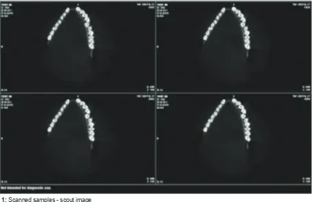
The advent of CBCT technology has paved way for the development of relatively small and inexpensive CT scanners dedicated for use in dentomaxillofacial imag-ing [Figure 1].
RESULTS
Tables 1a-e show mandibular central incisors, lateral incisors, and canines.
Morphological Measurements [Tables 1c-e, Figures 2 and 3]
Figure 2: Cone-beam computed tomography views - cor-onal, axial, and sagittal
DISCUSSION
Variation in root canal anatomy is common and is con-sidered as a normal phenomenon. For success in end-odontic treatment, a thorough understanding of the root canal morphology is necessary.
The studies on root canal anatomy by Hess and Zurcher[5] as earlyas 1924 reported that a root with a
graceful, tapering canal and a single apical foramen is an expectation rather than a rule Vertucci[18] in 1984
who classified root canal anatomy broadly into 8 types. CBCT now provides the clinician with the ability to observe an area in three different planes and thus to acquire 3D information. The combination of sagittal, coronal, and axial CBCT images eliminates the superim-position of anatomic structures.
CBCT scanning or digital volume tomography uses an extraoral imaging scanner to produce 3D scans of the maxillofacial skeleton at a considerably lower radiation dose than conventional CT scanning. CBCT scanning has been shown to be more accurate than digital radio-graphs in determining root canal systems.
When the root canal configuration deviates from the normal and expected anatomy, it is always difficult for an endodontist to adequately and effectively clean, shape, and obturate it 3D. The awareness of these pos-sible deviations that might occur in the root canal mor-phology would greatly enhance the quality of treatment. Many studies have reported that root canal systems vary according to race.[3,6,7,8] Literature reveals that
very few studies have been done on root canal mor-phology of teeth in an Indian population. Studies on
Table 1b: Root canal pattern (Vertucci’s classification)
Tooth (n=100) Type I Type II Type III Type IV Type V Type VI Type VII Type VIII C‑shaped canal
Central incisor 73 1 13 10 3 - - -
-Lateral incisor 67 3 17 8 5 - - -
-Canine 79 3 6 9 1 2 - -
-Table 1c: Crown/root length
Tooth (n=100) Crown length Root length Overall length
Central incisor 9.28 mm 12.9 mm 22.2 mm
Lateral incisor 8.63 mm 12.83 mm 21.4 mm
Canine 10.3 mm 14.8 mm 25 mm
Table 1d: Distance from the incisal edge to roof of the pulp chamber
Tooth (n=100) Mean (mm) Standard deviation
Central incisor 4.4171 1.2384
Lateral incisor 4.8516 1.0498
Canine 5.6022 1.2943
Table 1e: Relation of CEJ to roof of the pulp chamber Tooth
(n=100) CEJ coinciding with roof (%) the roof (%)CEJ above the roof (%)CEJ below
Central incisor 24 1 75
Lateral incisor 33 - 67
Canine 31 4 65
CEJ: Cementoenamel junction
Figure 2: Cone-beam computed tomography views - coronal, axial, and sagittal
Figure 3: Morphological measurements in mandibular teeth
Lateral incisor 100 74 11
-Root canal morphology of human permanent mandibular anterior teeth
IJOCR
morphologic variations have been reported on the man-dibular first premolars by Velmurugan et al. in 2009,[4]
mandibular first molar by Reuben et al.[2] in 2008, and
mandibular second molars by Prasanna et al. in 2010 on the Indian populations. Hence, this study was under-taken to extensively assess the root canal morphology of all the mandibular teeth.
The overall average length of the mandibular cen-tral incisor is 22.2 cm with an average crown length of 9.28 mm and an average root length of 12.9 mm. The mandibular lateral incisor with an average length is 21.4 mm ,with the crown length of 8.63 mm and with an average root length of 12.83 mm.
The mean distance from the incisal edge to the roof of the pulp chamber in mandibular central and lateral incisor was 4.41 mm and 4.85 mm, respectively.
The CEJ coincided with the roof of pulp chamber in 24% and 33% coincides with the cases, and it was lower than the roof of pulp chamber in 39 and 33% of the sam-ples, respectively.
Most common root canal pattern in central incisor and lateral incisor was type I pattern (89% and 74%). These results are similar to the results of a previous study done in Turkish population[9] but contrary to the
results reported by Uma et al. who reported that type III pattern was more prevalent. However, in this study, type III pattern was seen only in 13% and 17% of central incisors and lateral incisors, respectively.
In 2010, Boruah and Lalith[12] using staining
tech-nique reported that the majority of mandibular incisors in north-east Indian population had a single root canal type I (63.75%). Although 36.25% of roots possessed two canals only, 6.25% had two separate apical foramina.
In the present study, the incidence of second canal was found to be only 11%. Madiera and Hetenn have also reported similar incidence of 11.3% of two canals in Turkish population, whereas Miyashita M et al. in 1997 reported 15% (1085 samples) of two canals in mandib-ular incisors in Japanese population. Our result is con-trary to the results reported by Kartal and Yanicoglu[10]
(43%) and Benjamin and Dowson[11] (41.4%) who had
reported very high incidence of two canals.
CONCLUSION
Overall mandibular central incisors (73%), lateral inci-sors (67%), and canines (78%) - majority of samples dis-played type I configuration in Indian population. Two canals were detected in 11% of the both incisors and 14% in canines. Type III pattern was seen in 13% and 17% of the central and lateral incisors. 6% canines had type III configuration and 3% had type II. CEJ was commonly found below the pulp chamber (75%, 67%, and 65%).
Mandibular Canine
The average length of mandibular canines in our study was found to be 25 mm. This is in accordance with the result reported by Pucci and Reig[19] in 1944. Distance
from the incisal edge to the pulp chamber roof showed a mean value of 5.6 mm. This distance could act as a guide during access cavity preparation. In 31% of the samples studies, the pulp chamber roof coincided with CEJ. CEJ was lower than the roof of pulp chamber in 65% of sam-ples and, in 4% of the cases, above the pulp chamber. Hence, CEJ cannot be used as a guide for the assessment of depth of pulp chamber.
All the samples were single rooted. In the present study, type I pattern (79%) was the most common fol-lowed by type IV (9%). This result is in accordance with the earlier in study done by Sikri et al. in 2003 in Indian population.
2% of samples showed type VI pattern and 1% showed type V pattern. The existence of two canals was seen in 14%. This is in agreement with results of Hession[13] (11%), Green[14] (13%), and Koffe et al.[15]
(13.75%). It is, however, higher than the result of Bellizzi and Hartwell[16] (4.11%) and Ingle et al.[17] (6%) but lower
than the result of Vertucci[18] (22%)
CONCLUSION
CBCT allows identification of anatomic features and variations of the root canal system. Therefore, within the limitations of this study, it can be concluded that:
Mandibular central incisors (73%), lateral incisors (67%), and canines (78%) - majority of samples dis-played type I configuration. Two canals were detected in 11% of the both incisors and 14% in canines. Type III pattern was seen in 13% and 17% of the central and lat-eral incisors. 6% canines had type III configuration and 3% had type II. CEJ was commonly found below the pulp chamber (75%, 67%, and 65%).
In a clinical scenario, to treat and retreat, dentists should be aware of the possible existence of additional canals and variations in canal anatomy before initiating endodontic treatment.
REFERENCES
1. Slowey RR. Root canal anatomy. Road map to successful endodontics. Dent Clin North Am 1979;23:555-73.
2. Reuben J, Velmurugan N, Kandaswamy D. The evaluation of root canal morphology of the mandibular first molar in an Indian population using spiral computed tomography scan: An in vitro study. J Endod 2008;34:212-5.
3. Hess W, Zurcher E. The Anatomy of the Root Canals of Teeth of the Permanent and Deciduous Dentition. New York: William Wood and Co.; 1925.
Traumatol 1988;4:226-8.
6. Haddad GY, Nehme WB, Ounsi HF. Diagnosis, classifica-tion, and frequency of C-shaped canals in mandibular second molars in the Lebanese population. J Endod 1999;25:268-71. 7. Weine FS, Hayami S, Hata G, Toda T. Canal configuration
of the mesiobuccal root of the maxillary first molar of a Japanese sub-population. Int Endod J 1999;32:79-87.
8. Sert S, Bayirli GS. Evaluation of the root canal configurations of the mandibular and maxillary permanent teeth by gender in the Turkish population. J Endod 2004;30:391-8.
9. Velmurugan N, Sandhya R. Root canal morphology of man-dibular first premolars in an Indian population: A labora-tory study. Int Endod J 2009;42:54-8.
10. Calişkan MK, Pehlivan Y, Sepetçioğlu F, Türkün M, Tuncer
SS. Root canal morphology of human permanent teeth in a Turkish population. J Endod 1995;21:200-4.
11. Boruah L. Root canal morphology of permanent mandibu-lar incisors in a North East Indian population-in vitro study using a root canal stainig technique. Qutissience Int Endo 2010;4:274-8.
human mandibular incisor teeth. Oral Surg Oral Med Oral Pathol 1974;38:122-6.
14. Pucci FM, Reig R. Conductors radicularis. In: Editorial Medico-Quirurgica. Vol. 1. Buenos Aries: Argentina Medical Press; 1944.
15. Hession RW. Endodontic morphology. II. A radiographic analysis. Oral Surg Oral Med Oral Pathol 1977;44:610-20. 16. Green D. Double canal in single roots. Oral Surg Oral Med
Oral Pathol 1973;35:689-96.
17. Kaffe I, Kaufman A, Littner MM, Lazarson A. Radiographic study of the root canal system of mandibular anterior teeth. Int Endod J 1985;18:253-9.
18. Bellizzi R, Hartwell G. Clinical investigation of in vivo end-odontically treated mandibular anterior teeth. J Endod 1983;9:246-8.