R E S E A R C H
Open Access
Evaluation of the ovarian reserve function in
patients with metabolic syndrome in relation to
healthy controls and different age groups
Fevzi Balkan
1, Nurcan Cetin
2, Celil Alper Usluogullari
3*, Oguz Kaan Unal
4and Betul Usluogullari
5Abstract
Objective:To evaluate the ovarian reserve function in female patients with metabolic syndrome (MetS).
Methods:This study evaluated 136 subjects, 67 with MetS and 69 controls. Subjects were divided into three age groups. Group I included 49 subjects aged 20–29 years, 22 with MetS and 27 controls; group II included 45 subjects aged 30–39 years, 22 with MetS and 23 controls; and group III included 42 subjects aged 40–49 years, 23 with MetS and 19 controls. Demographic characteristics, anthropometrics, blood biochemistry, and gonadotrophic hormones were compared as total ovarian volume and antral follicle count on ovarian transvaginal ultrasonography.
Results:Serum levels of FSH, LH, E2 and progesterone were similar in the MetS and control groups, while testosterone levels were significantly higher in MetS patients than controls, both in the overall population (p = 0.024) and in those aged 20–29 years (p = 0.018). Total ovarian volume was significantly lower in MetS patients than controls, in both the overall population (p = 0.003) and those aged 20–29 years (p = 0.018), while antral follicle counts were similar. Ovarian volume correlated positively with antral follicle count (AFC) (r = 0.37; p < 0.001) and negatively with age (r = 0.34; p < 0.001) and FSH concentration (r = 0.21; p = 0.013). AFC was negatively correlated with age (r = 0.36; p < 0.001).
Conclusion:Ovarian reserve function is significantly lower in MetS patients than in healthy control subjects, particularly in women aged 20–29 years.
Keywords:Metabolic syndrome, Ovarian reserve, Ovarian volume, Antral follicle, Obesity, Gonadotropins
Introduction
Female fertility, by spontaneous conception or assisted reproductive techniques, decreases significantly with age, a reduction that may be due to the reduction in the number of primordial follicles over time [1-3]. Ovarian reserve is a measure of the reproductive potential of a woman in relation to primordial follicle count and oo-cyte quality. Follicle response to stimulation and fertility promoting medications is reduced in parallel to the reduc-tion in ovarian reserve. Determinants of ovarian reserve include basal FSH and inhibin B levels (4), estradiol and LH concentrations, LH/FSH ratio, response to stimulation with gonadotropin releasing hormone (GnRH), ovarian
volume, total antral follicle count (AFC) and ovarian stro-mal blood flow [4,5]. A meta-analysis found that AFC was a better predictor of ovarian response than basal FSH level [6], and several studies have shown that AFC, as deter-mined by high-resolution transvaginal ultrasonography, is a significant predictor of ovarian response [4-7].
Insulin resistance plays a central role in metabolic syn-drome (MetS) [8,9]. Obesity has been associated with multiple adverse reproductive outcomes in both males and females [10], although the exact mechanisms are largely unknown. The complexity of the human repro-ductive system makes identification of the mechanisms linking obesity and adverse reproductive functioning challenging [11].
Despite MetS having a negative impact on fertility in women of reproductive age, cross-sectional evidence sug-gests that increased ovarian reserve is associated with a
* Correspondence:[email protected] 3
Endocrinology and Metabolic Disease, Dr. Ersin Arslan State Hospital, Gaziantep, Turkey
Full list of author information is available at the end of the article
healthier cardiometabolic risk factor profile [12]. To our knowledge, however, the effect of MetS on ovarian func-tions has not been previously investigated. Therefore the present study was designed to evaluate ovarian reserve function in female patients with MetS in Turkey in rela-tion to healthy controls and among different age groups.
Methods Study population
The study included 136 subjects, comprising 67 pa-tients with MetS and 69 healthy controls, who were evaluated at the Department of Endocrinology and Metabolic Diseases, Aksaray State Hospital, between January and July 2013. Subjects were divided into three groups by age. Group I consisted of 49 sub-jects aged 20–29 years, including 22 MetS patients and 27 controls; group II consisted of 45 subjects aged 30–39 years, including 22 MetS patients and 23 controls; and group III consisted of 42 subjects aged 40–49 years, including 23 MetS patients and 19 con-trols. Subjects were included if they had a history of spontaneous pregnancy, regularly menstruated at intervals of 21–35 days, had cycle length variations of less than 4 days and had both ovaries. Subjects were excluded if they had a history of chronic renal or liver failure, autoimmune or connective tissue disease, a known malignancy, a history of smoking, known infertility, gynecological abnormalities such as dysfunctional uterine bleeding or menorrhagia, a his-tory of ovarian surgery, or if they had used a hormo-nal preparation or dubious herbal product within the 3 months prior to enrollment.
Following a detailed explanation of the objectives and protocol of the study, written informed consent was ob-tained from each subject. The study was conducted in accordance with the ethical principles stated in the Dec-laration of Helsinki and was approved by the institu-tional ethics committee of Aksaray State Hospital.
Study parameters
Demographic and clinical characteristics were recorded for each subject, including anthropometric factors, such as weight, height, body mass index (BMI), waist circum-ference, and waist-to-hip ratio (WHR). Blood biochemis-try parameters included fasting blood glucose (FBG), insulin, high density lipoprotein-cholesterol (HDL-C) and triglyceride (TG) concentrations and homeostasis model assessment-IR (HOMA-IR). Serum concentrations of go-nadotrophic hormones were measured, including follicle stimulating hormone (FSH), luteinizing hormone (LH), es-tradiol (E2), progesterone, and testosterone. Total ovarian volume (cm3) and AFC were determined by ovarian trans-vaginal ultrasonography.
Assessment of MetS
A diagnosis of MetS was based on the criteria of the Na-tional Cholesterol Education Program (NCEP)-ATP III and included three of the following five factors: (i) abdominal obesity, defined as a waist circumference (WC) > 102 cm in men and > 88 cm in women; (ii) serum TGs≥150 mg/dl; (iii) serum HDL < 40 mg/dl in men and < 50 mg/dl in women; (iv) blood pressure > 130/85 mmHg; and (v) FBG > 110 mg/dl [13].
Assessment of insulin resistance
The estimate of insulin resistance by HOMA-IR score was calculated with the formula fasting serum insulin (lU/ml) × FBG (mmol/l)/22.5 as described by Matthews and coworkers [14]. The cut-off value was taken as 2.7 for HOMA-IR [15].
Anthropometric measurements
Height, weight, and WC were measured by a designated physician. WC was measured with a folding tape at the natural waistline (the level of the umbilicus) in a hori-zontal plane. BMI was calculated by dividing body weight (kg) by the square of height (m), and WHR was calculated by dividing waist by hip circumference.
Blood biochemistry analysis
Venous blood samples were taken from the antecubital regions of all subjects between 08:00 am and 09:00 am after an overnight fast of 8–12 hours. Serum FPG con-centrations were measured by the hexokinase method. Serum FSH, LH, total testosterone, E2, progesterone and insulin were measured with specific electrochemilu-minescence immunoassays (Elecsys 2010 Cobas, Roche Diagnostics, Mannheim, Germany). Totalcholesterol, HDL-C and TG concentrations were measured using en-zymatic colorimetric assays by spectrophotometry (Abbott Architect C16000).
Assessment of ovaries by transvaginal ultrasonography
Statistical analysis
Statistical analysis was performed using SPSS for Win-dows 17 software (SPSS Inc., Chicago. IL). Normality of distribution of continuous variables was evaluated using the Shapiro Wilk test. Means were compared using Stu-dent’s t tests or Mann–Whitney U test, as appropriate. Spearman’s rho test was used for correlation analysis of factors in the MetS groups. A p value <0.05 was consid-ered statistically significant.
Results
Demographic characteristics and anthropometric and laboratory findings
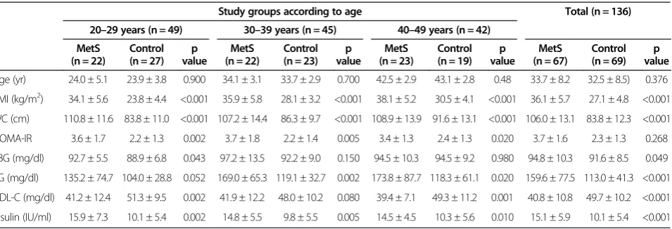
In the overall population, mean ± SDBMI (36.1 ± 5.7 vs. 27.1 ± 4.8 kg/m2, p < 0.001), WC (106.0 ± 13.1 vs. 83.8 ± 12.3 cm, p < 0.001), and insulin (15.1 ± 5.9 vs. 10.1 ± 5.4 IU/ml, p < 0.001), FBG (94.8 ± 10.3 vs. 91.6 ± 8.5 mg/dl, p = 0.049), and TG (159.6 ± 77.5 vs. 113.0 ± 41.3 mg/dl, p < 0.001) concentrations were significantly higher, while HDL-C concentrations (40.8 ± 10.8 vs. 49.7 ± 10.2 mg/dl, p < 0.001) were significantly lower among patients with MetS than in control subjects (Table 1). When grouped by age (20–29, 30–39, and 40–49 years), mean BMI (p < 0.001 each), WC (p < 0.001 each), HOMA-IR (p = 0.002, p = 0.005 and p = 0.02, respectively) and insulin concen-trations (p = 0.002, p = 0.005 and p = 0.01, respectively) were significantly higher in patients with MetS than in control subjects.
Gonadal hormone concentrations
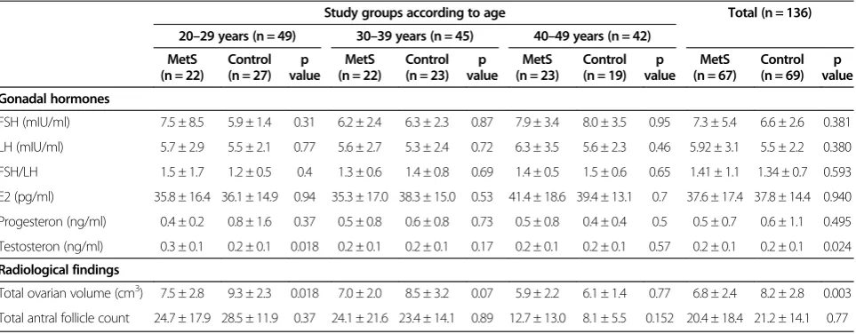
Serum concentrations of FSH, LH, E2 and progesterone were similar in overall patients and controls, as well as in each of the age groups (Table 2). Testosterone con-centrations, however, were significantly higher in MetS patients than in controls, both in the overall population
(0.2(0.1) vs. 0.2(0.1) ng/ml, p = 0.024) and in the 20–29 age group (0.3(0.1) vs. 0.2(0.1) ng/ml, p = 0.018).
Radiological findings
Total ovarian volume was significantly lower in MetS patients than in controls, both in the overall population (6.8 ± 2.4 vs. 8.2 ± 2.8 ng/ml, p = 0.003) and in the 20–29 age group (7.5 ± 2.8 vs. 9.5 ± 2.3 ng/ml, p = 0.018). AFCs were similar in patient and control groups, both in the overall population and the three age groups. Moreover, AFC did not correlate with age, either in the patient or control groups (p > 0.05 each).
Correlation of ovarian reserve function with clinical, laboratory and radiological findings
Ovarian volume correlated positively with AFC (r = 0.37; p < 0.001), while correlating negatively with age (r = 0.34; p < 0.001) and FSH concentration (r = 0.21; p = 0.013). AFC was correlated negatively with age (r = 0.36; p < 0.001).
Discussion
This study, the first to investigate ovarian reserve func-tion in women with MetS, found that ovarian reserve, as determined by ovarian volume, was significantly lower in patients with MetS than in healthy controls, in particular in women aged 20–29 years. Moreover, ovarian volume was found to be positively correlated with AFC, and ovar-ian volume and AFC were negatively correlated with age.
Ovarian reserve can be determined by measuring AFC and ovarian volume by ultrasonography during the early follicular phase. Although we found that ovarian volume was significantly lower in MetS patients than in controls, AFC tended to be lower and FSH concentrations higher in the former, a finding consistent with the reverse
Table 1 Demographic characteristics, anthropometrics and laboratory findings in study groups
Study groups according to age Total (n = 136)
20–29 years (n = 49) 30–39 years (n = 45) 40–49 years (n = 42)
MetS (n = 22)
Control (n = 27)
p value
MetS (n = 22)
Control (n = 23)
p value
MetS (n = 23)
Control (n = 19)
p value
MetS (n = 67)
Control (n = 69)
p value
Age (yr) 24.0 ± 5.1 23.9 ± 3.8 0.900 34.1 ± 3.1 33.7 ± 2.9 0.700 42.5 ± 2.9 43.1 ± 2.8 0.48 33.7 ± 8.2 32.5 ± 8.5) 0.376
BMI (kg/m2) 34.1 ± 5.6 23.8 ± 4.4 <0.001 35.9 ± 5.8 28.1 ± 3.2 <0.001 38.1 ± 5.2 30.5 ± 4.1 <0.001 36.1 ± 5.7 27.1 ± 4.8 <0.001
WC (cm) 110.8 ± 11.6 83.8 ± 11.0 <0.001 107.2 ± 14.4 86.3 ± 9.7 <0.001 108.9 ± 13.9 91.6 ± 13.1 <0.001 106.0 ± 13.1 83.8 ± 12.3 <0.001
HOMA-IR 3.6 ± 1.7 2.2 ± 1.3 0.002 3.7 ± 1.8 2.2 ± 1.4 0.005 3.4 ± 1.3 2.4 ± 1.3 0.020 3.7 ± 1.6 2.3 ± 1.3 0.268
FBG (mg/dl) 92.7 ± 5.5 88.9 ± 6.8 0.043 97.2 ± 13.5 92.2 ± 9.0 0.150 94.5 ± 10.3 94.5 ± 9.2 0.980 94.8 ± 10.3 91.6 ± 8.5 0.049
TG (mg/dl) 135.2 ± 74.7 104.0 ± 28.8 0.052 169.0 ± 65.3 119.1 ± 32.7 0.002 173.8 ± 87.7 118.3 ± 61.1 0.020 159.6 ± 77.5 113.0 ± 41.3 <0.001
HDL-C (mg/dl) 41.2 ± 12.4 51.3 ± 9.5 0.002 41.9 ± 12.2 48.0 ± 10.2 0.080 39.4 ± 7.1 49.3 ± 11.2 0.001 40.8 ± 10.8 49.7 ± 10.2 <0.001
Insulin (IU/ml) 15.9 ± 7.3 10.1 ± 5.4 0.002 14.8 ± 5.5 9.8 ± 5.5 0.005 14.5 ± 4.5 10.3 ± 5.6 0.010 15.1 ± 5.9 10.1 ± 5.4 <0.001
interaction between these two parameters. Nonethe-less, due to the positive correlation between ovarian volume with AFC and the negative correlations of both with age in our study population, our findings suggest that ovarian androgen production not only declines with menopause but also with older age [16].
The present findings, showing a significant reduction in ovarian volume in patients with MetS, are in agree-ment with results showing delayed menarche and irregu-lar menstruation in patients with type 1 diabetes [17] and significant reductions in ovarian volume and AFC in patients with type 2 diabetes aged 20–29 years [1], com-pared with age-matched controls. In contrast, ovarian volumes in subjects aged 20–39 and 40– 49 years were similar in those with MetS and healthy controls. Type I diabetes patients have also been shown to have a high risk of premature menopause [18].
A previous study of Turkish women aged 16 to 40 years found that ovarian volumes were significantly lower among patients with than without polycystic ovar-ian syndrome (12.5 vs. 5.4 cm3) [19]. Another study in-volving 62 infertile and 53 fertile women aged 35 to 45 years found that mean AFC counts and FSH levels were similar, while ovarian volume was significantly lower in infertile than in fertile women (1.8 vs. 6.1 cm3) [20].
Since ovarian volume is a parameter used in traditional in vitro fertilization methods [21], age and ovarian vol-ume were found to be negatively correlated with subject age [22]. Ovarian volume and AFC both decreased with age in our study population. A Brazilian study reported that mean ovarian volume was 7.1 cm3during the peri-menopausal period, decreasing 0.2cm3each year [23]. A study in China of 31 healthy volunteers aged 22 to 42 years, found that AFC declined at a rate of 0.95 follicles/ year or 60% [24], while another study of infertile Chinese
women showed that AFC declined 0.35 follicles/year or 3.8% [25,26].
Various mechanisms may be responsible for the reduc-tion in ovarian reserve among diabetic women. Chronic complications and prolonged hyperglycemia have been reported to negatively affect ovarian reserve, with regular blood glucose control suggested to improve fertility and menstruation anomalies [17].
Polycystic ovarian reserve and diabetes prevalence was reported to increase with increasing obesity, resulting in reductions in oocyte quality [27]. Diabetes related in-creases in menstruation anomalies and the risk of pre-mature menopause were shown to be associated with increased cardiovascular risks [28]. In agreement with the significantly higher testosterone concentrations we observed in MetS patients than in controls, a Chinese study of 719 women with polycystic ovary syndrome and 685 healthy volunteers found that patients with polycys-tic ovary syndrome and obesity had higher serum testos-terone and fasting insulin levels, lower LH levels, and enlarged ovarian follicles compared with control subjects [24]. In many studies on patients undergoing invitro fertilization procedures, increased BMI was found to have a negative impact on ovarian reserve [29]. More-over, inhibin B levels and AFC were significantly lower in overweight patients [29]. Indeed, we found that ovar-ian volume was greater in patients with BMI <30 kg/m2 than in subjects with higher BMI.
Although the impact of obesity on the female repro-ductive system remains unclear, obesity is considered a risk factor for poorer overall health, which, in turn, has negative effects on reproduction, menstrual function, ovulation, and GnRH regulation [30]. Obesity is also an independent risk factor for PCOS and plays important roles in the clinical, metabolic, and biochemical changes Table 2 Gonadal hormones and radiological findings in the study groups
Study groups according to age Total (n = 136)
20–29 years (n = 49) 30–39 years (n = 45) 40–49 years (n = 42)
MetS (n = 22)
Control (n = 27)
p value
MetS (n = 22)
Control (n = 23)
p value
MetS (n = 23)
Control (n = 19)
p value
MetS (n = 67)
Control (n = 69)
p value
Gonadal hormones
FSH (mIU/ml) 7.5 ± 8.5 5.9 ± 1.4 0.31 6.2 ± 2.4 6.3 ± 2.3 0.87 7.9 ± 3.4 8.0 ± 3.5 0.95 7.3 ± 5.4 6.6 ± 2.6 0.381
LH (mIU/ml) 5.7 ± 2.9 5.5 ± 2.1 0.77 5.6 ± 2.7 5.3 ± 2.4 0.72 6.3 ± 3.5 5.6 ± 2.3 0.46 5.92 ± 3.1 5.5 ± 2.2 0.380
FSH/LH 1.5 ± 1.7 1.2 ± 0.5 0.4 1.3 ± 0.6 1.4 ± 0.8 0.69 1.4 ± 0.5 1.5 ± 0.6 0.65 1.41 ± 1.1 1.34 ± 0.7 0.593
E2 (pg/ml) 35.8 ± 16.4 36.1 ± 14.9 0.94 35.3 ± 17.0 38.3 ± 15.0 0.53 41.4 ± 18.6 39.4 ± 13.1 0.7 37.6 ± 17.4 37.8 ± 14.4 0.940
Progesteron (ng/ml) 0.4 ± 0.2 0.8 ± 1.6 0.37 0.5 ± 0.8 0.6 ± 0.8 0.73 0.5 ± 0.8 0.4 ± 0.4 0.5 0.5 ± 0.7 0.6 ± 1.1 0.495
Testosteron (ng/ml) 0.3 ± 0.1 0.2 ± 0.1 0.018 0.2 ± 0.1 0.2 ± 0.1 0.17 0.2 ± 0.1 0.2 ± 0.1 0.57 0.2 ± 0.1 0.2 ± 0.1 0.024
Radiological findings
Total ovarian volume (cm3) 7.5 ± 2.8 9.3 ± 2.3 0.018 7.0 ± 2.0 8.5 ± 3.2 0.07 5.9 ± 2.2 6.1 ± 1.4 0.77 6.8 ± 2.4 8.2 ± 2.8 0.003
Total antral follicle count 24.7 ± 17.9 28.5 ± 11.9 0.37 24.1 ± 21.6 23.4 ± 14.1 0.89 12.7 ± 13.0 8.1 ± 5.5 0.152 20.4 ± 18.4 21.2 ± 14.1 0.77
that occur throughout PCOS [31]. The finding of signifi-cantly higher BMI and waist circumference among pa-tients with MetS and type II diabetes [1,32] accords with thehigh prevalence of infertility and low chance of preg-nancy of such patients, even using assisted reproductive technologies [33].
In conclusion, we found that ovarian reserve was lower in patients with MetS than in healthy control subjects, especially in those aged 20–29 years. We also observed a positive correlation between ovarian volume and AFC and negative correlations of both with age. Since a greater ovarian reserve has been associated with a healthier car-diometabolic risk factor profile [12], future larger scale studies are needed to clarify the role of obesity in the as-sociation between MetS and ovarian reserve, along with other likely determinants of this interaction.
Competing interests
The authors declare that they have no competing interests.
Authors’contributions
FB collected patients, and wrote the article, NC made USG to patients, CAU wrote article and made statistics, drafted the manuscript, OKU collected patients, BU collected patients, designed study. All authors read and approved the final manuscript.
Author details
1Endocrinology and Metabolic Disease, Aksaray State Hospital, Aksaray,
Turkey.2Radiology Department, Aksaray State Hospital, Aksaray, Turkey. 3Endocrinology and Metabolic Disease, Dr. Ersin Arslan State Hospital,
Gaziantep, Turkey.4Endocrinology and Metabolic Disease, Aksaray State Hospital, Aksaray, Turkey.5Gynecology and Obstetrics Department, Cengiz Gokcek State Hospital, Gaziantep, Turkey.
Received: 12 April 2014 Accepted: 4 June 2014 Published: 10 June 2014
References
1. Isik S, Ozcan HN, Ozuguz U, Tutuncu YA, Berker D, Alimli AG, Akbaba G, Karademir MA, Guler S:Evaluation of ovarian reserve based on hormonal parameters, ovarian volume, and antral follicle count in women with type 2 diabetes mellitus.J Clin Endocrinol Metab2012,97:261–269. 2. Bulun SE, Adashi EY:The physiology and pathology of the female reproduction axis.InWilliams textbook of endocrinology.10th edition. Edited by Larson PR, Kronenberg HM, Melmed S, Polonsky KS. Philadelphia, PA: Elsevier Science; 2003:587–664.
3. Templeton A, Morris JK, Parslow W:Factors that affect outcome of in vitro fertilisation treatment.Lancet1996,348:1402–1406.
4. Lass A, Skull J, McVeigh E, Margara R, Winston RM:Measurement of ovarian volume by transvaginal sonography before ovulation induction with human menopausal gonadotrophin for in vitro fertilization can predict poor response.Hum Reprod1997,12:294–297.
5. Hendriks DJ, Kwee J, Mol BW, te Velde ER, Broekmans FJ:Ultrasonography as a tool for the prediction of outcome in IVF patients: a comparative meta-analysis of ovarian volume and antral follicle count.Fertil Steril
2007,87:764–775.
6. Hendriks DJ, Mol BW, Bancsi LF, TeVelde ER, Broekmans FJ:Antral follicle count in the prediction of poor ovarian response and pregnancy after in vitro fertilization: a meta-analysis and comparison with basal follicle stimulating hormone level.Fertil Steril2005,83:291–301.
7. Broekmans FJ, Kwee J, Hendriks DJ, Mol BW, Lambalk CB:A systematic review of tests predicting ovarian reserve and IVF outcome.Hum Reprod Update2006,12:685–718.
8. Reaven GM:Banting lecture. Role of insulin resistance in human disease.
Diabetes1988,37:1595–1607.
9. Hu G, Qiao Q, Tuomilehto J:The metabolic syndrome and cardiovascular risk.Curr Diabetes Rev2005,1:137–143.
10. Masheshwari A, Stofberg L, Bhattacharya S:Effect of overweight and obesity on assisted reproductive technology–a systematic review.
Hum Reprod Update2007,13:433–444.
11. Jungheim ES, Travieso JL, Carson KR, Moley KH:Obesity and reproductive function.Obstet Gynecol Clin North Am2012,39:479–493.
12. Bleil ME, Gregorich SE, McConnell D, Rosen MP, Cedars MI:Does accelerated reproductive aging underlie premenopausal risk for cardiovascular disease?Menopause2013,20:1139–1146. 13. Expert Panel on Detection, Evaluation, and Treatment of High Blood
Cholesterol in Adults:Executive Summary of the Third Report of the National Cholesterol Education Program (NCEP) Expert Panel on Detection, Evaluation, and Treatment of High Blood Cholesterol in Adults (Adult Treatment Panel III).J Am Med Assoc2001,285:2486–2497. 14. Matthews DR, Hosker JP, Rudenski AS, Naylor BA, Treacher DF, Turner RC:
Homeostasis model assessment: insulin resistance and beta-cell function from fasting plasma glucose and insulin concentrations in man.
Diabetologia1985,28:412–419.
15. Gokcel A, Ozsahin AK, Sezgin N, Karakose H, Ertorer ME, Akbaba M, Baklaci N, Sengul A, Guvener N:High prevalence of diabetes in Adana, a southern province of Turkey.Diabetes Care2003,26:3031–3034. 16. Shifren JL, Schiff I:The aging ovary.J Womens Health Gend Based Med
2000,1:S3–S7.
17. Yeshaya A, Orvieto R, Dicker D, Karp M, Ben-Rafael Z:Menstrual characteris-tics of women suffering from insulin-dependent diabetes mellitus.Int J Fertil Menopausal Stud1995,40:269–273.
18. Dorman JS, Steenkiste AR, Foley TP, Strotmeyer ES, Burke JP, Kuller LH:
Familial Autoimmune and Diabetes (FAD) Study. Menopause in type 1 diabetic women: is it premature?Diabetes2001,50:1857–1862. 19. KöşüşN, KöşüşA, Turhan NÖ, Kamalak Z:Do threshold values of ovarian
volume and follicle number for diagnosing polycystic ovarian syndrome in Turkish women differ from western countries?Eur J Obstet Gynecol Reprod Biol2011,154:177–181.
20. Erdem M, Erdem A, Biberoglu K, Arslan M:Age-related changes in ovarian volume, antral follicle counts and basal follicle stimulating hormone levels: comparison between fertile and infertile women.Gynecol Endocrinol2003,17:199–205.
21. Syrop CH, Willhoite A, Van Voorhis BJ:Ovarian volume: a novel outcome predictor for assisted reproduction.Fertil Steril1995,64:1167–1171. 22. Kupesic S, Kurjak A, Bjelos D, Vujisic S:Three-dimensional ultrasonographic
ovarian measurements and in vitro fertilization outcome are related to age.Fertil Steril2003,79:190–197.
23. Oppermann K, Fuchs SC, Spritzer PM:Ovarian volume in pre- and peri-menopausal women: a population-based study.Menopause2003,
10:209–213.
24. Malhotra N, Bahadur A, Singh N, Kalaivani M, Mittal S:Does obesity compromise ovarian reserve markers? A clinician’s perspective.Arch Gynecol Obstet2013,287:161–166.
25. Ng EH, Yeung WS, Fong DY, Ho PC:Effects of age on hormonal and ultrasound markers of ovarian reserve in Chinese women with proven fertility.Hum Reprod2003,18:2169–2174.
26. Chang MY, Chiang CH, Chiu TH, Hsieh TT, Soong YK:The antral follicle count predicts the outcome of pregnancy in a controlled ovarian Hyperstimulation/intrauterine insemination program.J Assist Reprod Genet
1998,15:12–17.
27. Metwally M, Cutting R, Tipton A, Skull J, Ledger WL, Li TC:Effect of increased body mass index on oocyte and embryo quality in IVF patients.Reprod Biomed Online2007,15:532–538.
28. Van der Schouw YT, van der Graaf Y, Steyerberg EW, Eijkemans JC, Banga JD:Age at menopause as a risk factor for cardiovascular mortality.Lancet
1996,347:714–718.
29. Zhang HY, Guo CX, Zhu FF, Qu PP, Lin WJ, Xiong J:Clinical characteristics, metabolic features, and phenotype of Chinese women with polycystic ovary syndrome: a large-scale case–control study.Arch Gynecol Obstet
2013,287:525–531.
30. Brewer CJ, Balen AH:The adverse effects of obesity on conception and implantation.Reproduction2010,140:47–364.
Goodarzi MO, Strauss JF 3rd, McCarthy MI, Malecki MT, GIANT Consortium:
Impact of FTO genotypes on BMI and weight in polycystic ovary syndrome: a systematic review and meta-analysis.Diabetologia2012,55:636–2645. 32. Ding EL, Song Y, Malik VS, Liu S:Sex differences of endogenous sex
hormones and risk of type 2 diabetes: a systematic review and meta-analysis.JAMA2006,295:1288–1299.
33. Wang JX, Davies M, Norman RJ:Body mass and probability of pregnancy during assisted reproduction treatment: retrospective study.BMJ2000,
321:1320–1321.
doi:10.1186/1757-2215-7-63
Cite this article as:Balkanet al.:Evaluation of the ovarian reserve function in patients with metabolic syndrome in relation to healthy controls and different age groups.Journal of Ovarian Research20147:63.
Submit your next manuscript to BioMed Central and take full advantage of:
• Convenient online submission
• Thorough peer review
• No space constraints or color figure charges
• Immediate publication on acceptance
• Inclusion in PubMed, CAS, Scopus and Google Scholar
• Research which is freely available for redistribution