R E V I E W
Preventive and improvement effects of exercise training
and supplement intake in white adipose tissues on obesity
and lifestyle-related diseases
Takuya Sakurai
•Junetsu Ogasawara
•Takako Kizaki
•Yoshinaga Ishibashi
•Yoshikazu Sumitani
•Kazuto Takahashi
•Hitoshi Ishida
•Hiromi Miyazaki
•Daizoh Saitoh
•Shukoh Haga
•Tetsuya Izawa
•Hideki Ohno
Received: 6 October 2011 / Accepted: 5 February 2012 / Published online: 24 February 2012
ÓThe Japanese Society for Hygiene 2012
Abstract
Recent increases in the number of obese
indi-viduals and indiindi-viduals suffering from lifestyle-related
diseases, such as type 2 diabetes, that accompany obesity
have become a serious social problem. White adipose
tis-sue (WAT) is more than a mere organ for storage of
energy; it is also a highly active metabolic and endocrine
organ that secretes physiologically active substances
col-lectively known as adipokines, including tumor necrosis
factor-
a
and adiponectin. Dysregulated expression of
adi-pokines in WAT that is hypertrophied by obesity has been
closely associated with the phenomenon of insulin
resis-tance. Therefore, WAT is currently considered to be one of
the tissues that promote lifestyle-related diseases.
Reduc-tion of excess WAT that results from obesity is seen as an
important strategy in preventing and improving
lifestyle-related diseases. This review shows that exercise training
as well as intake of supplements, such as polyphenols, is
one strategy for this, because this regimen can result in
reduction of WAT mass, which affects the expression and
secretory response of adipokines.
Keywords
Exercise training
Polyphenol
White
adipose tissue
Obesity
Lifestyle-related diseases
Introduction
Recent changes in human lifestyles, including high-calorie
diets and a lack of physical exercise, have resulted in
increases in the occurrence of obesity [1]. Obesity
increa-ses the risk of so-called lifestyle-related diseaincrea-ses such as
type 2 diabetes and hyperlipidemia, and it is widely known
that obesity and type 2 diabetes are responsible for
arte-riosclerosis [2,
3]. Therefore, strategies for prevention and
improvement of obesity and lifestyle-related diseases are
thought to be an extremely important public health issue.
Many epidemiological studies have shown the
pre-ventive effects of exercise training (TR) and dietary
supplements, including polyphenols, on obesity and
life-style-related diseases; For example, Helmrich et al. [4]
tracked 5,990 male alumni of the University of
Pennsyl-vania and found that, for each 500 kcal increase in energy
expenditure during exercise per week, the risk of
devel-oping type 2 diabetes decreased by 6%. Another study on
21,271 male physicians in the USA over 5 years
demon-strated that the group that exercised to the point of breaking
into a sweat once per week had a lower risk of developing
diabetes [5]. On the other hand, regarding intake of
poly-phenols, such as catechin and procyanidin, subjects who
T. Sakurai (&)J. OgasawaraT. KizakiY. IshibashiH. Ohno
Department of Molecular Predictive Medicine and Sport Science, School of Medicine, Kyorin University, 6-20-2, Shinkawa, Mitaka, Tokyo 181-8611, Japan e-mail: [email protected]
Y. SumitaniK. TakahashiH. Ishida Third Department of Internal Medicine, School of Medicine, Kyorin University,
6-20-2, Shinkawa, Mitaka, Tokyo 181-8611, Japan H. MiyazakiD. Saitoh
Division of Traumatology,
National Defense Medical College Research Institute, 3-2, Namiki, Tokorozara, Saitama 359-8513, Japan S. Haga
University of Tsukuba, 1-1-1, Tennodai, Tsukuba, Ibaraki 305-8577, Japan T. Izawa
maintained habitual catechin-rich tea consumption for
more than 10 years reportedly had a lower percentage of
body fat and a lower waist-to-hip ratio than consumers who
did not [6]. Moreover, a Netherlands cohort study of 4,280
adults showed an inverse relationship between polyphenol
intake over a 14-year period and body mass index (BMI)
increase [7], although BMI varied only slightly with green
tea consumption in 2 Japanese epidemiological studies in
which a negative association was observed between tea
consumption and serum levels of total cholesterol [8,
9]. In
addition, the fact that polyphenols have powerful
antioxi-dative effects implies that they would be effective in
reducing cardiovascular diseases [10,
11]. Reportedly,
polyphenols also inhibit the oxidation of low-density
lipoprotein, identified as a leading cause of arteriosclerosis
[12–14].
The main role of subcutaneous and visceral white
adi-pose tissue (WAT) is supply and storage of energy, which
is carried out by adipocytes within the tissue. Much of the
ingested excess energy is stored within adipocytes in the
form of triglycerides consisting of fatty acids bonded to
glycerol. During exercise, triglycerides within adipocytes
are broken down due to the secretion of catecholamines
(adrenaline and noradrenaline) from the adrenal medulla
and sympathetic nerve peripheries, and the resultant fatty
acids are transported to tissues such as skeletal muscle
(Fig.
1) [15]. However, apart from adipose tissues
sup-plying and storing energy, they also secrete various
humoral factors called ‘‘adipokines’’ that are actively
involved in metabolic reactions. The dysregulated
expres-sion of adipokines due to obesity is known to be closely
related to the onset of lifestyle-related diseases [16–18].
Therefore, WAT is considered to be very important as one
of the tissues that cause lifestyle-related diseases.
This article reviews the effect of TR and the intake of
polyphenols on WAT mass and the expression of
adipo-kines in WAT.
The effect of TR in reducing the amount of WAT
TR is widely known to reduce the weight of subcutaneous
and visceral WAT in experimental animals and humans.
A dose–response relationship has been observed between
1, 2, 3 -AR 2-AR
Gs
cAMP G i2
HSL G s
Mg ATP
PKA
TG HSL
Glycero
FFA AC
PI3K
IRSPhosphodiesterase 3B Insulin
Insulin receptor
TR
TR
α
α α
β
Fig. 1 Effects of exercise training on the adrenergic receptor-mediated, adipocyte lipolytic response. Whenb-adrenergic receptor (b-AR) binds to an agonist and associates with a stimulatory G protein (Gs), adenylate cyclase (AC) is activated by Gsa subunit (Gas) and cyclic AMP (cAMP) is produced. cAMP stimulates cAMP-dependent kinase (PKA), and hormone-sensitive lipase (HSL) is phosphorylated and activated. The activated HSL hydrolyzes triglyc-erides (TG). On the contrary, TG degradation is suppressed by the
inhibition of cAMP production bya2-AR through the inhibitory G protein a subunit (Gai). Insulin inhibits the lipolytic response via downregulation of cAMP production by activated phosphodiester-ase 3B. Exercise training (TR) increphosphodiester-ases the association efficiency of theb-AR and Gs, and leads to a reduction in the amount of Gi protein. As a result, sensitivity and reactivity tob-AR agonists increase.FFA
the amount of TR and the amount of WAT mass reduction;
that is, relatively vigorous exercise produces a greater
effect [19]. However, detailed mechanisms that regulate
the dose–response relationship with exercise are not well
understood.
One of the reasons for a TR-induced reduction in WAT
mass might be hypotrophy of mature adipocytes [20–26],
which has been associated with greater adipocyte lipolysis
in exercise-trained subjects than in sedentary controls
[27,
28]. TR increases the association efficiency of the
b
-adrenergic receptor (
b
-AR) and stimulatory G protein,
and decreases the amount of inhibitory G protein in the
lipolytic response under the influence of catecholamines
(Fig.
1) [29,
30]. Therefore, sensitivity and reactivity to
b
-AR agonists increase. However, the results of the effects
of TR on the number of adipocytes contained in WAT are
controversial. Some studies, including ours, have shown
that WAT in exercise-trained rodents contains significantly
fewer cells than that in sedentary controls [21,
22,
26] or
tends to do so [20], but some have found no significant
difference in adipocyte numbers [23–25]. The difference in
the starting age for TR appears to influence the effects of
TR on adipocyte numbers, because when TR was started at
a very early stage of a rat’s life (0.7–1.1 weeks of age), it
reduced both adipocyte number and size; however, when
TR was started at 9–27 weeks of age, only adipocyte size
was reduced [21–24]. In fact, in our study, the starting age
of the rats (5 weeks old), corresponding to childhood, was
thought to be important for the reduction of adipocyte
numbers by TR [26]. Nevertheless, additional research is
necessary, because reports state that, even when TR is
started at 5 weeks of age, it does not affect adipocyte
number [25]. The TR-induced reduction in the number of
adipocytes appeared not to be due to apoptosis of mature
adipocytes, but rather to suppressed adipocyte
differentia-tion of stromal-vascular fracdifferentia-tion (SVF) cells containing
adipose tissue-derived stem cells (ADSC), which are
present in WAT for differentiation into adipocytes,
vas-cular endothelial cells, and the like [26,
31,
32]. Some of
the possible mechanisms for the TR-induced inhibition of
adipogenesis may be enhanced expression of pre-adipocyte
factor-1, which inhibits adipocyte differentiation [33], and
decreased expression of peroxisome proliferator-activated
receptor
c
, the master regulator of adipocyte differentiation
[34,
35], through upregulation of the protein level of
hypoxia-inducible factor-1
a
, which is also reported to
inhibit adipogenesis [36], in SVF cells [26] (Fig.
2). Unlike
the case of adipocyte differentiation, the ability of SVF
cells containing ADSC to differentiate into vascular
endothelial cells appears to be retained after TR [37]
(Fig.
2).
Efficacy of polyphenol intake in reducing WAT mass
In recent years, procyanidin has been reported to increase
the lipolytic response and has been found to possess
fat-burning effects [38]. We have also found that
lychee-derived polyphenol stimulates the lipolytic response in
isolated adipocytes from rats through activation of
TR
Apoptosis
Adipocytes
PPARγ Pref-1 HIF1 SVF cells
(ADSC)
TR-induced reduction in the number of adipocytes
HIF1-α ()
Vascular endothelial
cells
Fig. 2 Schematic model for a proposed mechanism of TR-induced reduction of adipocyte numbers in white adipose tissue. TR suppresses adipocyte differentiation of stromal-vascular fraction (SVF) cells containing adipose tissue-derived stem cells (ADSC) in white adipose tissue (WAT). Possible mechanisms for the TR-induced inhibition of adipogenesis may be enhanced expression of
extracellular signal-regulated kinase [39–41]. There are
also reports that polyphenols inhibit adipogenesis [42–44].
This is reflected in animal studies where polyphenol intake
seems to inhibit the increase of WAT that results from a
high-fat diet (HFD) [43–45]. However, as catechin is
reported to promote adipocyte differentiation in human
bone marrow mesenchymal cells [46], the effect of
poly-phenols on the differentiation of adipocytes remains
debatable. Regarding the effects of polyphenol intake on
exercise, combining TR with intake of catechin-laden tea
extract in animal studies seems to additively inhibit
HFD-induced obesity, increase the duration of endurance
exercise, and increase the utilization of lipids [47].
Fur-thermore, there are reports that have confirmed an
increased lipid oxidation rate in humans during exercise
with the intake of tea extract [48].
The influence of TR on the WAT inflammatory
response
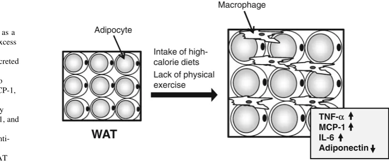
Recently, obesity has been closely associated with
low-grade chronic inflammatory response [16–18]. Indeed, an
increased number of infiltrating macrophages and
dysreg-ulated expression of some inflammation-related
adipo-kines, such as tumor necrosis factor-
a
(TNF-
a
) and
monocyte chemoattractant protein-1 (MCP-1), have been
observed in the WAT of obese mice [49–51]. Adipose
TNF-
a
expression in genetically obese (ob/ob) mice and
obese patients is increased [51,
52] along with the secretion
of TNF-
a
, which induces insulin resistance in skeletal
muscles and adipocytes [53–55]. In addition, expression of
the gene for MCP-1 is upregulated in the WAT of ob/ob
mice and HFD-induced obese mice, and overexpression of
MCP-1 in adipocytes promotes macrophage infiltration in
WAT [56,
57]. On the other hand, decreased levels of
adiponectin in addition to decreased expression of the gene
for adiponectin in WAT have been observed in the plasma
of obese patients [58,
59]. Because adiponectin induces
fatty acid oxidation and decreases the concentration of
triglycerides in skeletal muscle and liver, reduction of
adiponectin levels is implicated in the onset of type 2
diabetes [60,
61].
This section mainly describes the effect of TR on the
expression of representative adipokines in WAT (Fig.
3).
TNF-
a
The effect of TR on the expression of TNF-
a
in WAT is
controversial; For example, the increased expression of the
gene for TNF-
a
in the mesenteric visceral WAT of
HFD-induced obese mice is reported to have been inhibited by
self-propelled running exercise in a rotating cage over a
period of 6 weeks [62]. Furthermore, in a similar report
combining HFD with TR by treadmill running, there was
no difference in TNF-
a
gene expression in visceral WAT
between the HFD group that continued TR for 6 weeks and
the group with HFD alone. After a further 6 weeks,
how-ever, the increased expression of the gene for TNF-
a
due to
HFD was attenuated by TR [63]. We have reported a
decrease in the TNF-
a
protein content in the epididymal
WAT of lean rats as a result of treadmill running for
9 weeks [31,
64]. In contrast, Nara et al. [65] subjected
mice to the combination of a high-sucrose diet and TR that
consisted of self-propelled running, and reported an
increase in TNF-
a
gene and protein expression in
mesen-teric visceral WAT in comparison with a group that had
only a high-sucrose diet. Recently, Lira et al. [66] observed
an increased amount of TNF-
a
in the mesenteric visceral
WAT of rats subjected to 8 weeks of treadmill TR. In
addition to these animal studies, decreased TNF-
a
gene
expression has been observed in investigations of
subcu-taneous WAT in morbidly obese humans with average BMI
of 45.8 kg/m
2who underwent a combination of dietary
therapy and TR such as walking 5 times a week for
15 weeks [67]. However, in another study, no change in
Macrophage
Adipocyte
Intake of high-calorie diets Lack of physical exercise
TNF-αα MCP-1 IL-6
Adiponectin
WAT
expression of the gene for TNF-
a
was observed in the
subcutaneous WAT of obese adults even when body mass
and body fat were reduced after 12 weeks of aerobic
exercise [68].
MCP-1
Similar to TNF-
a
, conflicting results have been obtained with
regards to MCP-1. An increase in the gene expression of
visceral WAT in mice with obesity caused by HFD was
reportedly inhibited by TR that consisted of running [62,
63].
We have observed a decrease in MCP-1 protein content in
epididymal, retroperitoneal, and subcutaneous WAT of lean
rats as a result of treadmill running for 9 weeks [64].
Nev-ertheless, in other studies, changes in MCP-1 gene
expres-sion were observed neither in the subcutaneous or visceral
WAT in rats subjected to 4 weeks of self-propelled running
exercise, nor in the subcutaneous WAT of obese humans
subjected to 12 weeks of aerobic exercise [68,
69]. In
addi-tion, there are also cases in which no significant change in
MCP-1 gene expression was seen in WAT as a result of TR
despite a decrease in TNF-
a
gene expression [67].
Adiponectin
Increased expression of the gene for adiponectin was observed
in the subcutaneous WAT of obese patients who underwent a
combination of dietary therapy and TR such as walking 5
times a week for 15 weeks [67] or aerobic exercise for
12 weeks [68]. However, no effect of TR on adiponectin gene
expression in subcutaneous WAT was observed in an
exam-ination of obese women who were subjected to exercise on an
exercise bike 5 times a week for 12 weeks despite a reduction
in body fat [70]. We observed an increased expression of the
gene for adiponectin in the visceral and subcutaneous
adipo-cytes of rats as a result of 9 weeks of treadmill running [71].
Unexpectedly, Gollisch et al. [69] reported that short-term
(4 weeks) TR by self-propelled running inhibits
HFD-induced upregulation of the adiponectin gene expression in
the subcutaneous WAT of rats. Interestingly, in the present
study, no effect of TR on expression of the gene for
adipo-nectin in visceral WAT was observed.
The effect of TR on macrophage infiltration
into adipose tissue
Macrophage infiltration has been examined by macrophage
marker gene expression in WAT. In a study of mice
sub-jected to HFD and TR by treadmill running for 16 weeks,
Kawanishi et al. [72] reported that the increased expression
of the macrophage marker F4/80 gene observed in the
group with HFD alone was attenuated by TR. Moreover,
decreased gene expression of CD68 and CD14, also
macrophage markers, as a result of a 15-week combination
of TR and dietary therapy has also been observed in human
studies [67]. Nevertheless, in another study, no changes in
the expression of genes for CD68 and CD14 were observed
in the subcutaneous WAT of obese humans subjected to
12 weeks of aerobic exercise [68].
As explained above, conclusions have not necessarily
been reached regarding the influence of TR on the
expression of adipokines in WAT. Therefore, further
studies are required.
The effect of polyphenol intake on the WAT
inflammatory response
Animal and human studies have shown that the intake of
polyphenols improves insulin sensitivity [73–75]. To
clar-ify the reason for this, the effects of polyphenols on the
dysregulated expression of inflammation-related
adipo-kines in the WAT of obese animals and humans are being
examined. The intake of acacia-derived polyphenols by
diabetic-model KKAy mice subjected to HFD suppressed
the increase of visceral WAT mass and the dysregulated
expression of TNF-
a
and adiponectin in epididymal WAT
[76]. Similarly, we have also reported an attenuation of the
increase in epididymal WAT mass and the abnormal
expression of inflammation-related adipokines (TNF-
a
and
MCP-1) in mice with the intake of lychee-derived
poly-phenols [44]. Recently, tests where adipocytes and
mac-rophages were co-cultured have suggested that an increase
in TNF-
a
in the WAT of obese mice is a result of increased
expression of TNF-
a
in infiltrated macrophages rather than
in adipocytes [77]. We have shown that grape seed-derived
polyphenols attenuate the increase in TNF-
a
and MCP-1
resulting from this interaction with adipocytes and
mac-rophages [78]. A mechanism for the increased expression
of adiponectin from polyphenols has also been identified;
For example, when resveratrol, a type of polyphenol, is
added to 3T3-L1 adipocytes, the expression of adiponectin
increases with signaling through AMP-activated protein
kinase and Akt/forkhead transcription factor 1 [79].
How-ever, further studies are required, since no improvements in
the features of metabolic syndrome and no changes in
serum adiponectin values have been observed by green tea/
green tea extract intake in studies targeting the obese [80].
Is the effect of TR on adipokines a result of the decrease
in the amount of WAT?
their volume, and investigated the relationship between
adipocyte size and adipokine secretion. As a result,
adi-pocyte size showed a positive correlation with the ability to
secrete some adipokines, including leptin, interleukin-6,
and MCP-1. Therefore, one of the main reasons for
TR-induced change in adipokine production is thought to be a
reduction in adipocyte size. However, as for adipokine
production, it is unclear whether TR has effects other than
the reduction of adipocyte size.
Adipocytes have been shown to be reactive oxygen
species (ROS) producing cells, and the increased oxidative
stress in WAT resulting from obesity is suggested to be one
of the causes for the dysregulated expression of
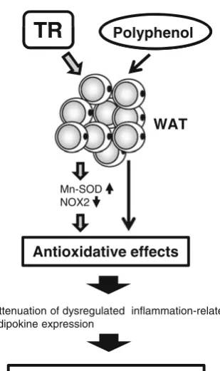
inflamma-tion-related adipokines [82]. We have observed that TR
decreases oxidative stress in visceral WAT through
enhanced protein expression of the antioxidant enzyme
manganese-containing superoxide dismutase and reduced
protein expression of the nicotinamide adenine dinucleotide
phosphate (NADPH) oxidase 2 (NOX2) [31,
64]. There are
many reports that acute exercise enhances the generation of
ROS, and that TR establishes a defense system against
oxidative stress in various tissues [83,
84]. Therefore, we
propose that the adaptation caused by exposure to oxidative
stress that is generated during exercise—TR-induced
enhancement of the antioxidant defense system in WAT—
diminishes oxidative stress, which is another reason for
attenuation of the dysregulated expression of
inflammation-related adipokines (Fig.
4). Furthermore, as the intake of
polyphenols attenuates the abnormal expression of
inflam-mation-related adipokines and increased oxidative stress in
WAT resulting from HFD [44] (Fig.
4), the combination of
TR and polyphenol intake can be an effective tool that
suppresses the adipose tissue inflammatory response.
Conclusions
Due to differences in the results experienced by
experi-mental subjects, the effects of TR and polyphenol intake on
WAT are not consistent among reports found in the
liter-ature. On balance, however, the influence is considered to
be positive; thus, the synergistic effects of TR and
poly-phenol intake as preventive and improvement tools for
obesity and lifestyle-related diseases will probably grow in
importance.
Acknowledgments This work was partially supported by Grants-in-Aid for Specific Project Research from the Ministry of Education, Culture, Sport, Science, and Technology of Japan. We are also grateful for financial support from the Nakatomi Foundation, Tokyo, Japan, and all contributors.
Conflict of interest We do not have any conflict of interest.
References
1. Ro¨ssner S. Obesity: the disease of the twenty-first century. Int J Obes Relat Metab Disord. 2002;26:S2–4.
2. Miranda PJ, DeFronzo RA, Califf RM, Guyton JR. Metabolic syndrome: definition, pathophysiology, and mechanisms. Am Heart J. 2005;149:33–45.
3. Wellen KE, Hotamisligil GS. Inflammation, stress, and diabetes. J Clin Invest. 2005;115:1111–9.
4. Helmrich SP, Ragland DR, Leung RW, Paffenbarger RS Jr. Physical activity and reduced occurrence of non-insulin-depen-dent diabetes mellitus. N Engl J Med. 1991;325:147–52. 5. Manson JE, Nathan DM, Krolewski AS, Stampfer MJ, Willett
WC, Hennekens CH. A prospective study of exercise and inci-dence of diabetes among US male physicians. JAMA. 1992;268: 63–7.
6. Wu CH, Lu FH, Chang CS, Chang TC, Wang RH, Chang CJ. Relationship among habitual tea consumption, percent body fat, and body fat distribution. Obes Res. 2003;11:1088–95. 7. Hughes LA, Arts IC, Ambergen T, Brants HA, Dagnelie PC,
Goldbohm RA, et al. Higher dietary flavone, flavonol, and cate-chin intakes are associated with less of an increase in BMI over time in women: a longitudinal analysis from the Netherlands Cohort Study. Am J Clin Nutr. 2008;88:1341–52.
8. Kono S, Shinchi K, Wakabayashi K, Honjo S, Todoroki I, Sak-urai Y, et al. Relation of green tea consumption to serum lipids and lipoproteins in Japanese men. J Epidemiol. 1996;6:128–33. 9. Tokunaga S, White IR, Frost C, Tanaka K, Kono S, Tokudome S,
et al. Green tea consumption and serum lipids and lipoproteins in
TR
TR
Polyphenol
WAT
Mn-SOD NOX2
Antioxidative effects
Attenuation of dysregulated inflammation-related adipokine expression
Anti-inflammatory effects
a population of healthy workers in Japan. Ann Epidemiol. 2002;12:157–65.
10. Stoclet JC, Chataigneau T, Ndiaye M, Oak MH, El Bedoui J, Chataigneau M, et al. Vascular protection by dietary polyphe-nols. Eur J Pharmacol. 2004;500:299–313.
11. Vita JA. Polyphenols and cardiovascular disease: effects on endothelial and platelet function. Am J Clin Nutr. 2005;81:292S– 7S.
12. Mangiapane H, Thomson J, Salter A, Brown S, Bell GD, White DA. The inhibition of the oxidation of low density lipoprotein by (?)-catechin, a naturally occurring flavonoid. Biochem Pharma-col. 1992;43:445–50.
13. Aviram M. Modified forms of low density lipoprotein and ath-erogenesis. Atherosclerosis. 1993;98:1–9.
14. Hayek T, Fuhrman B, Vaya J, Rosenblat M, Belinky P, Coleman R, et al. Reduced progression of atherosclerosis in apolipoprotein E-deficient mice following consumption of red wine, or its polyphenols quercetin or catechin, is associated with reduced susceptibility of LDL to oxidation and aggregation. Arterioscler Thromb Vasc Biol. 1997;17:2744–52.
15. Horowitz JF. Fatty acid mobilization from adipose tissue during exercise. Trends Endocrinol Metab. 2003;14:386–92.
16. Tilg H, Moschen AR. Adipocytokines: mediators linking adipose tissue, inflammation and immunity. Nat Rev Immunol. 2006;10: 772–83.
17. Rabe K, Lehrke M, Parhofer KG, Broedl UC. Adipokines and insulin resistance. Mol Med. 2008;14:741–51.
18. Lumeng CN, Saltiel AR. Inflammatory links between obesity and metabolic disease. J Clin Invest. 2011;121:2111–7.
19. Slentz CA, Houmard JA, Kraus WE. Exercise, abdominal obes-ity, skeletal muscle, and metabolic risk: evidence for a dose response. Obesity (Silver Spring). 2009;17(Suppl 3):S27–33. 20. Askew EW, Huston RL, Plopper CG, Hecker AL. Adipose tissue
cellularity and lipolysis. Response to exercise and cortisol treat-ment. J Clin Invest. 1975;56:521–9.
21. Oscai LB, Spirakis CN, Wolff CA, Beck RJ. Effects of exercise and of food restriction on adipose tissue cellularity. J Lipid Res. 1972;13:588–92.
22. Oscai LB, Babirak SP, Dubach FB, McGarr JA, Spirakis CN. Exercise or food restriction: effect on adipose tissue cellularity. Am J Physiol. 1974;227:901–4.
23. Owens JL, Fuller EO, Nutter DO, DiGirolamo M. Influence of moderate exercise on adipocyte metabolism and hormonal responsiveness. J Appl Physiol. 1977;43:425–30.
24. Bukowiecki L, Lupien J, Follea N, Paradis A, Richard D, Le-Blanc J. Mechanism of enhanced lipolysis in adipose tissue of exercise-trained rats. Am J Physiol. 1980;239:E422–9.
25. Oscai LB, Caruso RA, Wergeles AC, Palmer WK. Exercise and the cAMP system in rat adipose tissue. I. Lipid mobilization. J Appl Physiol. 1981;50:250–4.
26. Sakurai T, Endo S, Hatano D, Ogasawara J, Kizaki T, Oh-ishi S, et al. Effects of exercise training on adipogenesis of stromal-vascular fraction cells in rat epididymal white adipose tissue. Acta Physiol (Oxf). 2010;200:325–38.
27. Enevoldsen LH, Stallknecht B, Fluckey JD, Galbo H. Effect of exercise training on in vivo lipolysis in intra-abdominal adipose tissue in rats. Am J Physiol Endocrinol Metab. 2000;279:E585–92. 28. Nomura S, Kawanami H, Ueda H, Kizaki T, Ohno H, Izawa T. Possible mechanisms by which adipocyte lipolysis is enhanced in exercise-trained rats. Biochem Biophys Res Commun. 2002;295: 236–42.
29. Izawa T, Komabayashi T, Shinoda S, Suda K, Tsuboi M, Kosh-imizu E. Possible mechanism of regulating adenylate cyclase activity in adipocyte membranes from exercise-trained male rats. Biochem Biophys Res Commun. 1988;151:1262–8.
30. Izawa T, Mochizuki T, Komabayashi T, Suda K, Tsuboi M. Ca2? potentiates corticotropin-induced, but not isoproterenol-induced, [3H]guanosine diphosphate release in rat adipocyte membranes. Metabolism. 1992;41:462–4.
31. Sakurai T, Takei M, Ogasawara J, Watanabe N, Sanpei M, Yoshida M, et al. Exercise training enhances tumor necrosis factor-a-induced expressions of anti-apoptotic genes without alterations in caspase-3 activity in rat epididymal adipocytes. Jpn J Physiol. 2005;55:181–8.
32. Zuk PA, Zhu M, Mizuno H, Huang J, Futrell JW, Katz AJ, et al. Multilineage cells from human adipose tissue: implications for cell-based therapies. Tissue Eng. 2001;7:211–8.
33. Smas CM, Sul HS. Pref-1, a protein containing EGF-like repeats, inhibits adipocyte differentiation. Cell. 1993;73:725–34. 34. Gregoire FM, Smas CM, Sul HS. Understanding adipocyte
dif-ferentiation. Physiol Rev. 1998;78:783–809.
35. Rosen ED, Spiegelman BM. Molecular regulation of adipogen-esis. Annu Rev Cell Dev Biol. 2000;16:145–71.
36. Yun Z, Maecker HL, Johnson RS, Giaccia AJ. Inhibition of PPARc gene expression by the HIF-1-regulated gene DEC1/ Stra13: a mechanism for regulation of adipogenesis by hypoxia. Dev Cell. 2002;2:331–41.
37. Hatano D, Ogasawara J, Endoh S, Sakurai T, Nomura S, Kizaki T, et al. Effect of exercise training on the density of endothelial cells in the white adipose tissue of rats. Scand J Med Sci Sports. 2011;21:e115–21.
38. Pinent M, Blade´ MC, Salvado´ MJ, Arola L, Arde´vol A. Intra-cellular mediators of procyanidin-induced lipolysis in 3T3-L1 adipocytes. J Agric Food Chem. 2005;53:262–6.
39. Ogasawara J, Kitadate K, Nishioka H, Fujii H, Sakurai T, Kizaki T, et al. Oligonol, a new lychee fruit-derived low-molecular form of polyphenol, enhances lipolysis in primary rat adipocytes through activation of the ERK1/2 pathway. Phytother Res. 2009;23:1626–33.
40. Ogasawara J, Kitadate K, Nishioka H, Fujii H, Sakurai T, Kizaki T, et al. Oligonol, an oligomerized lychee fruit-derived poly-phenol, activates the Ras/Raf-1/MEK1/2 cascade independent of the IL-6 signaling pathway in rat primary adipocytes. Biochem Biophys Res Commun. 2010;402:554–9.
41. Ogasawara J, Kitadate K, Nishioka H, Fujii H, Sakurai T, Kizaki T, et al. Comparison of the effect of Oligonol, a new lychee fruit-derived low molecular form of polyphenol, and epigallocatechin-3-gallate on lipolysis in rat primary adipocytes. Phytother Res. 2011;25:467–71.
42. Pinent M, Blade´ MC, Salvado´ MJ, Arola L, Hackl H, Quacken-bush J, et al. Grape-seed derived procyanidins interfere with adipogenesis of 3T3-L1 cells at the onset of differentiation. Int J Obes (Lond). 2005;29:934–41.
43. Wolfram S, Wang Y, Thielecke F. Anti-obesity effects of green tea: from bedside to bench. Mol Nutr Food Res. 2006;50:176–87. 44. Sakurai T, Nishioka H, Fujii H, Nakano N, Kizaki T, Radak Z, et al. Antioxidative effects of a new lychee fruit-derived poly-phenol mixture, Oligonol, converted into a low-molecular form in adipocytes. Biosci Biotechnol Biochem. 2008;72:463–76. 45. Bose M, Lambert JD, Ju J, Reuhl KR, Shapses SA, Yang CS. The
major green tea polyphenol, (-)-epigallocatechin-3-gallate, inhibits obesity, metabolic syndrome, and fatty liver disease in high-fat-fed mice. J Nutr. 2008;138:1677–83.
46. Shin DW, Kim SN, Lee SM, Lee W, Song MJ, Park SM. (-)-Catechin promotes adipocyte differentiation in human bone marrow mesenchymal stem cells through PPARctransactivation. Biochem Pharmacol. 2009;77:125–33.
48. Venables MC, Hulston CJ, Cox HR, Jeukendrup AE. Green tea extract ingestion, fat oxidation, and glucose tolerance in healthy humans. Am J Clin Nutr. 2008;87(3):778–84.
49. Weisberg SP, McCann D, Desai M, Rosenbaum M, Leibel RL, Ferrante AW Jr. Obesity is associated with macrophage accu-mulation in adipose tissue. J Clin Invest. 2003;112:1796–808. 50. Xu H, Barnes GT, Yang Q, Tan G, Yang D, Chou CJ, et al.
Chronic inflammation in fat plays a crucial role in the develop-ment of obesity-related insulin resistance. J Clin Invest. 2003; 112:1821–30.
51. Fantuzzi G. Adipose tissue, adipokines, and inflammation. J Allergy Clin Immunol. 2005;115:911–9.
52. Hotamisligil GS, Shargill NS, Spiegelman BM. Adipose expression of tumor necrosis factor-a: direct role in obesity-linked insulin resistance. Science. 1993;259:87–91.
53. Peraldi P, Hotamisligil GS, Buurman WA, White MF, Spiegelman BM. Tumor necrosis factor (TNF)-a inhibits insulin signaling through stimulation of the p55 TNF receptor and activation of sphingomyelinase. J Biol Chem. 1996;271:13018–22.
54. Plomgaard P, Bouzakri K, Krogh-Madsen R, Mittendorfer B, Zierath JR, Pedersen BK. Source tumor necrosis factor-ainduces skeletal muscle insulin resistance in healthy human subjects via inhibition of Akt substrate 160 phosphorylation. Diabetes. 2005;54:2939–45.
55. Steinberg GR, Michell BJ, van Denderen BJ, Watt MJ, Carey AL, Fam BC, et al. Tumor necrosis factora-induced skeletal muscle insulin resistance involves suppression of AMP-kinase signaling. Cell Metab. 2006;4:465–74.
56. Kamei N, Tobe K, Suzuki R, Ohsugi M, Watanabe T, Kubota N, et al. Overexpression of monocyte chemoattractant protein-1 in adipose tissues causes macrophage recruitment and insulin resistance. J Biol Chem. 2006;281:26602–14.
57. Kanda H, Tateya S, Tamori Y, Kotani K, Hiasa K, Kitazawa R, et al. MCP-1 contributes to macrophage infiltration into adipose tissue, insulin resistance, and hepatic steatosis in obesity. J Clin Invest. 2006;116:1494–505.
58. Hu E, Liang P, Spiegelman BM. AdipoQ is a novel adipose-specific gene dysregulated in obesity. J Biol Chem. 1996;271: 10697–703.
59. Arita Y, Kihara S, Ouchi N, Takahashi M, Maeda K, Miyagawa J, et al. Paradoxical decrease of an adipose-specific protein, adiponectin, in obesity. Biochem Biophys Res Commun. 1999; 257:79–83.
60. Kadowaki T, Yamauchi T. Adiponectin and adiponectin recep-tors. Endocr Rev. 2005;26:439–51.
61. Kadowaki T, Yamauchi T, Kubota N, Hara K, Ueki K, Tobe K. Adiponectin and adiponectin receptors in insulin resistance, dia-betes, and the metabolic syndrome. J Clin Invest. 2006;116: 1784–92.
62. Bradley RL, Jeon JY, Liu FF, Maratos-Flier E. Voluntary exer-cise improves insulin sensitivity and adipose tissue inflammation in diet-induced obese mice. Am J Physiol Endocrinol Metab. 2008;295:E586–94.
63. Vieira VJ, Valentine RJ, Wilund KR, Antao N, Baynard T, Woods JA. Effects of exercise and low-fat diet on adipose tissue inflammation and metabolic complications in obese mice. Am J Physiol Endocrinol Metab. 2009;296:E1164–71.
64. Sakurai T, Izawa T, Kizaki T, Ogasawara JE, Shirato K, Imaiz-umi K, et al. Exercise training decreases expression of inflam-mation-related adipokines through reduction of oxidative stress in rat white adipose tissue. Biochem Biophys Res Commun. 2009;379:605–9.
65. Nara M, Kanda T, Tsukui S, Inukai T, Shimomura Y, Inoue S, et al. Running exercise increases tumor necrosis factor-a secret-ing from mesenteric fat in insulin-resistant rats. Life Sci. 1999;65:237–44.
66. Lira FS, Rosa JC, Yamashita AS, Koyama CH, Batista ML Jr, Seelaender M. Endurance training induces depot-specific changes in IL-10/TNF-a ratio in rat adipose tissue. Cytokine. 2009; 45:80–5.
67. Bruun JM, Helge JW, Richelsen B, Stallknecht B. Diet and exercise reduce low-grade inflammation and macrophage infil-tration in adipose tissue but not in skeletal muscle in severely obese subjects. Am J Physiol Endocrinol Metab. 2006;290: E961–7.
68. Christiansen T, Paulsen SK, Bruun JM, Pedersen SB, Richelsen B. Exercise training versus diet-induced weight-loss on metabolic risk factors and inflammatory markers in obese subjects: a 12-week randomized intervention study. Am J Physiol Endocri-nol Metab. 2010;298:E824–31.
69. Gollisch KS, Brandauer J, Jessen N, Toyoda T, Nayer A, Hirshman MF, et al. Effects of exercise training on subcutaneous and visceral adipose tissue in normal- and high-fat diet-fed rats. Am J Physiol Endocrinol Metab. 2009;297:E495–504.
70. Polak J, Klimcakova E, Moro C, Viguerie N, Berlan M, Hejnova J. Effect of aerobic training on plasma levels and subcutaneous abdominal adipose tissue gene expression of adiponectin, leptin, interleukin 6, and tumor necrosis factor a in obese women. Metabolism. 2006;55:1375–81.
71. Miyazaki S, Izawa T, Ogasawara JE, Sakurai T, Nomura S, Kizaki T, et al. Effect of exercise training on adipocyte-size-dependent expression of leptin and adiponectin. Life Sci. 2010;86:691–8.
72. Kawanishi N, Yano H, Yokogawa Y, Suzuki K. Exercise training inhibits inflammation in adipose tissue via both suppression of macrophage infiltration and acceleration of phenotypic switching from M1 to M2 macrophages in high-fat-diet-induced obese mice. Exerc Immunol Rev. 2010;16:105–18.
73. Wu LY, Juan CC, Ho LT, Hsu YP, Hwang LS. Effect of green tea supplementation on insulin sensitivity in Sprague–Dawley rats. J Agric Food Chem. 2004;52:643–8.
74. Potenza MA, Marasciulo FL, Tarquinio M, Tiravanti E, Co-lantuono G, Federici A. EGCG, a green tea polyphenol, improves endothelial function and insulin sensitivity, reduces blood pres-sure, and protects against myocardial I/R injury in SHR. Am J Physiol Endocrinol Metab. 2007;292:E1378–87.
75. Brasnyo´ P, Molna´r GA, Moha´s M, Marko´ L, Laczy B, Cseh J, et al. Resveratrol improves insulin sensitivity, reduces oxidative stress and activates the Akt pathway in type 2 diabetic patients. Br J Nutr. 2011;106:383–9.
76. Ikarashi N, Toda T, Okaniwa T, Ito K, Ochiai W, Sugiyama K. Anti-obesity and anti-diabetic effects of acacia polyphenol in obese diabetic KKAy mice fed high-fat diet. Evid Based Com-plement Alternat Med. 2011;2011:952031.
77. Suganami T, Nishida J, Ogawa Y. A paracrine loop between adipocytes and macrophages aggravates inflammatory changes: role of free fatty acids and tumor necrosis factora. Arterioscler Thromb Vasc Biol. 2005;25:2062–8.
78. Sakurai T, Kitadate K, Nishioka H, Fujii H, Kizaki T, Kondoh Y, et al. Oligomerized grape seed polyphenols attenuate inflamma-tory changes due to antioxidative properties in coculture of adi-pocytes and macrophages. J Nutr Biochem. 2010;21:47–54. 79. Wang A, Liu M, Liu X, Dong LQ, Glickman RD, Slaga TJ, et al.
Up-regulation of adiponectin by resveratrol: the essential roles of the Akt/FOXO1 and AMP-activated protein kinase signaling pathways and DsbA-L. J Biol Chem. 2011;286:60–6.
80. Basu A, Du M, Sanchez K, Leyva MJ, Betts NM, Blevins S, et al. Green tea minimally affects biomarkers of inflammation in obese subjects with metabolic syndrome. Nutrition. 2011;27:206–13. 81. Skurk T, Alberti-Huber C, Herder C, Hauner H. Relationship
82. Furukawa S, Fujita T, Shimabukuro M, Iwaki M, Yamada Y, Nakajima Y, et al. Increased oxidative stress in obesity and its impact on metabolic syndrome. J Clin Invest. 2004;114:1752–61. 83. Gomez-Cabrera MC, Domenech E, Vin˜a J. Moderate exercise is an antioxidant: upregulation of antioxidant genes by training. Free Radic Biol Med. 2008;44:126–31.