www.fm.viamedica.pl O R I G I N A L A R T I C L E
Address for correspondence: Kilarkaje Narayana, Department of Anatomy, Centre for Basic Sciences, Kasturba Medical College, Bejai, Mangalore, 575004 Karnataka, India, tel: +91 824 2211746 (o), fax: +91 824 2428183, e-mail: narayana68@yahoo.com
The incidence of a superficial arterial pattern
in the human upper extremities
Sujatha D’Costa, Bhamy Mangala Shenoy, Kilarkaje Narayana
Department of Anatomy, Centre for Basic Sciences, Kasturba Medical College, Bejai, Mangalore, Karnataka, India
[Received 23 July 2004; Revised 5 October 2004; Accepted 5 October 2004]
The incidence of superficial arteries was studied in 68 (38 right and 30 left) upper extremities. One right limb of an adult male presented a superficial arte-rial pattern (2.63%, total 1.47%) resembling a superficial brachio-ulno-radial artery (SBUR). The median nerve crossed the superficial brachial artery (SBA) from the posterior to the medial side and again posterior to the same at the cubital fossa. The superficial brachial artery divided into superficial radial and superficial ulnar arteries, which coursed distally superficial to the muscles but deep to the deep fascia. The superficial radial artery passed deep to the extensor tendons of the thumb. The superficial ulnar artery gave only muscular branches in the forearm. The superficial radial artery gave origin to the radial recurrent artery and the common interosseous trunk. The latter gave origin to a palmar type of median artery, muscular branches, and an artery that divided into ante-rior and posteante-rior ulnar recurrent arteries. It also gave origin to the anteante-rior and posterior interosseous arteries. The latter provided the interosseous recurrent artery and a branch that coursed towards the olecranon process of the ulna. The knowledge of this variation is important since it may be compromised in surgical procedures of the upper limb.
Key words: upper limb, superficial brachial artery, superficial radial artery, superficial ulnar artery, common interosseous artery
INTRODUCTION
The superficial arteries of the upper extremity may be mistaken for veins, thus becoming a basis for in-tra-arterial injections [4, 18]. They may also be en-countered during elevation of the forearm flaps [5, 6, 8, 17] or misinterpreted in contrast radiographs [14]. These arteries may be superficial or deep to the deep fascia but run superficially to the muscles of the arm and forearm [16]. These superficial arteries include the superficial brachial (SBA), superficial brachio-ra-dial, superficial brachio-ulnar, superficial brachio-ulno-radial (SBUR), superficial brachio-median, and super-ficial median and supersuper-ficial radial arteries.
The superficial brachio-radial artery (the high origin of the radial artery coursing over the
MATERIAL AND METHODS
In the present study, 68 upper extremities (38 right and 30 left) were studied for the incidence of superficial arteries. During routine dissection obser-vations were made for the presence or absence of any superficial arteries. If they were present, such cases were further dissected and studied in detail.
RESULTS
Of the 68 limbs observed, 1 right limb of an adult male presented a superficial arterial pattern (2.63%, total 1.47%) and this variation could be termed a SBUR. The brachial artery was superficial and in the cubital fossa, it was anterior to the bicipital tendon and the median nerve. The latter passed through a hiatus formed by the ulnar artery and the common in-terosseous trunk (CIT, vide infra) and reached the fore-arm, thence following a normal course. The superficial brachial artery gave origin to 6 branches (Fig. 1A). The first originated from the medial side 7.5 cm distal to the upper limit of the head of the humerus, supplying the triceps. The profunda brachii was a thin artery, which gave 2 muscular branches to the long head of the triceps brachii and continued as a single artery sup-plying the same muscle. The profunda brachii did not
give its terminal branches, the radial and middle col-lateral arteries. The 3rd and 4th branches were the
supe-rior ulnar collateral and infesupe-rior ulnar collateral arter-ies respectively. The latter originated from the lateral side of SBA and then coursed posteriorly to it to the medial side. Another 2 muscular branches originated from the lateral aspect of SBA (Fig. 1A). The superficial brachial then terminated in the radial and ulnar arter-ies, which were also superficial in the forearm (Fig. 1B). The superficial ulnar artery gave origin only to muscular branches from the medial aspect and coursed superficially to the forearm flexors, dividing at the wrist into superficial and deep branches (Fig. 2). The superficial palmar arch gave origin to 3 com-mon palmar digital branches. The 4th
branch sup-plied the adjacent sides of the thumb and the index finger. The radial artery was also superficial, lying on the brachio-radialis, but passing distally deep to the extensor tendons of the thumb. The common interosseous trunk was a branch from the superfi-cial radial artery originating 1 cm distal to the bifur-cation of SBA (Fig. 2A). The common interosseous trunk coursed disto-medially and provided a muscu-lar branch that supplied the flexor pollicis longus, while another branch supplied the flexor carpi
ul-Figure 1 A. The superficial brachial artery (SBA) and its branches. The median nerve (M) has been pulled medially to expose the branch-es of the SBA. The profunda brachii (PBA) was a thin artery. Superior ulnar collateral (SUCA) originated from the SBA considerably be-low the origin of the PBA. The inferior ulnar collateral artery (IUCA) originated from the lateral aspect of the SBA and then crossed poste-rior to the same. Other muscular branches are seen in the photograph. The ulnar nerve (UN), the radial nerve (R), the median nerve (M), the musculocutaneous nerve (MCN) and the muscles biceps brachii (BB) and brachialis (BR) are indicated. B. Photograph of forearm showing the superficial radial (R) and superficial ulnar (U) arteries. The R gave origin to the common interosseous trunk (CIT) and radial recurrent artery (RRA). The CIT gave origin to the palmar type of median artery (MA), anterior interosseous artery (AIA), and a common stem dividing into anterior (AUR) and posterior (PUR) ulnar recurrent arteries. The median nerve (M), the ulnar nerve (UN) and anterior interosseous nerve (AIN) are indicated.
A B
AIN&AIA
R
MA U
AUR&PUR UN
SBA M
PBA
UN
SUCA
M
BR BB
MCN R
SBA
IUCA
CIT
naris. The median artery originated from CIT and accompanied the median nerve up to the wrist (pal-mar type). Another branch originated from the prox-imal aspect of CIT, which immediately divided into 2 branches recurring towards the medial epicondyle (ulnar recurrent arteries). The superficial ulnar artery passed through a gap between these 2 ulnar recur-rent arteries. The common interosseous trunk then terminated in the anterior and posterior interosseous arteries. The latter gave origin to 1 branch which wound around the ulna and reached the olecranon process. The interosseous recurrent artery also had its origin slightly distal to the origin of the former and coursed towards the lateral epicondyle (Fig. 2A). The superficial radial artery then continued distally and formed the deep palmar arch. There was no superficial branch of the radial and hence the super-ficial palmar arch was incomplete (Fig. 2B). One branch originated from the deep branch of the su-perficial radial artery and supplied the thumb. The superficial radial artery also gave the arteria prin-ceps pollicis and arteria radialis indicis in the first inter-metacarpal space, supplying the thumb and the index finger respectively (Fig. 2B). A branch from the superficial palmar arch coursed laterally across the arteria radialis indicis and divided into 2 branches to supply the thumb and the index finger (Fig. 2B).
DISCUSSION
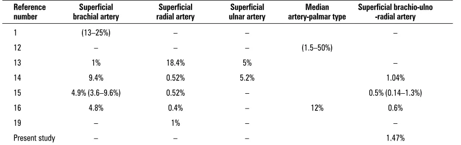
The arterial variation observed in 1 right limb (2.63%) out of a total of 68 limbs (1.47%) examined could be termed a SBUR, although there are quali-fications to be made, since it differs from a typical SBUR as defined in earlier studies [9, 16]. The incidence of SBUR reported by previous re-searchers ranges from 0.14–1.3% (Table 1) and the incidence observed in the present study is compa-rable. In the present case the axillary artery contin-ued as SBUR, unlike its usual formation as a branch of the former [15]. A normal brachial artery usually accompanies SBUR [15], although this was not ob-served in the present case. The brachial part of this variation could be compared to a SBA but there were differences. Firstly, the profunda brachii was very thin compared to its normal counterpart and, secondly, the distally located origin of the superior ulnar collateral artery was in contrast to its site of origin in normal conditions [7]. The absence of ter-minal branches of the profunda brachii is in agree-ment with the previous report on SBA [9]. Howev-er, the variations in SBA observed in this case con-trast with the observations that SBA does not show any further variations [16].
The radial artery was superficial only at its proxi-mal part and then coursed deep to the extensor ten-Figure 2 A. Photograph of forearm showing the superficial radial artery (R) and common interosseous trunk (CIT). Note that the ulnar ar-tery has been removed (arrow) to view the CIT and its branches. The CIT gave a branch that divided into anterior (AUR) and posterior (PUR) ulnar recurrent arteries. The posterior interosseous artery (PIA) coursed to the posterior compartment. It gave a branch (open ar-row), which coursed towards the olecranon process. The interosseous recurrent artery (IRA) originated slightly distal to that of the former and coursed towards the medial epicondyle. The muscular branch (M), the median artery (MA), and the anterior interosseous ar-tery (AIA) are indicated. B. Photograph of the hand showing superficial (SPA) and deep (DPA) palmar arterial arches. The SPA was in-complete and continued (open arrow) laterally over the radialis indicis (RI) and divided into two branches (arrows) to supply the thumb and index finger. The superficial radial artery (RA) gave origin to the princeps pollicis (PP) and RI separately. The superficial ulnar artery is indicated (UA).
CIT AUR
PUR
PIA M
AIA IRA
A B
R
MA
PP
MN
DPA
SPA
dons of the thumb, hence differing from a typical SRA [15]. Moreover, it showed further differences by giving off CIT and radial recurrent artery from its medial side, which then crossed it superficially. In around 41% of SRAs, an anastomosis has generally been observed with other arteries at the elbow [13], although that was absent in this case. The common interosseous trunk could be a branch of any artery of the forearm [2, 3], although in this case it origi-nated from SRA as a thick branch. When the varia-tion is a SBUR, CIT is formed as a continuavaria-tion of the co-existing normal brachial artery [15]. The common interosseous artery taking its origin from a high ra-dial artery or, very rarely, from the normal rara-dial ar-tery has been reported [2, 3]. The origin and course of the radial recurrent artery and the origin of a pal-mar type of median artery from CIT were other dif-ferences from the previous reports on this vessel [2]. The palmar type of median artery has been found to be present in around 12% of cases as a branch of any one of the forearm arteries [12] and in around 1% of the cases it was superficial [10]. An additional branch of the posterior interosseous artery coursing towards the olecranon process has not been men-tioned in previous reports [2]. This branch might have taken part in the anastomosis around the elbow joint. The arteries of the hand also differed from the nor-mal pattern [7]. The continuation of a superficial arch laterally to supply the thumb and the index finger was another interesting observation associ-ated with SBUR.
The mechanisms of development of this arterial variation are unknown. It may be that when the ar-teries of the upper limb develop during stages 12–23 [14] of human embryogenesis, some
alter-ations in enlargement, differentiation and the hae-modynamics of capillaries result in SBUR. This case is, however, clinically important since the superficial arteries are vulnerable to trauma and intra-arterial injections. Moreover, accurate knowledge of this type of arterial variation is important in reparative sur-gery of the upper limb.
REFERENCES
1. Anagnostopoulou S, Venieratos D (1999) An unusual branching pattern of the superficial brachial artery accompanied by an ulnar nerve with two roots. J Anat, 195: 471–476.
2. Bergman RA, Afifi AK, Miyauchi R (2004) Illustrated encyclopedia of human anatomic variations (http:// //www.vh.org/anatomic variants/html).
3. Brash JC (1951) Cunninghams’ text book of anatomy. 9th Ed. Oxford University Press, London pp. 1285–1292. 4. Deligonul U, Gabliani G, Kern MJ, Vandormel M (1988) Percutaneous brachial catheterization: the hidden ha-zard of high brachial artery bifurcation. Catheter Car-diovasc Diag, 14: 44–45.
5. Devansh MS (1996) Superficial ulnar artery flap. Plas-tic Reconstr Surg, 97: 420–426.
6. Funk GF, Valentino J, McCulloch TM, Graham SM, Hoff-man HT (1995) Anomalies of forearm vascular anato-my encountered during elevation of the radial fore-arm flap. Head and Neck, 17: 284–292.
7. Gabella (1995) Cardiovascular system. In: Williams PL, Bannister LH, Berry MM, Collins P, Dyson M, Dussek JE, Ferguson MWJ (eds.). Gray’s anatomy. Churchill Livingstone, London pp. 1538–1544.
8. Heden P, Gylbert L (1990) Anomaly of the radial artery encountered during elevation of the radial forearm flap. J Reconstr Microsurg, 6: 139–141.
9. Jurjus AR, Sfeir RB, Bezirdjian R (1986) Unusual varia-tion of the arterial pattern of the human upper limb. Anat Rec, 215: 82–83.
10. Lippert H, Pabst R (1985) Arterial variations in man. JF Bergman Verlag. Munchen pp. 71–73.
Table 1. Incidence of variations in upper limb arteries related to the present study
Reference Superficial Superficial Superficial Median Superficial brachio-ulno number brachial artery radial artery ulnar artery artery-palmar type -radial artery
1 (13–25%) – – –
12 – – – (1.5–50%)
13 1% 18.4% 5% –
14 9.4% 0.52% 5.2% 1.04%
15 4.9% (3.6–9.6%) 0.52% – 0.5% (0.14–1.3%)
16 4.8% 0.4% – 12% 0.6%
19 – 1% – –
Present study – – – 1.47%
11. Rodriguez-Baeza A, Nebot J, Ferreira B, Reina F, Perez J, Sanudo JR (1995) An anatomical study and ontogenic explanation of 23 cases with variations in the main pattern of the human brachio-antebrachial arteries. J Anat, 187: 473–479.
12. Rodriguez-Niedenfuhr M, Sanudo JR, Vazquez T, Nearn L, Logan B, Parkin I (1999) Median artery revisited. J Anat, 195: 57–63.
13. Rodriguez-Niedenfuhr M, Sanudo JR, Vazquez T, Nearn L, Logan B, Parkin I (2000) Anastomosis at the level of elbow joint connecting the deep, or normal, brachial artery with major arterial variations of the upper limb. J Anat, 196: 115–119.
14. Rodriguez-Niedenfuhr M, Burton GJ, Deu J, Sanudo JR (2001) Development of the arterial pattern in the upper limb of staged human embryos: normal de-velopment and anatomic variations. J Anat, 199: 407–417.
15. Rodriguez-Niedenfuhr M, Vazquez T, Nearn L, Ferrei-ra B, Parkin I, Sanudo JR (2001) Variations of the arte-rial pattern in the upper limb revisited: a morphologi-cal and statistimorphologi-cal study with a review of the literature. J Anat, 199: 547–566.
16. Rodriguez-Niedenfuhr M, Vazquez T, Parkin IG, Sanu-do JR (2003) Arterial patterns of the human upper limb: update of anatomical variations and embryological development. Eur J Anat, 7 (Suppl 1): 21–28. 17. Thoma A, Young JEM (1992) The superficial ulnar
ar-tery “trap” and the free forearm flap. Ann Plastic Surg, 28: 370–372.
18. Thomas R, Newell R (1995) Anomalous arteries in the upper limb. Clin Anat, 7; 57.