• No results found

PI3K-AKT-mTOR-Signaling and beyond: the Complex Network in Gastroenteropancreatic Neuroendocrine Neoplasms

N/A
N/A
Protected

Academic year: 2020

Share "PI3K-AKT-mTOR-Signaling and beyond: the Complex Network in Gastroenteropancreatic Neuroendocrine Neoplasms"

Copied!
30
0
0

Loading.... (view fulltext now)

Full text

(1)

T

T

h

h

e

e

r

r

a

a

n

n

o

o

s

s

t

t

i

i

c

c

s

s

2014; 4(4):336-365. doi: 10.7150/thno.7851

Review

PI3K-AKT-mTOR-Signaling and beyond: the Complex

Network in Gastroenteropancreatic Neuroendocrine

Neoplasms

Franziska Briest

1,2

, Patricia Grabowski

1,3

1.

Medizinische Klinik 1 CBF, Dept. of Gastroenterology, Infectious Diseases, Rheumatology CC13, Charité Berlin, Germany;

2.

Freie Universität Berlin, Institute for Chemistry and Biochemistry, Berlin, Germany;

3.

Department of Hematology and Internal Oncology and Center for Neuroendocrine Tumours Bad Berka – ENETS Center of Excellence,

Zentralklinik Bad Berka, Germany.

Corresponding author: Franziska Briest, Phone: +49 30 8445 4579 e-mail: franziska.briest@charite.de.

© Ivyspring International Publisher. This is an open-access article distributed under the terms of the Creative Commons License (http://creativecommons.org/ licenses/by-nc-nd/3.0/). Reproduction is permitted for personal, noncommercial use, provided that the article is in whole, unmodified, and properly cited.

Received: 2013.10.09; Accepted: 2013.12.16; Published: 2014.01.29

Abstract

Gastroenteropancreatic neuroendocrine neoplasms are heterogeneous in their clinical behavior

and require therapies specially tailored according to staging, grading, origin and expression of

peptide receptors. Despite extensive scientific efforts, the therapy options are still not satisfactory.

The main reasons are due to the lack of a broad mechanistic knowledge, an insufficient

classifica-tion of specific diagnostic sub-groups, and predictive markers. GEP-NEN tumors evade early

di-agnosis because of slow asymptomatic growth behavior and are frequently not detected until

metastasized. How signaling networks contribute to tumor progression and how these networks

interact remains unclear in large parts. In this review we summarize the knowledge on the growth

factor responsive non-angiogenetic pathways in sporadic GEP-NENs, highlight promising

mecha-nistic research approaches, and describe important therapy targets.

Key words: Gastroenteropancreatic neuroendocrine neoplasms, signal transduction, growth

fac-tors, kinases, biotherapy, molecular biology, inhibitor.

Introduction

GEP-NENs (Gastroenteropancreatic

crine neoplasms) emerge from various

neuroendo-crine cells of the gastroenteropancreatic system and

represent the largest subgroup of neuroendocrine

neoplasms. This heterogenic entity of solid tumors,

formerly termed GEP-NETs or GEP-NECs

(gastroen-teropancreatic neuroendocrine tumors and

carcino-mas) or “carcinoids”, displays a broad spectrum of

characteristics concerning behavior during growth

and differentiation, functional aspects, localization

and prognosis.

Although they are ranked among rare neoplastic

diseases in general, their incidence has increased

ex-ponentially throughout the last decade. Currently,

GEP-NENs state the second most common

gastroin-testinal malignancy after colorectal cancer [1].

The majority of GEP-NENs is characterized by

slow proliferating, well differentiated G1 phenotypes

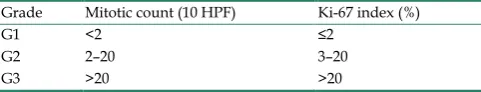
(WHO/ENETS classification 2010, refer to table 1),

which are often diagnosed late in the developmental

course by the occurrence of metastases (NEN G1,

previously termed WDNET: well differentiated

neu-roendocrine tumor). In contrast, a small G3 subgroup

of rapidly growing and poorly differentiated

GEP-NENs display a behavior that is comparable to

those of prevalent solid carcinoma entities (NEN G3

or NEC, previously called PDNEC: poorly

differenti-ated neuroendocrine carcinoma). The third group,

characterized by an intermediate malignancy or

un-clear behavior, NEN G2, approximates the former

Ivyspring

(2)

description of a well differentiated neuroendocrine

carcinoma. The tumor grade is dependent on the

pro-liferative behavior marked by mitoses and by the

Ki-67 antigen, which is a proliferation-related antigen

and is immunohistochemically analyzed during

di-agnosis by default (refer to table 1). In contrast to

previous classifications or the TNM staging, the

dif-ferentiation of the tumor cells is not involved in

grading decisions, which still hampers the evaluation

of slowly growing poorly differentiated or highly

proliferating well differentiated tumors and does not

provide insights regarding invasive behavior.

How-ever, the main advantage of the current grading is the

possibility to predict proliferation behavior and thus

facilitate decisions regarding surgical resection,

chemo-, radio- and biotherapy or monitoring.

Table 1.

WHO 2010 grading for (neuro-)endocrine tumors.

Grade

Mitotic count (10 HPF)

Ki-67 index (%)

G1

<2

≤2

G2

2–20

3–20

G3

>20

>20

Several signaling cascades influence malignant

transformation, progression and metastasis in

neu-roendocrine cancers including RTKs (receptor

tyro-sine kinases) and GPCRs (G-protein coupled

recep-tors) downstream signaling, which regulate Ras/Raf,

MAPK, PI3K-Akt-mTOR and JNK and lead to DNA

synthesis and cell proliferation. Pancreatic NENs are

highly vascularized and nourished. Accordingly, they

exhibit a vast expression of growth factors such as

VEGF (vascular endothelial growth factor), PDGF

(platelet-derived growth factor), IGF-1 (insulin-like

growth factor 1), bFGF (basic fibroblast growth

fac-tor), TGF-

α and

-

β

(transforming growth factor) and

PIGF (placental growth factor). Not surprisingly,

ab-errant receptor activity, including those of the IGF-1R

(IGF-1 receptor) and FGFR3 (FGF receptor 3), and

highly activated downstream signaling is frequent in

pNENs [2-5].

Analogous to the pancreatic subgroup,

gastro-intestinal NENs overexpress VEGF, bFGF, TGF α and

-

β, PDGF, IGF

-1 and its corresponding receptors

PDGFR (PDGF receptor), IGF-IR, EGFR (epidermal

growth factor receptor), VEGFR (VEGF receptor), and

c-kit (stem cell factor receptor). They thus exhibit

in-creased growth factor, pro-angiogenic and typically

pro-secretory signaling [6-16]. This work will focus on

the major growth-factor related signaling networks,

namely the PI3 kinase and the MAP kinase cascades

and their importance for GEP-NEN therapy and

di-agnosis today and for future therapy approaches.

Aberrant receptor activity induces

sus-tained growth factor signaling in

GEP-NENs and activates the PI3K and

MAPK signaling network.

IGF receptors

The

IGF-1R is one of the crucial RTKs in

gas-troenteropancreatic neuroendocrine tumor growth

factor biology. The intrinsic RTK activity of IGF-1R is

activated upon binding of its respective ligand and

leading to auto-phosphorylation of intracellular

tyro-sine residues in the juxtamembrane and C-terminal

domains. Those phosphorylated tyrosines serve as

docking stations for insulin receptor substrates, such

as IRS-1 and Src resulting in PI3K signaling via

Grb2/SOS and Ras and MAPK pathways [17].

NEN cells have been shown to secrete high

amounts of IGF-1. In gastrinomas, increased levels of

both IGF-1 and the corresponding IGF-1R were

asso-ciated with tumor growth, aggressiveness, and

pro-gression [18]. Furthermore, other studies have

dis-played that the expression of IGF-1R is decreased in

functionally inactive neuroendocrine tumors of

dif-ferent offspring in relation to their functional

ana-logues. These findings and further

in vitro

experi-ments suggest that IGF-1 is not only a major autocrine

regulator of neuroendocrine tumor growth but also of

neuroendocrine secretion itself. Inhibition of IGF-1R

activity, e.g. by direct inhibition or by blocking its

regulators, such as HSP90 (heat shock protein 90),

resulted in decreased PI3K and ERK1/2 (extracellular

signal-regulated kinase) signaling and induction of

cell cycle arrest and apoptosis [14, 18-26].

Addition-ally, an alternatively spliced IGF-1R mRNA transcript

could be detected with a higher abundance in

neuro-endocrine tumors of different offspring, suggesting

that post-transcriptional mechanisms may cause

reg-ulatory aberrations [19].

In addition to aberrant receptor and ligand

abundance, an important regulator of IGF signaling

was found to be significantly up-regulated in

meta-static NENs in two gene expression studies: IGFBP3

(IGF binding protein 3), which is considered to

main-tain the serum level of IGF-1 in a tissue specific pro- or

antiproliferative manner. IGFBP3 was overexpressed

in >80% of lymph node or distant metastases versus

<60% in primary pNEN lesions [27-29]. Those data

might indicate a stoma or tumor cell-controlled

regu-lation of a distinct IGF-1 homeostasis and allocation

even in target tissues with a completely different

composition. Adaptive and cooperative behavior of

metastasizing NEN cells in the context of circulation

and homing should be further explored in the future.

(3)

highly expressed in GEP-NENs with an altered

abundance which depends on IGF binding factors and

the relative ratio of specific receptor isoforms. IGF-1

has been shown to be a major autocrine regulator of

neuroendocrine tumor growth and of neuroendocrine

secretion.

EGF receptors and FGF

The

EGFR belongs to the HER receptor family

that consists of EGFR (HER1 or erbB1), erbB2 (HER2),

erbB3 (HER3) and erb4 (HER4). Gastrointestinal and

pancreatic NENs express and activate EGFRs. In

im-munohistochemical analyses of NENs located in

dif-ferent primary locations, 96% of the specimens were

positive for EGFR expression and 63% were positive

for phosphorylated EGFR [6]. Another study

demon-strated a significantly higher expression (> 91%) in

metastatic and non-metastatic gastrointestinal NENs

in contrast to <25% in primary and metastatic pNEN

[30]. A third study retrospectively evaluated the

ex-pression of EGFR and one of its ligands, TGF-

α

(transforming growth factor alpha), in pNENs,

demonstrating that 63% of the tumors were positive

for TGF-alpha and 65% were positive for the

intra-cellular and/or extraintra-cellular domain of EGFR, but

failed to prove a correlation with size, functional

sta-tus, secretory profile, or biologic behavior [31]. These

data were confirmed by Nilsson and colleagues, who

showed that several human neuroendocrine tumors

express both TGF-alpha and EGF receptors

in vivo

and

in vitro

, suggesting an autocrine mechanism [9].

Di Florio and colleagues recently demonstrated

that gastrointestinal hormones and neurotransmitters

stimulate the growth of the human BON, QGP-1 and

the rat Rin-14B-cell lines in an EGFR dependent

manner through activation of PKC and Src kinases,

matrix metalloproteinase activation and the

genera-tion of reactive oxygen species [32].

Although a recent analysis of pancreatic

neuro-endocrine tumors uncovered frequent single

nucleo-tide polymorphisms (SNPs) in

PDGFRA

and

EGFR

, no

druggable

EGFR,

KIT

or

PDGFRA

mutation could be

found in these receptors to date. Nevertheless,

ele-vated copy number of the EGFR and

HER-2/neu

loci

could be detected in 38% and 33% of the cases and

high expression of PDGFRA in 65%, respectively [21].

The non-secretory protein FGF13 has recently

been described as new progression marker in pNENs.

Although FGF13 is insufficient to stimulate FGF

re-ceptors, it was demonstrated to be an independent

predictor of a shorter progression-free survival

asso-ciated with positive Ki-67 staining. Furthermore FGF

overexpression correlates with the occurrence of liver

metastases and shortened disease-free survival in

patients that underwent complete tumor resection

[33]. FGF13 has recently been identified as

microtu-bule-stabilizing protein in neuronal cells that

pro-motes neuronal migration in the cerebral cortex.

The-se procesThe-ses might also facilitate disThe-semination of

neuroendocrine tumor cells that share at least some

characteristics with neuronal cells, but the underlying

mechanisms remain unknown [34].

Taken together, these studies have accounted for

high growth factor abundance in GEP-NENs.

Alt-hough SNPs in several growth factor receptors have

been demonstrated, no druggable mutation could be

found to date. Therefore growth factor receptors

might serve as targets for anti-growth receptor

ther-apy in patients with GEP-NENs, as several IGF-1R

and EGFR inhibitors are currently under clinical

as-sessment (refer to Supplementary Material: suppl. 1).

Nevertheless further subgroup-specific genetic and

post-transcriptional analyses are necessary to clarify

the role of the distinct receptor aberrations and

im-prove the therapeutic potential of growth factor

re-ceptors as therapeutic targets in GEP-NENs.

Somatostatin receptors mediate

an-ti-proliferative signals, inhibit PI3K and

MAPK signaling and are important

ther-apeutic targets in GEP-NENs

Somatostatin signaling

The cyclopeptide family of SSTs

(somato-statins), which function as somatotroprelease

in-hibiting factors, is distributed throughout the central

nervous system and peripheral organs and can be

found in endocrine, immune and neuronal cells, as

well as in certain tumors. Its preserved peptide

structure indicates a fundamental regulatory function

in vertebrate hormone homeostasis.

The human genome includes five non-allelic

genes that encode for five or six distinct

transmem-brane domain G-protein-coupled SSTRs (somatostatin

receptors). The gene encoding SSTR2 produces two

splice variants (SSTR2A and SSTR2B) in mouse and

presumably in humans as well [35-38], whereas the

other genes are intronless and generate one receptor

in each case [39-42]. The natural ligands of SSTR1-5

(SST-14, SST-28 and cortistatin) are bound with a high

affinity. Nevertheless the majority of (longer acting)

synthetic peptide analogues, namely MS201-995

(oc-treotide), RC-160 (vapreotide), BIM 23014 (lanreotide)

and MK 678 (Seglitide), only interact with the

sub-types SSTR 2, 3 and 5 to a satisfactory extent.

Moreo-ver, Pasireotide (SOM 230) shows higher binding

ca-pacity towards SSTR1 [43, 44].

(4)

endocrine target cells. Appropriately, binding of

so-matostatin and its analogues to SSTRs triggers a

number of intracellular signaling events, initiated by

specific G-Protein activation. These G-proteins

func-tion as transducers and in turn modulate the activity

of several key enzymes including adenylyl cyclase,

phospho-tyrosine phosphatases (PTPs) and MAPKs

(mitogen activated kinases) [45-47]. Inhibitory effects

of SST on adenylyl cyclase, cAMP production and

several distinct ion channels regulate both endocrine

and exocrine secretion [39, 48, 49]. Intracellular PTPs

exert anti-proliferative activity by deactivation of

in-tracellular PI3K and MAPK signaling. Phosphatases,

such as SHP-1 and SHP-2, mediate SST-induced cell

cycle arrest in many cell lines

in vitro

. SHP-2 is also

considered to inactivate insulin and epidermal

growth factor binding RTKs and to repress C-Raf and

MAPK signaling, whereas SHP-1 is involved in

dephosphorylation of the p85 PI3K regulatory subunit

and IRS-1 and thus, decreased PDK-1

(phosphoinosi-tide-dependent kinase 1) and Akt activity. It

further-more inhibits S phase entry through the

overexpres-sion of p27kip1 and increase of hypo-phosphorylated

retinoblastoma gene product (Rb) [47, 50-61]. A third

PTP is involved in the SST-induced proliferation

ar-rest: DEP-1 (density enhanced phosphatase-1, also

termed PTPη in rats) is a

negative regulator of

Src-mediated growth factor receptor activity and

par-ticipates in cellular differentiation [62-66].

The mechanism of SST signaling has been

con-firmed in different cellular models indicating that this

modular multi-effector pathway, which is induced by

several SSTRs, transmitted by similar kinases and

PTPs and resulting in the activation of final effector

PTPs, is a common mechanism in various cells [67,

68]. Furthermore the downstream signaling of the

distinct SSTRs differs in certain subtype specific

func-tions: e.g. whereas both SSTR2 and 5 regulate GH

secretion, insulin secretion is predominantly

con-trolled by SSTR5. Glucagon secretion and immune

response are SSTR2 dependent (reviewed in [39]).

Somatostatin receptors in GEP-NEN therapy

Gastroenteropancreatic neuroendocrine

neo-plasms express all five subtypes of SSTRs, although at

a variable extent and in various combinations. The

most prevalent receptor is SSTR2A, which has been

immunohistochemically detected in 84% of the

GEP-NEN specimens in a recent study. SSTR3, 4, 5

and 1 have been found expressed in 84, 44, 32 and

32%, respectively [69]. This data agree with previous

studies in insulinomas where the SSTR2, 1 and 3 have

been found to be predominantly expressed [70]. In

midgut NENs SSTR2 is the most prominent as well,

but followed by SSTR1 and SSTR5, and, with a less

frequency, by SSTR3 and SSTR4. [70-73]. Comparable

findings have been recently published in a cohort of

67 NENs, with an expression of SSTR1, 2a, 3, 4 and 5

in 42, 63, 6, 32 and 65% of the cases, respectively.

In-terestingly, the SSTR5 immunoreactivity was

corre-lated with the presence of metastases and

angioinva-sion [74]. Other studies have previously demonstrated

a positive correlation of SSTR2 and SSTR5 expression

with a better prognosis and higher differentiation

[75-80].

Although corresponding data is rare, some

studies have demonstrated a frequent co-expression

of SSTRs and the D2R (dopamine D2 receptor) in

GEP-NENs [38, 77, 81]. Co-expression of SSTR5 and

D2R is assumed to contribute to a

het-ero-oligomerization and creation of a novel receptor

with

enhanced

functional

activity [82].

A Co-expression

of SSTR2 and SSTR5 with D2R and its correlation with

low tumor grade has been described in mixed NENs

[77]. The high frequency of SSTR1,

SSTR2

, and SSTR5

expression and inverse correlation with COX2, a

cy-tochrome C oxidase and component of the respiratory

chain, was recently demonstrated. The study also

ac-counted for a better prognosis concomitant with a

generally high SSTR expression [76].

Additionally, one group was also able to

demonstrate a high expression of the human SSTR2B

which raises further questions, whether this splice

variant exists in humans [38].

Data concerning the SSTR receptor distribution

pattern in different subgroups was published for

SSTR2A, which was present in a high frequency in

carcinoids and gastrinomas, but detectable in only

half of the analyzed insulinomas, suggesting one

reason for the high number of octreotide insensitive

insulinomas [83, 84]. Another study demonstrated a

higher SSTR expression in non-pancreatic NENs

ver-sus pancreatic NENs for all receptor isoforms [78].

This high heterogeneity of data, beyond the fact

that SSTR2 exerts to be the most prevalent of the

generally highly expressed SSTRs in GEP-NENs, is

summarized in table 2 which highlights the need for

better defined criteria. For example, a discussion

about scoring systems as criteria in the use of mono-

versus polyclonal antibodies is still ongoing [69,

85-88].

(5)

increasingly focused on this fundamental aspect of

SSTR regulation. [89-95].

Throughout two decades, efforts have already

been made to translate the knowledge of

neuroendo-crine SSTR expression into effective GEP-NEN

thera-pies. During this process a multitude of somatostatin

peptide agonists and antagonists and non-peptide

agonists and antagonists have been developed, of

which the agonists have found their way into clinical

applications (refer to Supplementary Material: suppl.

2).

The clinical use of SST analogues is limited in

clinical application due to a poor oral bioavailability,

short half-life and immunogenicity. Non-peptide

an-alogues are considered to be more advantageous as

synthesis can be directed upon a higher specificity,

bioavailability and less immunogenicity.

Further-more, somatostatin analogue-conjugated radioligands

are applied in PRRT (peptide receptor radionuclide

therapy) and scintigraphy-based diagnosis (Reviewed

in [96-106]).

The two octapeptide analogues octreotide and

lanreotide, which are also available as long-acting

repeatable (LAR) depot formulation, bind to SSTR2

with a high and to SSTR5 with a moderate affinity.

They inhibit the release of neuroendocrine hormones

and have been shown to prolong disease stability in

>50% of patients with progressive GEP-NEN disease.

Although no objective tumor responses were

ob-served in several studies, including the randomized,

prospective Phase III PROMID trial in patients with

metastatic neuroendocrine midgut tumors, patients

profit from prolonged time to progression [107-117].

The results of the Phase III CLARINET-study using

lanreotide in progressive neuroendocrine tumors of

various sites are awaited later on this year. Further

SST analogues, binding to the other SSTRs, such as

Pasireotide, which binds to SSTR1, 2, 3 and 5, have

been clinically assessed so far and chimeric molecules

directed towards SSTRs and DRs (dopastatins) are

under development and preclinical assessment in

GEP-NENs [45, 118-123].

In summary, the SSTRs are highly expressed in

GEP-NENs. They have evolved into establishing

therapy targets, although to date the individual

ex-pression patterns of the patients have not been related

to therapeutic outcome.

Table 2.

SSTR expression in GEP-NENs. Several studies have been conducted to date, but with very heterogeneous results. SSTR2 is

presumed to be the most prevalently expressed isoform.

Reference

Tissue

Method

SSTR1

SSTR2

SSTR3

SSTR4

SSTR5

Papotti 2002 [73]

GEP-NENs

RT-PCR

90,1%

84,8%

78,8%

24,2%

42,4%

IHC pAb

68,2%

36,4%

63,6%

Reubi 2003 [70]

Midgut NENs

receptor auto-radiography 50-60%

90-100%

10-20%

< 10%

~ 50%

Srirajaskanthan 2009 [77] Low grade

IHC pAb

100%

100%

Interm. gr.

94,4%

94,4%

High grade

66,7%

66,7%

Corleto 2009 [75]

functioning NEN

RT-PCR

73%

100%

64%

9%

64%

Zamora 2010 [78]

GEP-NENs

IHC

46%

86%

26%

24%

62%

Kim 2011 [76]

GEP-NENs

IHC m/pAb

84%

72%

55%

Diakatou 2011 [38]

GEP-NENS

IHC pAb

39%

62% (2A)

49% (2B)

38%

15%

38%

Kaemmerer 2012 [69]

GEP-NENs

IHC mAb

32%

84%

84%

44%

32%

Schmid 2012 [74]

GEP-NENs

IHC mAb

42%

63%

6%

32%

65%

The PI3K-pathway is frequently

deregu-lated in many human cancers entities

in-cluding neuroendocrine neoplasms of the

gastroenteropancreatic system

PI3 Kinases

The lipid kinase family of PI3Ks

(Phosphati-dylinositol 3-kinases)

consists of three classes (I-III)

that promote the phosphorylation of

3-hydroxyl-phosphoinositides. Whereas

heterodi-meric class I PI3Ks influence cellular proliferation,

insulin signaling and inflammation, the monomeric

class II PI3Ks determine the regulation of membrane

trafficking. The sole class III member is involved in

autophagy. The most important subclass in human

cancers is formed by class IA PI3Ks [124, 125].

Extracellular growth factor signaling such as by

VEGF, PDGF, IGF-1, FGF and TGF-

α and

-

β is

(6)

into proximity and thus functions as a docking site for

Akt and PDK-1, allowing the latter to phosphorylate

Akt at T308 (Threonin-308) [125, 126].

Phosphoryla-tion on both, T308 by PDK-1 and on S473 (Serin-473)

by mTORC2 is required for the full activation of Akt

kinase activity [127].

The

PIK3CA

gene, which encodes for p110α (the

catalytic subunit of class I PI3K) and is considered as

the only relevant catalytic subunit in the context of

cancer associated mutations, was found mutated in

only 1.4% and 8% of pNENs, respectively [128, 129].

Data about PI3K-

p85α subunit mutation nor PI3K

amplification in NENs have not been published to

date.

The regulatory impact of PI3K could be

vali-dated by preclinical studies with PI3K inhibitors.

LY294002

, a quercetin analogue and PI3K inhibitor,

decreased cell proliferation in non-gastrointestinal

neuroendocrine cell lines when applied as single

agent or combined with rapamycin [130, 131]. Studies

with

LY294002 treatment of

rat-derived GEP-NEN cell

lines propose an inhibitory effect of

LY294002

on the

VEGF secretion by neoplastic endocrine cells [132].

The mTORC2-PI3K-mediated activation of the ERK

cascade during mTOR inhibition of NENs was

demonstrated through stimulation of human

neuro-endocrine BON (pNEN), GOT-1 (ileal NEN), KRJ-I

(

ileal NEN)

, H-STS (hepatic metastasis

of ileal NEN)

and NCI-H727 (bronchial carcinoid) cell lines with

single and dual inhibitors [133-135]. Previous studies

on BON cells have demonstrated that LY294002

blocks the constitutive activation of PI3K and ERKs,

respectively. PI3K, but not the ERK cascade, regulates

expression of cyclin D1 and p27kip1, induced by an

autocrine IGF-I loop, in BON cells [136]. Not least,

PI3K signaling is negatively involved in NE secretion,

as demonstrated by PI3K subunit p110α

-inhibition

in

vitro. Li et al. demonstrated that inhibition of p110α

increases neurotensin granule

trafficking

by

up-

regulating α

-tubulin acetylation and regulating

Ras-related protein Rab27A in BON and QGP-1 cells

[137].

A vast number of agents and inhibitors

interfer-ing with PI3 kinases and upstream receptors have

been developed so far and are currently in different

stages of clinical testing (refer to Supplementary

Ma-terial: suppl. 3). The majority have not been assessed

in GEP-NENs thus far.

The physiological inhibition and termination of

PI3K signaling by degradation of PI(3,4,5)P3 is

medi-ated by two major types of phosphatases. The SH2

domain-containing inositol phosphatases SHIP1 and

SHIP2 dephosphorylate position 5 of the inositol ring

and produce PI (3,4)P2. The loss of SHIP2 results in a

significant increase of insulin sensitivity, indicating

that this phosphatase is a crucial regulator of PI3K

signaling downstream of insulin [138].

In summary, PI3 Kinases show up to be rarely

mutated in GEP-NENs but contribute to several

feedback loops as signal transduction mediators. They

are therefore prominent targets for dual and

com-bined targeting therapy approaches but their

im-portance for GEP-NEN disease remains to be further

explored in clinical and in vivo contexts.

The tumor suppressor PTEN

Another crucial phosphatase and tumor

sup-pressor protein involved in growth factor signaling is

PTEN (phosphatase and tensin homologue). After

recruitment from cytosol to plasma membrane, PTEN

dephosphorylates position 3 of PI(3,4,5)P3 and

pro-duces PI(4,5)P2. Loss of PTEN is frequently observed

in a wide variety of human cancers, with the highest

incidence found in endometrium, central nervous

system, skin, and prostate cancers [139, 140]. Beyond

its function as upstream regulator of PI3K signaling,

loss of PTEN was linked to occurrence of metastases

and is regarded as critical marker for therapy

re-sistance and sensitivity towards mTOR inhibition

[139, 141-144].

The activity of PTEN is lost through diverse

mechanisms in many different entities of cancer, but

the majority of

PTEN

mutations induce truncations of

the protein.

PTEN

is often mutated in tumor-prone

germ line diseases and in cancer-associated somatic

mutations [145-147].

The impact of PTEN towards cellular integrity is

not limited to its cytoplasm-located lipid phosphatase

activity. PTEN is localized in the nucleus under

vari-ous conditions, such as cell differentiation and cell

cycle arrest under stress and apoptotic stimuli, e.g. by

regulating the APC/C (anaphase-promoting

com-plex/cyclosome) [148-152].

Nuclear localization has been linked to

mainte-nance of chromosome stability. The absence of nuclear

PTEN is thus linked to tumor aggression, which

sug-gests that the nuclear localization of PTEN is

tu-mor-suppressive [148, 153-155]. PTEN also influences

cytoskeletal remodeling processes and thereby

con-trols cell size, cell invasion and migration [156, 157].

(7)

sig-naling.

In analogy to many other cancers, PTEN is

fre-quently affected in pancreatic neuroendocrine

neo-plasms (pNENs).

PTEN

gene mutations are rare

events in 7 and 9% of the cases respectively, but

re-duced PTEN expression is a frequently observed

phenomenon in sporadic pNENs. Allelic loss of

het-erozygosity of

PTEN

, which is located at 10q23.3, was

identified in one-third of sporadic pNENs. In 25% of

pNENs a somatic deletion of 10q occurs [33, 128, 154,

159-162].

In nonfunctioning endocrine tumors of the

pan-creas alterations in microRNA expression have been

related to endocrine and acinar neoplastic

transfor-mation and progression of malignancy. Assuming

that MicroRNA-21 regulates the expression of PTEN

in hepatocellular cancer, this scenario is as well

dis-cussed as a potential inhibitory mechanism of

PTEN

down-regulation in pNENs [163-165].

In primary pNENs, low expression of PTEN

correlates with advanced WHO phenotype and

high-er prolifhigh-eration index. Moreovhigh-er, hyphigh-er-phosphoryla-

hyper-phosphoryla-tion of mTOR is associated with significantly lower

5-year overall survival [166].

Several studies have demonstrated that the

sub-cellular localization of PTEN is altered in both

spo-radic and VHL-associated pNENs, although there are

contradictory conclusions whether cytoplasmic or

nuclear localization is sufficient to predict therapy

outcome or whether an increased or decreased

nu-clear to cytoplasmatic ratio is associated with worse

prognosis [33, 154, 167, 168].

In vitro

data concerning the response of

neuro-endocrine pancreatic (BON) and insulinoma (CM) cell

lines towards triciribine inhibition suggest that PTEN

might also influence the sensitivity of pNENs cells to

Akt inhibition [169-171].

In general, in GEP-NENs the expression of PTEN

was found to correlate with response to

streptozoto-cin-based cytostatic therapy and to systemic therapy

with streptozotocin and doxorubicin [168]. As

ob-served in the pNENs subclass study, PTEN

expres-sion in overall GEP-NENs was prevalently effected in

tumors with lower differentiation in contrast to those

with well differentiated phenotype [172].

PTEN might therefore serve as therapy response

and malignancy marker, especially in combination

with TSC2 expression analyses (see below). The

mechanisms that lead to the down regulation of PTEN

remain almost unexplored.

The role of Akt

The

Akt

family of serine/threonine kinases

(Akt1/2/3, also known as protein kinase B,

PKBα/β/γ)

is the key mediator of PI3K signaling and

connector to several interrelated pathways. It is

in-volved in the majority of cellular processes, such as

protein synthesis and cell growth, survival,

prolifera-tion and metabolism. Presuming that these processes

constitute the backbone of cancer development and

progression [173], Akt isoforms represent a highly

prominent target for GEP-NET therapy research and

for drug development in general.

The most prevalent member of the Akt family of

protein kinases is Akt1, which is implicated in cell

growth and survival. Akt2 is predominantly

ex-pressed in muscle and adipocytes and is in involved

in the insulin-mediated regulation of glucose

homeo-stasis. The distribution of Akt3 is almost limited to

testes and brain [174-178].

Full Akt activation depends on the concomitant

phosphorylation of two distinct sites that can be

acti-vated independently: the PDK-1-catalyzed T308

phosphorylation inside of the activation loop serves

as readout of PI3K activation. In contrast, the

phos-phorylation of S473 in the hydrophobic motif of the

C-terminal tail indicates a mTORC2 to Akt feedback

signaling activity or is induced by PIKK (PI3

ki-nase-related kinase) superfamily or DNA-PK. Activity

of Akt is detected by a fivefold increase upon S473

phosphorylation [179-184]. Furthermore the role of

the Akt phosphorylation site is not limited to

activa-tion enhancement, but also required for target

speci-fication. One of the most prevalent downstream

tar-gets of Akt T308 is TSC2 (tuberous sclerosis complex

2). Akt mediated phosphorylation of the TSC2 subunit

hinders TSC1/2 complex formation and activates the

GAP function of TSC2 toward the small GTPase Rheb.

Hereupon Rheb activates mTORC1 at S2448, leading

to the phosphorylation of the downstream effectors

4EBP1 (Eukaryotic translation initiation factor

4E-binding protein 1) and p70S6K (70 kDa ribosomal

protein S6 kinase 1, also termed S6K) [185].

Phos-pho-(T308)-Akt mediated PI3K signaling also

deter-mines cellular metabolism, cell cycle progression and

insulin signaling by affecting the subcellular

localiza-tion of GSK3 (glycogen synthase kinase 3) [125,

186-189]. Interestingly,

in vitro

and

in vivo

studies on

deletion of Rictor, mSIN1 (stress-activated map kinase

interacting protein 1) or mLST8 (mammalian lethal

with SEC13 protein 8) suggest, that the inhibition of

mTORC2 mediated S473 phosphorylation selectively

affects Akt substrates FOXO1 and FOXO3a (O-Family

Forkheadbox proteins), with little effect on Akt

sub-strates GSK3 and TSC2 [183, 190]. The discrepancy

between S473 phosphorylation and resulting Akt

ac-tivity indicates that S473 phosphorylation might not

be the major regulator of Akt activity [191].

(8)

as BIM (Bcl-2 interacting mediator of cell death) and

BAD (BCL2-associated agonist of cell death) by

phosphorylation, leading to their sequestration and

degradation. Akt also inhibits the expression of

pro-apoptotic genes and cell cycle inhibitors such as

p21cip1 and p27kip1 by targeting their transcription

factors and stimulates cyclin D1-

and

c-Myc-controlled cell cycle progression by inhibiting

their negative regulators, e.g. FOXOs and GSK3.

Relatively few studies are available on Akt

phosphorylation and expression in GEP-NENs. Weak

expression of Akt or presence of the PI3-signaling

antagonist PTEN was associated with response to

systemic chemotherapy and Akt overexpression has

been correlated to shorter median survival rates for

patients with well-differentiated and poorly

differen-tiated tumors [168].

Shah and colleagues demonstrated a

phos-pho-S473 activation of Akt in 76% of 98 differently

graded NEN-tissue samples, indicating that activation

of Akt may contribute to GEP-NEN tumorigenesis.

Unfortunately the data failed to correlate Akt S473

phosphorylation to tumor grade by multi-variant

sta-tistical analysis [6]. These findings were supported by

further immunohistochemical analysis of

entero-pancreatic NENs respecting Akt S473

phosphoryla-tion. In a subsequent study S473-activated Akt was

found in 61 % of the tissues, but also failed to correlate

with tumor grade, tumor size or the presence of

me-tastases [192].

Nevertheless, NEN patients treated with

evero-limus and octreotide in an open-label phase II trial

(NCT00113360) showed a significant longer PFS in the

case of T308-phosphorylated Akt in pretreatment and

on-treatment tumor biopsies. Even partial responders

were more likely to have an increased Akt T308

phosphorylation in comparison to non-responders.

Therefore, although baseline Akt activation is

associ-ated with a more aggressive clinical course, increase

of Akt activation has been considered a predictor of

rapamycin response. These data have been supported

by several preclinical studies on rapamycin treated

cell lines of various offspring

in vitro

and xenografted

[193-195].

In vitro

experiments on neuroendocrine cell

lines have furthermore evaluated an isoform specific

impact of Akt on NEN cancerogenesis that might be

important for therapeutic approaches. Only the

di-rected down-regulation of Akt1 and Akt3 decreased

the phosphorylation of classical Akt downstream

targets such as GSK3α/β, MDM2 and p70S6K and

suppressed cell viability. Inhibition and knockdown

of Akt1 and Akt3 resulted in decreased ERK1/2

phosphorylation and Akt3 ablation induced apoptosis

in BON cells, indicating that this isoform is

particu-larly relevant to neuroendocrine cell survival. Akt1

and Akt3 seem to be important for NEN cell viability

while Akt2 may have antitumor activity as

demon-strated by pan and isoform-selective knockdown of

Akt. The data suggest a particular role for these Akt

isoforms in NEN activity [196].

Several specific inhibitors of pan-Akt, Akt

isoforms and PDK-1, an Akt regulator, are

commer-cially available. Only a few of them are already under

clinical and preclinical investigation in GEP-NENs

(refer to Supplementary Material: suppl. 4).

In conclusion, the Akt proteins serve as crucial

feedback mediators within the growth factor response

network. Nevertheless, the sole focus on pan-Akt S473

phosphorylation might not suffice to understand the

complex interplay between various isoforms,

phos-phorylation sites and scaffold regulators. Akt

activa-tion and subtype distribuactiva-tion has a high potential to

predict therapy response, but the importance of the

specific patterns in vitro and in clinical settings

re-mains to be elucidated.

mTOR complexes and feedback activation

The most important downstream mediator of

PI3K-Akt signaling is the serine/threonine kinase

mTOR mammalian target of rapamycin. It is

encod-ed by the

FRAP 1

gene and contributes to a multitude

of cancer associated cellular processes. It forms two

distinct protein complexes: mTORC1 and mTORC2.

Whereas mTORC1 is sensitive to rapalogues such as

rapamycin, everolimus and temsirolimus, mTORC2 is

considered resistant to rapamycin and insensitive to

nutrient signals [197].

The mTOR complex 1 is formed by the mTOR

protein itself, associated with RAPTOR (Regulatory

associated protein of mTOR), and two negative

regu-lators, PRAS40 (proline rich Akt substrate 40 kDa) and

DEPTOR (DEP domain containing mTOR interacting

protein) [198-201]. RAPTOR functions as a scaffold

and positively modulates the mTOR kinase reaction

towards the mTORC1 key targets 4EBP1 and p70S6K

in vivo

[198]. The second subunit of the mTORC1

(9)

mTORC2-mediated signaling back to Akt, monitored

by S473 phosphorylation [201, 204].

PI3K-mTORC1 signaling controls the

transcrip-tion of many genes, some of which are involved in

metabolic pathways and regulate nutrient-responsive

transcription programs [205-208]. Its major

down-stream effectors, including p70S6K and 4EBP1 are

regulated by phosphorylation [209]. Subsequently,

activated p70S6K phosphorylates S6 (40S ribosomal

protein S6) and other ribosomal proteins and

elonga-tion factors and therefore enhances the translaelonga-tion of

mRNAs and promotes proteins synthesis [210].

Alt-hough p70S6K can also be activated by mTOR

inde-pendent pathways such as PDK-1, MAPK and SAPK

(stress-activated protein kinase), the

mTORC1-mediated phosphorylation at T389 is

re-quired for its complete activation [211].

Hypo-phosphorylated 4EBP1 inhibits the

initia-tion of protein translainitia-tion by binding and inactivating

eIF4E (eukaryotic translation initiation factor 4E).

Phosphorylation of 4EBP1 by mTORC1 promotes

dissociation from eIF4E and thereby facilitates

eIF4E-dependent translation initiation [212].

Furthermore, mTORC1 controls the activity of

many other proteins, such as ODC (ornithine

decar-boxylase), glycogen synthase, HIF-

1α (hypo

x-ia-

inducible factor 1α), eEF2 kinase (eukaryotic elo

n-gation factor 2 kinase), PKCδ and PKC

ɛ

(protein

ki-nases C delta and epsilon), PP2A (protein

phospha-tase 2A), p21cip1 and p27Kip1 cyclin-dependent

ki-nase inhibitors and STAT3 (signal transducer and

activator of transcription 3) [213-223]. Accordingly,

the impact of the mTORC1 complex spans a multitude

of cellular processes that modulate cellular behavior

in response to local circumstances and links

availabil-ity of growth factors, nutrients and energy to cell

growth, survival, proliferation, angiogenesis and

mo-tility.

The rapamycin-insensitive mTOR Complex 2

consists of mTOR mLST8, mSin1, PROTOR1/PRR5

and 5 PROTOR2/PRR5L (proline-rich protein),

HSP70 (heat shock 70 kDa protein) and DEPTOR.

The mSin1 protein contains a Ras binding

do-main and a PH-dodo-main, which allows localizing the

protein near the plasma membrane. It is therefore

considered to be important for the assembly and

dy-namic localization of mTORC2 and to the

phosphor-ylation of Akt at S473. HSP70 assures proper assembly

of the protein complex under physiological conditions

and following heat shock [190, 224-227]. HSP70,

which has various cellular functions beyond

main-taining proper protein structures, has been found to

exist in a neuroendocrine tumor specific truncated

isoform in NENs. The authors conclude, that the

al-tered HSP70 isoform equilibrium might contribute to

apoptosis inhibition or might be based on similarities

with neuronal cells, as protein folding and protection

against aggregate formation is of particular

im-portance in the nervous system [228].

The mTOR complex 2 is activated by growth

factors, G protein-coupled receptor ligands and

cyto-kines. It phosphorylates PKC-

α and paxillin (a focal

adhesion-associated adaptor protein) and regulates

the activity of the small GTPases Rac and Rho which

control motility, invasion and cytoskeletal assembly.

Beside motility aspects that play distinct roles in

tastasis, the Akt S473 feedback activation, that is

me-diated by mTORC2 following mTORC1 inhibition, is

one of the crucial mechanisms for PI3K cancer

sig-naling [181, 229-231].

In summary, two major feedback loops control

PI3K-signaling, which could potentially impact

ther-apy approaches, especially with regard to

monother-apy (refer to figure 1): Activation of p70S6K by

mTORC1 causes feedback inhibition of IGF-1/insulin

signaling by phosphorylating IRS-1 (insulin receptor

substrate 1), causing IRS-1 degradation, and leads to

decreased PI3K signaling and reduced Akt T308

phosphorylation. Reciprocally, rapalogue-induced

inhibition of mTORC1 consequently inhibits p70S6K

phosphorylation, but relieves this feedback and

duces Akt T308 re-phosphorylation and thus

in-creased mTORC2 activation. Subsequent Akt S473

phosphorylation follows in an mTORC2-dependent

manner, which attenuates the therapeutic effects of

rapalogues in tumors, in model systems and in

pa-tients as well [204, 232, 233]. This feedback activation

could be obviated in serum free

in vitro

conditions,

due to the known requirement for growth factors for

mTORC1 to PI3K feedback loops [204]. For some

cancer entities it is evident that even inhibition of

both, mTORC1 and mTORC2, e.g. by

ATP-competitive mTOR kinase inhibitor AZD8055,

did not result in a persistent inhibition of PI3K

sig-naling: although sustained inhibition of mTORC2

activity and AKT S473 phosphorylation was

detecta-ble, a distinct activation of RTK signaling occurred,

which induced PI3K signaling and reinduction of

T308 phosphorylation [234].

(10)

rate determined by MIB-1 (Ki-67 antibody for

paraf-fin-embedded tissue specimens) [235]. Another

im-munhistochemical analysis detected high levels of

S2448 phosphorylated mTOR in 67% of poorly

dif-ferentiated neuroendocrine carcinomas (including

positive staining of all large cell neuroendocrine

car-cinomas) in contrast to 27% mTOR activation in

well-differentiated tumors and carcinomas (WHO

2000 classification). Further statistical validation failed

due to the low number of analyzed patients [236].

Figure 1:

PI3K signaling feedbacks: PI3K signaling is highly activated and

de-regulated in GEP-NENs. Several feedback loops contribute to this effect

[330, 331]. RTK activation by ligand binding triggers a phosphorylation

cascade onto IRS-1 and PI3K. The latter phosphorylates PIP2 and thereby

activates PDK-1 and Akt at T308. Akt in turn inhibits TSC2 by

phos-phorylation, which leads to mTOR activation. The mTOR complex 1

activates translation via eIF4A and p70S6K and cell cycle progression,

whereas mTORC2 relieves a feedback loop by fully-activating Akt at S273.

S6K promotes a negative feedback onto IRS-1 and thereby counteracts

mTORC1 inhibition. The two major negative regulators, PTEN and TSC2

are frequently down regulated in GEP-NENs which lead to a highly

de-regulated Akt activation [33, 128].

Significant correlation of mTOR expression with

primary tumor location and metastatic status in

GEP-NENs could be demonstrated by Kasajima and

colleagues. Expression of mTOR, 4EBP4 and

phos-phorylated p70S6K was found to be higher in foregut

than in midgut tumors. Furthermore, higher

prolifer-ation indices (indicated by Ki-67) were associated

with significantly higher mTOR and 4EBP1

expres-sion, as well as with higher activation of the mTOR

targets 4EBP1, p70S6K and eIF4E (indicated by

phosphorylation) [237].

One of the major regulatory proteins of mTOR

activation, TSC2, is frequently down-regulated in

pNENs and correlates with worse prognosis

con-cerning overall survival, time to progression and

dis-ease-free survival. Low levels of TSC2 significantly

correlate with functional tumor status and

aggres-siveness [33]. Although neither TSC2 nor PTEN were

significant independent prognostic indicators

re-garding occurrence of metastases in a multivariate

analysis by Missiaglia and colleagues, conjoint low

levels of PTEN and TSC2 are assumed to be related to

liver metastasis and might predict response to

PI3K-pathway inhibitors. While somatic mutation of

TSC2

and

PTEN

was found in only less than 10% of

pNENs, 85% of primary pNENs showed altered

pro-tein levels of TSC2, PTEN or both. [33, 128].

Germline mutations in

TSC1

and

TSC2

tumor

suppressor genes, resulting in the activation of the

mTORC1 pathway, have been described in patients

with tuberous sclerosis complex. TSC is an autosomal

dominant genetic disorder and most of the occurring

neoplasms are benign with an early onset [238]. A

study on 219 TSC patients revealed that the incidence

of pNENs was 1.8% as compared to the rate of 0,002%

in the general population. Therefore it seems likely

that pNENs are a dominant pancreatic pathology in

the setting of TSC and aberrant TSC1 and TSC2

pro-teins may play a crucial role in the development of

pancreatic neuroendocrine lesions [238, 239, 114,

240-242].

In summary, several studies have highlighted

the importance of high mTOR expression and

activa-tion, activation of its targets as well as the functional

impact of the low expression of its direct negative

regulator, TSC2. Relevant somatic mutations that

in-fluence the expression of either TSC2 or mTOR have

not been detected thus far. Additional regulatory

mechanisms such as transcriptional or

post-transcriptional gene silencing might be involved

in their regulation and require further investigation.

Inhibition of mTOR as therapeutic strategy

(11)

2011.

Studies on genetic determinants of rapalogue

response have demonstrated that drug-resistant cells

(such as HT-29, HCT116, and DLD-1) carried

muta-tions in both

PIK3CA

and

KRAS/BRAF

.

Everoli-mus-sensitive cells displayed PI3K pathway

altera-tions but no mutaaltera-tions in the

KRAS/BRAF

genes,

ren-dering neuroendocrine tumors a favorable target for

everolimus treatment [239]. This effect is assumed to

be dependent on mTOR-Ras/Raf crosstalk and its

outcome depends on Raf mutational status

(paradox-ical activation, see below).

Nevertheless in preclinical studies of everolimus,

inhibition of mTORC1 in neuroendocrine cell lines

was demonstrated to induce growth inhibition and

induction of apoptosis [240]. However it also led to a

global upregulation of upstream PI3K signaling and

to cross-activation of Ras/Raf/Erk signaling via

mTORC1-p70S6K-IRS-1 mediated negative feedback

loops. This cross-activation resulted in upregulation

of VEGF secretion, through a raise of NF-

κB (nucl

ear

factor-

κB)

-mediated VEGF expression and HIF-

induction [134, 135, 195].

In vivo

, Rapamycin

mono-therapy was notably efficacious in pNEN bearing

transgenic mice and prolonged survival concomitant

with stable disease. Nevertheless, the tumors

devel-oped resistance [241]. Preclinical xenograft studies on

mouse cells that mimic PDNEC revealed a significant

deduction of tumor mass and Ki-67 in response to

everolimus treatment, suggesting further exploration

in poorly differentiated neuroendocrine cancers, that

particularly lack therapy options to date [242].

Three human phase II or III studies have been

conducted, including 600 patients with advanced

pNENs [114, 243, 244]. The response rate as assessed

by conventional Response Evaluation Criteria in Solid

Tumors (RECIST) criteria has been shown to be very

low; however, everolimus significantly affected

pro-gression-free survival. The RADIANT-3 clinical

study, which led to FDA approval, resulted in a stable

disease of 73% and 51% in the everolimus and placebo

arms respectively, rather than a partial response with

5% and 2%.

Concluding, everolimus delayed tumor

progres-sion without changing the pattern of progresprogres-sion

among patients with advanced pNENs [244, 245].

Recent therapeutic approaches have therefore focused

on inhibitors of alternative components of the PI3K

pathway, dual target inhibitors and effective

combi-natory bio/chemotherapies.

Outlook: PI3 signaling in diagnosis and therapy

The multiple results of PI3K signaling analyzes

in GEP-NENs are summarized in table 3. Noticeably,

with few exceptions, no study detected serious

acti-vating mutations of PI3K-pathway mediators. In

con-trast to other cancer entities, where activating

muta-tions in receptors or the PI3 kinase itself are frequent,

GEP-NENs thus lack appropriate druggable targets.

Nevertheless, a high level of deregulation (as

indi-cated by extensive activation of kinases) triggers

pro-liferation, neoangiogenesis and a secretory

pheno-type. The complex interactions within the

autoregu-latory network bypass the current therapeutic

ap-proaches. It is therefore all the more important to

identify bottle neck factors that might not have been

recognized as key players to date. Evaluating the role

of regulator non-coding RNAs might also answer

some of the questions, why the neuroendocrine

growth factors pathways are deregulated to that high

extent although almost no appreciable mutations

could be detected responsible to date. Furthermore,

the tumor stroma and tumor-associated cells also

contribute to a growth factor-saturated

microenvi-ronment in general. Studying their importance for

GEP-NEN cancerogenesis and metastasis might also

reveal a high potential of prognostic factors and

therapeutic interventions.

Table 3.

Summary of studies that analyzed the role of PI3K

signaling in GEP-NENs.

Study results

Reference

Expression of multiple growth factors is very high in

GEP-NENs

[2-16].

Expression of IGF-1 and IGF-1R is elevated in GEP-NENs

[14, 18-26]

Elevated copy number of the EGFR and HER-2/neu loci

might contribute to high receptor expression

[21, 246]

EGFR expression is high in GEP-NENs and correlates

with metastasis in pNENs

[6, 9, 30, 31]

PIK3CA

mutations are rare events

[128, 129].

PI3K signaling stimulates the ERK pathway of NE cells

in

vitro

[133-135]

PI3K signaling triggers secretion

in vitr

o

[132, 136,

137]

PTEN mutations are rare events, but LOH may

contrib-ute to malignant transformation in

(pancreatic) NENs

[33, 128, 154,

161, 162]

Low PTEN expression correlates with prognosis and

response to therapy

[166, 168, 169,

172]

Subcellular localization of PTEN might serve as

progno-sis marker

[33, 154, 167,

168]

Akt overexpression associated with shorter median

survival in with well- and poorly differentiated tumors

[168]

Phospho-T308 Akt might serve as marker of rapamycin

response

[193-195]

Phospho-S473 Akt might not serve as a valuable marker

[6, 192]

Subtype specific activity: Akt1 and Akt3 pro-survival;

Akt2 pro-apoptotic

[196]

Expression and activation of mTOR, 4EBP4 and p70S6K

is associated with higher proliferation index and might

correlate with low differentiation

[235-237]

Low level of TSC2 is correlated with tumor status and

(12)

The MAPK pathways promotes growth

and cancer progression as well as

feed-back bypasses to PI3K signaling

The MAPK cascades

Alteration of the Ras-MAPK cascades has

fre-quently been described in human cancer. These

pathways comprise several kinases that transmit

ex-tracellular signals from growth factors, chemokines

and ECM signals and regulate cell growth,

differenti-ation, proliferdifferenti-ation, apoptosis and migration. They are

classified in several interconnected branches

con-structed of functional analogues of MAPKs

(mito-gen-activated kinases), their MAPK kinases and the

latter’s MAPKK kinases. The four most important

branches are (1) the ERK/MAPK, (2) the JNK (c-Jun

amino-terminal kinase)/SAPK pathway, (3) the p38

pathway and (4) the BMK (big mitogen-activated

protein kinase)/ERK 5 pathway [247]. We will

there-fore focus on the classical ERK/MAPK cascade and

glance to other important members.

The classical cascade is induced by ligand

bind-ing to RTKs, such as VEGFR or EGFR, leadbind-ing to its

dimerization and auto-phosphorylation of the

intra-cellular c-terminal region. Thereby binding sites for

adaptor proteins are generated that in turn recruit

GEFs (guanine nucleotide exchange factors) to the

plasma membrane. GEFs facilitate the binding of GTP

to Ras proteins. GTP-bound Ras recruits Raf kinases

and activates the latter’s serine/threonine kinase

function to phosphorylate MEK and induce the

phosphorylation of ERK effector kinases [248].

Abnormal activation of receptor tyrosine kinases

or gain-of-function mutations in the

RAS

and

BRAF

genes have been frequently identified in a vast

num-ber of cancers. Multiple cross links and feedback loops

to other mitogen pathways induce therapy resistance

and bypass inhibitory approaches. The MAPK

path-way contributes to neuroendocrine cancerogenesis,

although many aspects in GEP-NENs remain to be

explored.

The role of Ras

The Ras superfamily is divided into five main

families which in total comprise more than 150

members in humans: Ras, Rho, Rab, Arf, and Ran. The

Ras family members are the most intensively studied.

Three human

RAS

genes encode four Ras protein

isoforms, designated as

H-Ras, N-Ras

,

K-Ras4A

and

K-Ras4B

. Ras proteins exhibit GTPase function and a

structure that is related to the G

α

subunit of

hetero-trimeric G proteins. G proteins are molecular switches

that oscillate from inactive GDP-bound to active

GTP-bound states [249]. The cyclic process of

GDP/GTP is facilitated by GEF and GAP (GTPase

activating proteins) classes of regulatory proteins that

build a three-protein-complex with Ras.

The Ras proteins are anchored in close proximity

to adaptor proteins such as Grb2 (growth factor

re-ceptor bound protein 2) and GEFs in the plasma

membrane by farnesylation. The SOS family (son of

sevenless) of RasGEFs, facilitates the exchange of Ras

bound GDP with GTP and thereby activates Ras by

conformational change [250].

Activating point mutations of the three Ras

fam-ily genes are common in human cancers (30%) with a

very high incidence in pancreatic adenocarcinoma,

colorectal and lung cancers. Mutations in other Ras

superfamily GTPases are rare and thus, their

hy-per-activation requires alternative induction

mecha-nisms [251-255]. Deregulation of their regulators is a

common event.

Aberrant signaling from growth factor receptors,

in particular, RTKs and GPCRs (G protein-coupled

receptors), or up-regulated gene expression can lead

to aberrant GEF regulation and thus to enhanced

ac-tivity of small GTPase proteins of the Ras

superfami-ly.

GAPs, which return the GTPase to its

GDP-bound inactive state, have been shown to exert

crucial roles in curtailing GTPase activity in cancer.

Since activation of Ras superfamily GEFs has been

frequently described in human cancers, loss of GAP

activity permits uncontrolled GTPase activity and can

promote cancer [256-262].

In contrast to other entities such as pulmonary

neuroendocrine carcinomas, mutations in the Ras

genes are uncommon and rarely documented in

GEP-NENs. Early studies could demonstrate that Ras

mutations are virtually absent in

gastroenteropancre-atic neuroendocrine neoplasms [21, 263-265]. H-Ras

and K-Ras expression could be detected in 65% and

10%, respectively. However, further information

re-garding its activity has not been generated to date

[266], but much more comprehensive data is available

for the Ras downstream targets, especially for B-Raf.

Wild type Raf and the importance of

paradoxical activation

Activation of Raf family members (A-Raf, B-Raf

and C-Raf or Raf-1) is initiated by binding of their Ras

binding domain to Ras-GTP and release from a 14-3-3

dimer bound to the N-terminal phosphorylation site.

Concomitant conformational changes stimulate their

serine/threonine kinase activity, dimerization and

trigger sequential phosphorylation and activation of

their targets MEK and ERK [267, 268].

(13)

and dephosphorylation of negative regulatory

resi-dues, to reach maximal activation [269, 270].

Fur-thermore the kinase activity of B-Raf is higher than

those of the other family members [271].

Recent studies discovered that besides its

in-volvement in oncogenic Ras signaling,

BRAF

itself is

also mutated at a high frequency in human cancers,

especially in melanoma (30-60%), thyroid cancer

(30-50%) and ovarian cancer ( 30%) [272]. A very

common

BRAF

mutation,

B-RAFV600E

, is involved in

the expression of hypoxia-inducible factor-1α and

VEGF and thus contributes to neoangiogenesis in

those entities [273-275].

Overexpression or gain of function mutation of

full-length Raf (or the truncated catalytic domain)

leads to the activation of the ERK pathway and

in-creases proliferation and tumor growth. Although

there is no evidence that Raf activation participates in

senescence evasion to date, it has been demonstrated

to retrain apoptosis by regulating the expression

and

/

or the activity of Bcl-2 family members [276].

Activated Raf is also involved in EMT (epithelial

to mesenchymal transition), invasion and metastasis

by promoting the production of TGFβ. Additionally,

both B-Raf and C-Raf antagonistically control cell

contractility and migration: B-Raf increases

Rho-dependent contractility and opposes migration in

an ERK-dependent manner, whereas C-Raf reduces

contractility and increases migration by interfering

with the activity of the cytoskeleton-based Rho

effec-tor ROCK2 (Rok-

α)

[277-284].

The C-Raf protein appears as part of a

multi-protein complex composed of HSP90, p50, and several

scaffold proteins, such as 14-3-3. This complex is

re-quired for controlling its stability and activation

sta-tus as well as its activity [285-287]. The

phosphoryla-tion state of C-Raf is influenced by multiple further

protein kinases, including Src, PKC (protein kinase C)

family members, the p21cip1-activated protein kinase

PAK, and Akt [288-291].

In the case of the ubiquitous C-Raf, other targets

potentially contributing to tumor progression have

been identified such as the NF-

κB, Rb and BAD

[292-294]. C-Raf also contributes to genomic

instabil-ity. Loss of RKIP (Raf kinase inhibitor protein) or

C-Raf overexpression lowers the activity of the

Au-rora-B kinase, allowing cells to bypass the spindle

assembly checkpoint [295].

The serine/threonine phosphorylation of MEK

proteins as an intermediate step of the MAPK cascade

features two special objectives: to enhance the

coop-erativity of activation of the MAPK and to allow

modulation by other signaling events. In the case of

the ERK1/2 pathway, amplification occurs at the

Raf-MEK step, because MEK1 is much more abundant

than Raf. Another controlling feature of this step in

the MAPK cascade depends on the dual

phosphory-lation of the MAPK by MEK. The tyrosine residues of

ERK1/2 are phosphorylated with a higher affinity

than threonine, leading to a nonprocessive

phos-phorylation and to the establishment of a threshold.

The tyrosine phosphorylated proteins remain in an

inactive state and accumulate until the threshold is

reached. Subsequently the kinases are rapidly

con-verted to the active state by threonine

phosphoryla-tion [296-303].

MEK mutations are rare events in human cancers

with an incidence of 3% in melanomas and 2% in

co-lon carcinomas [304].

The data concerning Raf and MEK downstream

signaling and the discussion whether raf inhibition is

reasonable or not is very contradictory and is still in

the focus of preclinical investigation. Several further

inhibitors of Raf and MEK are under preclinical

as-sessment in GEP-NENs however with not very

promising results to date (refer to Supplementary

Material: suppl. 6).

Genetic analyses demonstrated that B-Raf

muta-tions are rare in GEP-NENs [263, 305-307], with the

exception of colorectal NENs, which have been

demonstrated to harbor ~ 60%

KRAS

and

BRAF

muta-tions, but presumably due to its high content of

ade-noma and/or an adenocarciade-noma cells [308].

Nevertheless the small GTPase Rap1 and B-Raf

are highly expressed in GEP-NEN specimens, and

both contribute to ERK1/2 and E26-like kinase (Elk-1)

activation in NE cell lines [309]. Disrupting

Raf–MEK–Erk signaling by the B-Raf inhibitor Raf265

significantly decreased Bcl-2 level and sensitized to

TRAIL signaling in neuroendocrine cell lines. Raf265

inhibited Erk1/2 phosphorylation but in turn induced

Akt phosphorylation and VEGF secretion, suggesting

the existence of a compensatory feedback loop onto

PI3K-Akt signaling [135, 310, 311].

In vitro

experiments demonstrated the crucial

Figure

Table 1. WHO 2010 grading for (neuro-)endocrine tumors.
Table 2. SSTR expression in GEP-NENs. Several studies have been conducted to date, but with very heterogeneous results
Figure 1: cascade onto IRS-1 and PI3K. The latter phosphorylates PIP2 and thereby activates PDK-1 and Akt at T308
Table 3. Summary of studies that analyzed the role of PI3K signaling in GEP-NENs.
+5

References

Related documents

We demonstrate that regulation of blood pressure by extrarenal AT 1A receptors cannot be explained by altered aldosterone generation, which suggests that AT 1 receptor actions

Strep- PHF7 PHDmut RINGmut is a PHF7 protein carrying both mutant RING (Cys3-His-Cys4 mutated Ala3-Ala-Ala4) and PHD (Cys4-His-Cys3 mutated to Ala4-Ala-Ala3) domains in its

The key idea of the proposed scheme is to exploit the excess of degrees-of-freedom (DoF) offered by equip the BS with a large number of antennas and to jointly

We developed an automated approach in which integrated version of parametric active contour models is applied to extract left and right edges from MLS data [ 4 ].. The implementation

The tangential radius of curvature ( rt ) and perpendicular distance from the point to optical axis ( y ) of all data point on a semi-meridian and vertex radius of curvature ( r

The Diagnostic and Statistical Manual of Mental Disorders (known as the D.S.M.) is published by the American Psychiatric Association (A.P.A.), and is the main classification of

Context proximity as a paradigm for connecting artefacts is of particular interest in our project, as artefacts enabled with Smart-Its have built-in context awareness.. Context

In this paper, an investigation on the generalization of the most common classical iterative methods used for the solution of linear systems (like Gauss–Seidel, SOR,