R E V I E W
Open Access
Do alterations in muscle strength, flexibility,
range of motion, and alignment predict
lower extremity injury in runners: a
systematic review
Shefali M. Christopher
1,2*, Jeremy McCullough
3, Suzanne J. Snodgrass
2and Chad Cook
4Abstract
Background:Injury is common in running and seen to impact up to 94% of recreational runners. Clinicians often use alterations from normal musculoskeletal clinical assessments to assess for risk of injury, but it is unclear if these assessments are associated with future injury.
Objectives:To identify alterations in muscle strength, flexibility, range of motion, and alignment that may predict lower extremity injury in runners.
Methods:Articles were selected following a comprehensive search of PubMed, Embase, CINAHL, and SPORTDiscus from database inception to May 2018. Included articles were prospective cohort studies, which specifically analyzed musculoskeletal impairments associated with future running-related injury. Two authors extracted study data, assessed the methodological quality of each study using the Critical Appraisal Tool and assessed the overall quality using the GRADE approach.
Results:Seven articles met the inclusion criteria. There was very low quality of evidence for the 7 identified clinical assessment alteration categories. Strong hip abductors were significantly associated with running-related injury in one study. Increased hip external-to-internal rotation strength and decreased hip internal range of motion were protective for running injury, each in one study. Decreased navicular drop in females had a protective effect for running-related injury in one study.
Conclusions:Due to very low quality of evidence for each assessment, confounders present within the studies, a limited number of studies, different measurement methods among studies, measurement variability within clinical assessments, inconsistent definitions of injury and runner, different statistical modeling, and study bias, caution is suggested in interpreting these results.
Keywords:Running, Examination, Injury
Background
Injury in runners is common, affecting 19.4 to 94.4%
of runners annually [1, 2]. A high incidence of lower
extremity running injuries such as Achilles tendino-pathy, anterior and/or lateral knee pain, hamstring injury, stress fractures, or medial tibial stress syndrome, is
reported commonly in the scientific literature [1,3].
Des-pite widespread research on running injuries and their treatment, there are few long-term strategies or guidelines
for preventing injuries in runners [4]. Alterations in
ob-jective musculoskeletal clinical assessments that predict whether a runner is at risk of injury might potentially form the basis of long-term prevention strategies.
A method for identifying those at risk for future running-related injuries is necessary in clinical or community wellness settings. Recently, researchers have focused on developing models to predict running-related injury (RRI) by examining the interaction of factors such as
training related characteristics (i.e. work load) [5] and
acute to chronic workload ratios (i.e. changes in weekly
running distance) [6,7]. Several studies [8–15] have
inves-tigated running gait and formally evaluated kinematic and * Correspondence:[email protected]
1Department of physical therapy Education, Elon University, Elon, NC 27244, USA 2School of Health Sciences, The University of Newcastle, Callaghan, Australia
Full list of author information is available at the end of the article
© The Author(s). 2019Open AccessThis article is distributed under the terms of the Creative Commons Attribution 4.0
International License (http://creativecommons.org/licenses/by/4.0/), which permits unrestricted use, distribution, and
measures used in laboratories are not readily transferable to clinical practice, as they require complex equipment such as force plates and motion analysis systems.
In practice, clinicians use objective assessments to deter-mine alterations in muscle strength, muscle flexibility, joint range of motion, and alignment during evaluation of runners. Clinicians use results of these tests to explain
RRI to patients [16] as these assessments have been
hy-pothesized to be associated with running injuries [17–19].
They often rely on the results of single studies reporting individual tests as well as studies that use cross sectional designs. To our knowledge, alterations in objective mus-culoskeletal clinical assessments have not been formally investigated for their ability to predict injury in runners in a systematic review. Therefore, the objective of this review is to identify alterations in muscle strength, flexibility, joint range of motion, and alignment that may predict lower extremity injury in runners in order to improve fu-ture statistical modeling for injury risks in runners.
Syn-theses of clinical assessments’utility may assist clinicians
who commonly use stand-alone findings from single cross-sectional studies to evaluate risk in athletes.
Methods
Study design
This study used thePreferred Reporting Items for
System-atic Reviews and Meta-Analyses (PRISMA) statement
during the search and reporting phase of this systematic
review [20]. The systematic review was also registered
with PROSPERO International prospective register of systematic reviews (CRD42016020087).
Search strategy
PubMed, Cumulative Index of Nursing and Allied Health Literature (CINAHL), Embase, and SPORTDis-cus databases were searched in consultation with a bio-medical librarian to identify studies reporting the use of objective musculoskeletal clinical assessments predicting lower extremity injury in runners from database incep-tion to May 2018. Keywords and standardized vocabu-lary (e.g. medical subject headings (MeSH) for PubMed) were combined with Boolean operators to build the searches. The search terms for PubMed are included in
Appendix 1. The searches for CINAHL, Embase, and
SPORTDiscus were built from the PubMed search using controlled vocabulary for each database. A detailed hand search involving references from the selected articles and gray literature was conducted, as computerized searches can occasionally omit relevant articles. Searches were limited to humans.
dinal designs examining the relationship between musculo-skeletal clinical assessments of the lower extremity assessed in a baseline cohort of runners who were uninjured and were followed over time to identify occurrence of an RRI. This inclusion criteria assisted our aim of predictive
model-ing, as the included studies “predict the output value for
new observations given their input value”[21]. We only
in-cluded studies that reported on strength of association (i.e., odds, hazard, or risks ratios in either bivariable or multivar-iable models) to assist predictive modelling. Odds ratio is used to compare the odds of an outcome when exposed to
the variable of interest [22], hazard ratio measures the risk
of complication given different event rates [23], and risk
ra-tio measures risk of an event happening in one group
com-pared to another group [24].
Running-related injury was operationally-defined in this review by at least one of the following: 1) diagnosed by a medical physician, athletic trainer or physical therapist, 2) presence of pain with duration of symptoms > 24 h, 3) de-creased running mileage, or 4) missed workouts. Lower ex-tremity was defined as any anatomic structure caudal to the lumbar spine. Included studies had to report on RRI. We excluded studies that did not mention clinical assessments, as well as studies using 3D analysis (camera/video) for in-terpretation. We excluded studies investigating 3D running kinematics (3D biomechanical risk factors) as this review focused on factors evaluated by clinicians. Due to time and expense, 3D is not regularly used by clinicians. We also excluded 2D video analysis as the validity and reliability of this evaluative method is still being established and the focus of this review was objective assessments that are
frequently used by clinicians [25–27]. We also excluded
military studies as the running conditions (e.g. footwear, carrying load, clothing) are usually different from recre-ational or competitive runners that would be seen in a community-based setting. Our inclusion criteria allowed for a variety of runner characteristics and follow-up points.
Study selection
Two authors (SC and JM) reviewed abstracts and se-lected full text articles independently. Disagreements on whether to include an article were resolved by consult-ing a third author (CC).
Data extraction
in the manuscript; therefore, no authors were contacted for further information.
Quality of studies
Included full text articles were each assessed independently by two authors (SC and JM) using the Critical Appraisal Tool
(CAT), adapted form of theCritical Appraisal Form for
Quan-titative Studies to evaluate the methodological quality of the
selected papers [28,29]. This tool was chosen because a
simi-lar study investigating biomechanical risk factors in runners
with defined injuries also used the adapted CAT [29]. The tool
is designed to evaluate study quality based on the sample,
measures, methods, and outcomes. Items that met criteria,‘+’,
were added to the total score, with the best quality score of 16.
A CAT score of > 75% was deemed good quality, 50–75%
moderate quality, and lower than 50% poor quality [29].
To evaluate the overall quality of evidence and strength of the findings for of the each clinical assessment alteration cat-egory, the GRADE approach (Grading of Recommendations
Assessment, Development and Evaluation) [30] was used.
The quality of each specific clinical assessment alteration cat-egory (Low or very low, as these were observational studies) was based on the performance of the studies against five do-mains: Risk of bias (methodological quality of each clinical
assessment test alteration) [31], inconsistency (heterogeneity
within assessment test categories) [32], indirectness
(applic-ability of the findings in terms of population and outcomes)
[33], imprecision (the number of participants and events and
width of confidence level for each assessment) [34], and
pub-lication bias (the probability of selective pubpub-lication) [35].
Results
Search results
Initially, before 189 duplicates were removed, the search yielded 916 results (PubMed 317, Embase 379,
SPORT-Discus 33, CINAHL 179, and 8 via hand search)(Fig. 1).
After the first screening, 50 full-text articles were re-trieved. Following a consensus meeting, seven articles were included in this review. Reference checking did not find any additional studies.
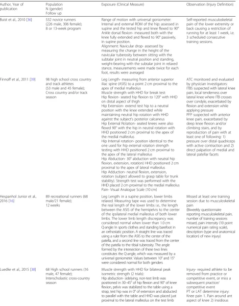
A Patient, Exposure, Outcomes (PEO) table, which de-scribes attributes of each study (author, population, exposure,
and injury definition) is included inAppendix 2. Descriptions
of the objective musculoskeletal clinical assessments identified in the included studies and their methods of measurement
have been outlined inAppendix 2. The number of runners
included in each study sample ranged from 59 to 532.
Quality of studies
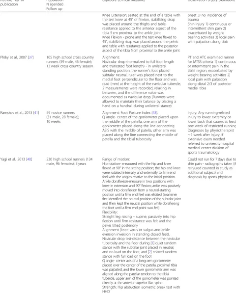
The results of the assessment of quality of each study
using the critical appraisal tool are reported in Table1.
Among the seven studies included in this review, per the CAT, two were of good methodological quality (>
75%) [36, 37] and five were of moderate quality (50–
75%) [16,38–41]. The majority of methodological
short-comings were observed in the following items: sample
bias (7/7 studies) [16, 36–41], reporting validity of
mea-sures (5/7 studies) [16, 38–41], justification of sample
size (5/7 studies) [16,38–41], and reporting reliability of
measures (5/7 studies) [16,38–41].
The included studies in this review were all observational design, and therefore per the GRADE approach were
consid-ered of low quality of evidence overall [31]. When evaluating
each domain, the clinical assessment alterations categories were downgraded either for imprecision, indirectness, incon-sistency or all three, resulting in very low quality evidence for each clinical assessment alteration investigated in this review
[33,34,42]. Publication bias refers to the probability of
select-ive publishing and due to the limited amount of studies for each the clinical assessment alterations(up to three) this item
was not used to downgrade evidence in this review [35]. The
results of GRADE are reported in Table2.
Objective musculoskeletal clinical assessments (Table2)
Hip strength
Evidence for hip strength was of very low quality (hip abduc-tion strength downgraded due to indirectness, inconsistency, and imprecision whereas the rest were downgraded due to in-directness and imprecision). Of the two studies investigating
hip abduction strength, one study [39] reported that stronger
hip abduction strength was significantly associated with injured
runners (OR = 5.35, 95% CI= 1.46, 19.53)whereas the other
study [38] found no significant association. Finnoff et al. [39],
also reported a significant protective association with increased hip external rotation to internal rotation strength ratio RRI (OR = 0.01, 95% CI= < 0.01, 0.44). There were no significant associations between hip adduction, abduction to adduc-tion ratio, external rotaadduc-tion, internal rotaadduc-tion, flexion,
ex-tension, flexion-to-extension strength ratio and RRI [39].
Hip joint range of motion
Evidence for hip joint range of motion was of very low quality (downgraded due to indirectness and
inconsist-ency). Two studies [36, 40] investigated hip internal and
external range of motion, of which one study [40] found
that increased hip internal rotation was protective against RRI in females that developed medial tibial stress
syn-drome (aOR = 0.91, 95% CI= 0.85, 0.99) [40].
Hip alignment
Evidence for hip alignment was of very low quality (Q angle downgraded for indirectness and inconsistency, and leg
length downgraded for imprecision). Two studies [16, 40]
investigated Q angle and one study [16] investigated leg
Hip flexibility
Evidence for hip flexibility was of very low quality
(down-graded for indirectness and imprecision). One study [40]
investigated straight leg raise and did not find significant as-sociation between straight leg raise test and RRI.
Knee strength
Evidence for knee strength was of very low quality
(down-graded for indirectness and imprecision). One study [38]
investigated knee strength using a HHD and did not find a significant association between quadriceps strength or hamstring strength and RRI.
Ankle alignment
Evidence for ankle alignment was of very low quality (navicu-lar drop downgraded for indirectness and inconsistency, and foot posture index downgraded for indirectness and
impreci-sion). Three studies [36, 37, 40] investigated navicular drop
Table 1Quality assessment of included studies–adapted from the Critical Appraisal Form (CAT) for Quantitative Studies [28,29]
Author I-1 I- 2 I-3 I-4 I-5 I- 6 I- 7 I-8 I-9 I-10 I-11 I-12 I-13 I-14 I-15 I-16 T.S T.%
Buist et al., 2010 [36] + + – + + + – + + – + + + + + + 13 81.25
Finnoff et al., 2011 [39] + + – + + + – + + – – + + + + + 12 75.0
Hespanhol Junior et al., 2016 [16] + + – + + + – + + – – + + + + + 12 75.0
Luedke et al., 2015 [38] + + – + + – + + + + – + – + – + 11 68.75
Plisky et al., 2007 [37] + + – + + + + + + + + + + + + + 15 93.75
Ramskov et al., 2013 [41] + + – – + + – + + – – + + + + + 11 68.75
Yagi et al., 2013 [40] + + – + + + – + + – – + + + + + 12 75.0
Note. Item 1: Purpose of the study was clearly stated, Item 2: Study design was appropriate, Item 3: Study detected sample bias, Item 4: Measurement biases were detected in the study, Item 5: Sample size was stated, Item 6: The sample was described in detail, Item 7: Sample size was justified, Item 8: Outcomes were clearly stated and relevant, Item 9: Method of measurement was described sufficiently, Item 10: The measures used were reliable, Item 11: The measures used were valid, Item 12: The results were reported in terms of statistical significance, Item 13: The analysis methods used were appropriate, Item 14: Clinical importance was reported, Item 15: Missing data were reported when appropriate, Item 16: Conclusions were relevant and appropriate given methods and results of the study
Abbreviations I- Item, T.S- total score, T%- total CAT %, meets criteria‘+’, does not meet criteria‘-’
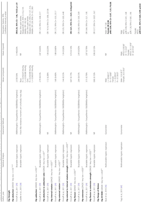
Table 2 Clinical measures and the reported predictive statistics in the 7 studies investigated in this review Author, ye ar Statistical Analysis Assessmen t Method Values (uninjured) Values (injured) Association Statistic, 95% Confidence Interval; p -value Hip Strength Hip abduction (GRADE -Very low +++O) b,c,d Finnoff et al., 2011 [ 39 ] Bivariable logistic regression (%BWxheight ) = Torq ue(Nxm)× 100/ [BW(N)x height(m)] 2.57(0.53)% 3.14(0.63)% OR:5.35, 95% CI= 1.4 6, 19.53; p :<.01 Luedke et al., 2015 [ 38 ] Bivariable logistic regression Force (N)x resistance moment arm (m)/body mass (kg).
Boys: R=
0.25(0.07) Nm/Kg L = 0. 25(0.08) Nm/Kg
Girls: R=
0.25(0.08) Nm/Kg L = 0. 26(0.07) Nm/Kg NR Boys: Shin pain tertiles Weakest:OR:1.25, 95% C=I 0.2, 9.9. Middle: OR 1.00, NA Girls: Shin pain tertiles Weakest OR:1.23, 95% CI= 0.7, 21.6, Middle: OR 2.28, 95% CI= 0.2, 28.0 Hip adduction (GRADE-Very low ++OO) c,d Finnoff et al., 2011 [ 39 ] Bivariable logistic regression (%BWxheight ) = Torq ue(Nxm)× 100/ [BW(N)x height(m)] 2.79 (0.61)% 2.87 (0.45)% OR: 1. 23, 95% CI= 0.48, 3.17 Hip abduction to adductio n ratio (GRADE-Very low ++O) c,d Finnoff et al., 2011 [ 39 ] Bivariable logistic regression NR 1.12 (0.28)% 1.06 (0.25)% OR: 14.14, 95% CI= 0.90, 221.06 Hip internal rotation (GRADE -Very low ++OO) c,d Finnoff et al., 2011 [ 39 ] Bivariable logistic regression (%BWxheight ) = Torq ue(Nxm)× 100/ [BW(N)x height(m)] 1.68 (0.40)% 1.88 (0.68)% OR: 2. 75, 95% CI= 0.33, 23.17 Hip external rotat ion (GRADE-Very low ++OO ) c,d Finnoff et al., 2011 [ 39 ] Bivariable logistic regression (%BWxheight ) = Torq ue(Nxm)× 100/ [BW(N)x height(m)] 1.44 (0.31)% 1.34 (0.26)% OR: 0. 35, 95% CI= 0.03, 4.48 Hip external to intern al rotation strength (GRADE -Very low ++OO) c,d Finnoff et al., 2011 [ 39 ] Bivariable logistic regression NR 0.87 (0.17)% 0.74 (0.13)% OR: 0.01, 95% CI= < 0.01 , 0.44;p:0.02 Hip flexion (GRADE -Very low ++OO) c,d Finnoff et al., 2011 [ 39 ] Bivariable logistic regression (%BWxheight ) = Torq ue(Nxm)× 100/ [BW(N)x height(m)] 2.84 (0.61)% 2.49 (0.92)% OR: 0. 40, 95% CI= 0.05, 3.09 Hip extension (GRADE-Very low ++OO) c,d Finnoff et al., 2011 [ 39 ] Bivariable logistic regression (%BWxheight ) = Torq ue(Nxm)× 100/ [BW(N)x height(m)] 3.15 (0.79)% 2.87 (0.79)% OR: 0. 64, 95% CI= 0.21, 1.90 Hip flexion to extensio n strength (GRADE-Very low ++OO ) c,d Finnoff et al., 2011 [ 39 ] Bivariable logistic regression NR 0.86 (0.15)% 0.96 (0.13)% OR: 0. 17, 95% CI= 0.021, 5.61 Hip Range of Motion Hip IR ROM (GRADE -Very low ++OO) b,c Buist et al., 2010 [ 36 ] Multivariable logistic regression Goniometer
Male L=
30.6(8.1)°
R
=
31.1(8.8)°
Female L=
35.9(9.5)° R = 37.7(8.3)° NR Male: HR: 1.00 Female HR 0.98 aHR: 0.99, 95% CI= 0.97 , 1.01; P:0.08 Yagi et al., 2013 [ 40 ] Multivariable logistic regression Goniometer Male: 12.4 (8.7)° Female: 25.5 (9.5)° Male: MTSS :12.9(5.8)° SF: 7.5 (3.5)° Fema le: MTSS : 31.1 (9.9)° SF: 20.7(7.6)°
Male MTSS: aOR:
0.99, 95% CI 0.91, 1.08 SF: aOR: 1.26, 95% CI 0.81, 1.96
Female MTSS: aOR
Table 2 Clinical measures and the reported predictive statistics in the 7 studies investigated in this review (Co ntinued) Author, ye ar Statistical Analysis Assessmen t Method Values (uninjured) Values (injured) Association Statistic, 95% Confidence Interval; p -value Hip ER ROM (GRADE -Very low ++OO) b,c Buist et al., 2010 [ 36 ] Multivariable logistic regression Goniometer
Male: L=
39.7(11.6)
°
R
=
40.2(12.9)°
Female L=
45.7(14.3) ° R = 45.8(13.9)° NR Male: HR: 1.01 Female: HR:1.00 Yagi et al., 2013 [ 40 ] Multivariable logistic regression Goniometer Male: 39.7(8.8)° Female: 35.1 (9.0)° Male: MTSS : 44.5(8.9)° SF: 40.0(14.1)° Fema le: MTSS : 37.4 (8.5)° SF: 43.3 (2.9)°
Male: MTSS: aOR:
0.96, 95% CI 0.88, 1.03 SF: aOR: 0.76, 95% CI 0.56, 1.03
Female MTSS: aOR:1.0,
95% CI 0.93, 1.08 SF: aOR:1.0, 95% CI 0.90, 1.11 Hip Alignment Q angle (GRADE -Very low ++OO) b,c Hespanhol junior et al., 2016 [ 16 ] Multivariable logistic regression Goniometer 10.1(5.1)° 11.8(5.0)° OR:0.9, 95% CI= 0. 8, 1.0 Ramskov et al., 2013 [ 41 ] Bivariable logistic regression Goniometer L = 11.1(4.4)° R = 11.1(5.0)° L = 8.2(4.5)° R = 9.1(4.5)° cRR: 1. 26, 95% CI= 0.49, 3.23 Leg length (GRADE-Very low +OOO) d Hespanhol junior et al., 2016 [ 16 ] Multivariable logistic regression Measuring Tape 0.5(0.6)cm 0.4(0.6)cm OR: 1. 3, 95% CI= 0.6, 2.7 Hip Flexibility Stra ight leg raise (GRADE-Very low ++OO ) c,d Yagi et al., 2013 [ 40 ] Multivariable logistic regression Goniometer Male:74.3(10 .4)° Female:76.1 (12.5)° Male: MTSS :77.6(8.5)° SF:60.0 (14.1)° Fema le: MTSS :77.7(11.0)° SF:78.3 (7.6)°
Male MTSS: aOR:
0.99, 95% CI= 0.60, 1.29 SF: aOR: 1.38, 95% CI= 1.04, 1.83
Female MTSS: aOR:
0.98, 95% CI= 0.92, 1.05 SF: aOR:1.00, 95% CI= 0.90, 1.11 Knee Strength Quadriceps strength (GRADE-Very low ++OO ) c,d Luedke et al., 2015 [ 38 ] Bivariable logistic regression Fo rc e (N)x re sistanc e mom e nt arm (m )/bod y m ass (kg) . B o ys :R = 0. 31 (0 .0 6) N m /k g L = 0. 30(0.05)Nm/kg
Girls: R=
Table 2 Clinical measures and the reported predictive statistics in the 7 studies investigated in this review (Co ntinued) Author, ye ar Statistical Analysis Assessmen t Method Values (uninjured) Values (injured) Association Statistic, 95% Confidence Interval; p -value Hamstring strength (GRADE -Very low ++ OO) c,d Luedke et al., 2015 [ 38 ] Bivariable logistic regression Fo rc e (N)x re sistanc e mom e nt arm (m )/bod y m ass (kg) .
Boys: R=
0.22(0.06) Nm/kg L = 0. 21(0.06) Nm/kg
Girls: R=
0.20(0.03) Nm/kg L = 0. 20(0.04) Nm/kg NR Boys: Shin pain Tertiles Weakest OR:1.20, 95% CI= 0.2, 8.8, Middle: OR: 0. 40, 95% CI= 0.1, 5.2 Girls: Shin pain Tertiles Weakest: OR: 1.33, 95% CI= 0.2,16.7 Middle: OR: 0.55, 95% CI= 0.1, 9.9 Ankle Alignm ent Navicular drop (GRADE -Very low ++OO) b,c Buist et al., 2010 [ 36 ] Multivariable logistic regression NR
Male: L=
6.
6(3.5)mm
R
=
6.7(3.5)mm
Female: L=
6. 0(3.1)mm R = 6.2(2.8)mm NR Male HR 1.02 Female HR 0.92 aHR-0.8 7, 95% CI= 0.77, 0.9 8; p:0.01 Plisky et al., 2007 [ 37 ] Bivariable logistic regression Ruler perpendicular to the floor > 10 mm N Boys: 20(43.5) N Girls:24(40.7) <1 0 m m N Boys:26(56. 5) N Girls:25(59.3) N 15.8 N 14.9 OR: 1. 0 OR: 0. 9, 95% CI= 0.3, 2.8 Yagi et al., 2013 [ 40 ] Multivariable logistic regression Goniometer Male: 4.5(3.4)mm Female:4.2(2.4)mm Male MTSS :4.9(3.0) mm SF: 2.4(3.1)mm Fema le MTSS :4.9(3.0)mm SF: 3.4(2.9)mm
Male MTSS: aOR:0.93,
95% CI= 0.75, 1.14 SF: aOR: 1.00, 95% CI= 0.71, 1.42
Female MTSS: aOR:
Table
2
Clinical
measures
and
the
reported
predictive
statistics
in
the
7
studies
investigated
in
this
review
(Co
ntinued)
Author,
ye
ar
Statistical
Analysis
Assessmen
t
Method
Values
(uninjured)
Values
(injured)
Association
Statistic,
95%
Confidence
Interval;
p
-value
Ankle
Rang
e
of
Motion
Ankle
dorsif
lexion
(GRADE-Very
low
+OOO)
c
Buist
et
al.,
2010
[
36
]
Multivariable
logistic
regression
Goniometer
Male: L=
KB-104.7(7.8)°
KS-99.2(8.2)° R= KB-104.6(7.5)° KS-99.2(7.8)° Female: L= KB-103.6(11.5)
°
KS-99.0(10.9)° R= KB-103.8(8.7)°
KS-99.1(9.2)°
NR
Male HR:
1.01(KB
)
HR:
1.01
(KS)
Female HR:
1.00(KB
)
HR:
1.00
(KS)
OR
odds
ratio,
aOR
adjusted
odds
ratio,
HR
Hazard
ration,
aHR
adju
sted
haza
rd
ratio,
RR
risk
ratio,
cRR
cumulative
relative
risk,
SF
stress
fracture,
MTSS
medial
tibial
stress
syndro
me,
KB
knee
bent,
KS
knee
straight
GRADE
workin
g
group
grade
s
of
evid
ence:
(bolded?
heading
for
below
items)
Low
quali
ty:
Further
rese
arch
is
likely
to
have
an
important
impact
on
our
findin
gs
Very
low
quali
ty:
We
are
unce
rtain
about
the
findings
a.
Item
was
downgraded
due
to
risk
of
bias
in
methods,
recruitment,
follow
up
or
selective
reporting
b.
Item
was
downgrad
e
due
to
inconsistency
such
as
difference
in
measurement
method,
pop
ulation,
injury
definit
ion
within
the
stud
ies
include
d
in
th
e
outcom
e
c.
Item
was
downgrad
ed
due
to
ind
irectness
and
therefore
applicabili
ty
of
findings
regard
ing
population
or
outcomes
d.
Item
was
downgrad
ed
due
to
imprec
ision
(i.e.
small
sample
size
<
and the development of running injuries. One study [36] found a significant protective relationship between decreased navicular drop amount in females and injury (HR = 0.92); two studies did not find a significant relationship between
navicu-lar drop and injured runners. One study [41] investigated the
Foot Posture Index [43] and did not find a significant
rela-tionship between foot posture and injured runners.
Ankle joint range of motion
Evidence for ankle range of motion was of very low
quality (downgraded for indirectness). One study [36]
in-vestigated ankle dorsiflexion range of motion and did not report a significant association between ankle dorsi-flexion (in knee straight and bent) and RRI.
Discussion
Findings within the studies
The goal of this study was to summarize the results of stand-alone studies that have investigated clinical assessment and risk of injury. Synthesizing the work should improve an understanding of which factors may be transferable to a clinical environment. Stand-alone findings such as increased hip external to internal rotation strength ratio and decreased navicular drop were protective of injury, but only in a few studies. We also found that increased hip abduction strength was predictive of injury and decreased hip internal rotation was protective of injury in runners, largely contradicting clinical thought and results from non-longitudinal studies of
association [44]. In no cases did we find compelling
evi-dence from multiple studies of common predictors of injury risk in running. Also, all clinical assessment alteration cat-egories had very low quality of evidence; therefore, clinicians should be very cautious interpreting the results below.
As stated, increased hip external to internal strength ratio was seen to be protective for injury in runners that developed patella femoral pain syndrome. This finding was reported in
one study by Finnoff et al. [39] Although the authors did not
operationally define this ratio, it is assumed that an increase in hip external rotator strength when compared to internal rotator strength would be protective for runners. The hip ex-ternal rotators muscles control femoral inex-ternal rotation and
a lack of control may be linked with running injury [45,46].
It is important to note there were several confounders in this study. The study did not report running distance per week (mileage) nor did it report any injury history, both of which have been associated as risk factors for injury. Because these athletes were high school runners, these factors could have
significantly influenced results [1].
Decreased navicular drop was seen to be protective of injury
in this review. This finding was reported in one study [36];
however, it was not significant among the two other studies
[25,28] that did investigate this measure. Excessive pronation
of the foot causes tibial rotation and has been seen to be
re-lated to medial stress syndrome in runners [47]. This finding
was investigated in novice runners participating in a 13-week training program for a 4-mile running event and therefore cannot be applied to all running populations in general.
Increased hip abduction strength was found to be predict-ive of injury in one cohort study. The finding that runners with stronger hip abductors were more associated with RRI may have been due to a number of confounders. The partic-ipants included in the study were high school athletes, pos-sibly novice runners. As mentioned before, weekly training mileage and injury history were not reported. Finnoff et al.
[39], theorized that the injured subjects in the group had
higher body mass index (BMI), which could have led to higher hip abduction moments. To compensate for these larger moments, the runners may have developed increased
hip abductor (eccentric) strength over time [39]. This
find-ing shows that some injured runners may have increased strength, specifically if they are younger or novice runners with a higher BMI. Caution should be used when interpret-ing this result with all runninterpret-ing populations.
Decreased hip internal rotation was found to be
protect-ive in one cohort study [40]. Excessive hip internal
rota-tion has been associated with injury during jump landing tasks and lack of control of the lower extremity in the frontal and transverse planes has also been hypothesized
as a cause for injury in runners [48,49]. Decreased
mobil-ity could therefore be beneficial and protective for run-ners, as it would require less neuromuscular control. This finding shows that stiffness in runners may not be an
im-pairment as previously thought [50,51], specifically if they
are young and may not have developed the neuromuscular control needed to stabilize the limb. Caution should be used while interpreting the findings of this study as partic-ipants were high school runners. Shin pain was the only injury reported. Mileage of the runners was not reported; however, frequency of training was. Experience was noted as national, state, or entry level, however no history of running injury or amount of running miles was reported.
Findings between the studies
The GRADE level of evidence quality was very low for all objective assessment alteration categories included in this review. Studies were downgraded for either indirectness, in-consistency, imprecision or all three. There were no com-mon predictors across a number of studies in this review. There may be several reasons for the lack of commonality or the occasional findings that are contradictory to clinical thought, such as differences in subject demographics, dif-ferent measurement methods, measurement variability within clinical assessments, inconsistent definitions of in-jury and runner, different statistical models, and study bias. These issues have been further addressed below.
drop [36, 37,40] or Foot Posture Index [41]. This lack of homogeneity between studies resulted in difficulties com-paring clinical assessments between studies, even when studies focused on a similar construct (e.g., alignment).
A variety of methods was used to define and report the clinical assessments, even when the same testing device was used. For instance, weakness in hip HHD assessment was often reported by asymmetry between left and right
sides [39,40]. However, another study [38] divided strength
into three tertiles (weakest, middle and strong) across par-ticipants and used the strongest strength values as the
com-parator. One study [38] multiplied the HHD reading by the
moment arm and then normalized it to the participant’s
body mass. The other studies normalized HHD values to
body mass and height [39]. This variability in the reporting
of muscle strength assessments made it difficult to compare studies, perform meta-analyses, or identify common pat-terns of muscle strength in included prospective studies.
Population and injury definitions were also heterogeneous among studies. Running populations in studies varied from novice to recreational, with more males than females in the
Q angle studies [13,29]. Running related injury has been
de-fined many ways in the literature, as evidenced by the wide variability of injury incidence rates reported in various
stud-ies [1,2,52]. When defining an injury, studies used: 1)
evalu-ation by a medical physician, athletic trainer or physical
therapist [39], 2) presence of pain with duration of
symp-toms > 24 h [37], 3) decrease in running mileage, 4) missed
workouts [16] or, 5) a combination of the variables listed [36,
38,40,41] all which were included in our study. Consistent
reporting about injury severity, the course of treatment, pre-vious injury, or whether the runner had sought assistance from a health care provider was lacking. Difference in levels of injury severity would likely alter associational modeling and influence the statistical significance of the findings.
Lastly, statistical modeling was different among studies. Three studies used a multivariable model, whereas four studies used a bivariable model. Among the three studies that used a multivariable model, measures of independent
variables such as age [36], other clinical tests [16] and BMI
[40] were also included in the regression analysis model.
This could have influenced the relationship between
singu-lar clinical test (such as navicusingu-lar drop) [36] and RRI.
Previous reviews investigating the risk of RRI have also
reported similar criticisms [53,54]. Winter et al. [53]
inves-tigated fatigue and RRI, and were unable to find conclusive patterns of associations due to a lack of homogeneity of the runners, small sample sizes, and the distances that were run to determine fatigue. A systematic review studying ver-tical ground reaction force and injury was also unable to make recommendations due to a lack of prospective studies investigating this variable and its association with injury
with iliotibial band syndrome (ITBS) may have associated increased peak knee internal rotation and peak hip adduc-tion during stance (based on one prospective cohort study), but because of limitations in effect size and the number of studies and methods, the authors did not make any add-itional recommendations. In the one review that investi-gated alterations to the musculoskeletal system, similar to the current study, i.e., plantar pressures, the authors con-cluded there was inconsistency among studies and
sug-gested improved methodology for future research [55].
Limitations
There are several limitations to this review. Studies with post-operative populations were excluded from the study, so it is possible the runners included in the selected studies had less severe injuries, which potentially influenced the clinical assess-ment alterations between baseline and future injury. This was performed to better generalize the results to the population of runners commonly seen in outpatient community-based
clinics, who often present without having seen a surgeon [56].
Conclusion
This review suggests that objective assessments that measure alterations in muscle strength, flexibility, alignment, and range of motion of the lower extremity had very low quality of evi-dence. Within the studies there were several confounders
such as participant’s experience, unknown injury history, and
unknown weekly running mileage, all of which have been
seen to be associated with RRI [1]. Among the studies, there
were a limited number of studies investigating each assess-ment, inconsistent results, different measurement methods among studies, measurement variability within clinical assess-ments, inconsistent definitions of injury and runner, different statistical modeling, and study bias. Future studies should aim to improve the quality of the studies as well as use standard-ized assessments and minimize confounders when conduct-ing clinical research to predict injury in runners.
Appendix 1
Search terms used in PubMed database
Injury[tiab] OR Injuries[tiab] OR“physiopathology”
[Sub-heading] OR “injuries” [Subheading] OR “Wounds and
Injuries”[Mesh]) AND (Runner[tiab] OR Runners[tiab] OR
Appendix 2
Table 3PEO (Population, Exposure, Observation) Table; description of included articles
Author, Year of publication
Population N (gender) Follow up
Exposure (Clinical Measure) Observation (Injury Definition)
Buist et al., 2010 [36] 532 novice runners (226 male, 306 female); 8 or 13-week program
Range of motion with universal goniometer: Internal and external ROM of the hip: assessed in supine and the tested hip and knee flexed to 90° Ankle dorsal flexion- measured both with the knee fully extended and flexed to 90° passively, in supine position.
Alignment: Navicular drop- assessed by measuring the change in the height of the navicular tuberosity between sitting with the subtalar joint in neutral position and standing, weight-bearing with the subtalar joint in relaxed stance, measurements were made twice for each foot, results were averaged
Self-reported musculoskeletal pain of the lower extremity or back causing a restriction of running for at least 1 week, i.e. 3 scheduled consecutive training sessions.
Finnoff et al., 2011 [39] 98 high school cross country and track athletes
(53 male and 45 female); Cross country and/or track season
Leg Length- measuring from anterior superior iliac spine (ASIS) to a point 2 cm proximal to the apex of medial malleolus
Muscle strength with HHD for break test: Hip flexion- seated hip flexion to 120° with HHD on distal aspect of thigh
Hip Extension- extend test hip to a neutral position with the knee extended while maintaining neutral hip rotation with HHD against the subject’s posterior calcaneus Hip External Rotation- seated knees were also flexed 90° with the hip in neutral rotation with HHD positioned 2 cm proximal to the apex of the medial malleolus
Hip Internal rotation- position identical to the one used for hip external rotation strength testing with HHD positioned 2 cm proximal to the apex of the lateral malleolus
Hip Abduction- 30° abduction with neutral hip flexion, extension, rotation) HHD positioned 2 cm proximal to the apex of lateral malleolus Hip Adduction- neutral flexion, extension, rotation (subject allowed to grasp table for trunk stability). Strength test was performed with the HHD placed 2 cm proximal to the medial malleolus Pain- Visual Analogue Scale (10 cm)
ATC monitored and evaluated by physician investigators: ITBS suspected with lateral knee pain, local tenderness over lateral knee where ITB crosses over condyle, exacerbated by flexion and extension while applying pressure
PFP suspected with anterior knee pain, exacerbated by deep knee flexion and/or climbing stairs, and by reproduction of pain with at least one of following: 1) pressure over distal quadriceps with active contraction and 2) direct palpation of medial and lateral patellar facets
Hespanhol Junior et al., 2016 [16]
89 recreational runners (68 male/21 female); 12 weeks
Leg Length: in a supine position, lower limbs relaxed. Measuring tape was used to determine the real length of the lower limbs i.e., the length between the ASIS of the hemipelvis to the center of the ipsilateral medial malleolus of both lower limbs. The lower limb length discrepancy was considered normal when lower than 1.0 cm Q-angle: In sports clothes and standing barefoot in an orthostatic position. A straight line was traced using a ruler from the ASIS to the center of the patella, and a second line was traced from the center of the patella to the tibial tuberosity. The angle formed by the intersection of these two lines constitutes the Q-angle, which was measured by a universal goniometer. Values between 10° and 15° were considered normal for both genders
Missed at least one training session due to musculoskeletal pain
(Biweekly questionnaire reporting musculoskeletal pain, number of training sessions missed, pain intensity (10 point numerical pain rating scale), description (type and anatomical location) of new injury)
Luedke et al., 2015 [38] 68 High school runners (16 male, 47 female);
Interscholastic cross-country season
Muscle strength with HHD for bilateral peak isometric strength (2 trials):
Hip abduction- sidelying, non-test limb was positioned in 30–45° of hip flexion and 90° of knee flexion, pelvis was stabilized to the table using a strap, test hip was in 0° of extension and abducted to parallel with the table and HHD was placed just proximal to the lateral malleolus on the test limb
publication N (gender) Follow up
Knee Extension: seated at the end of a table with the test knee at 45° of flexion, stabilizing strap was placed around the thighs and table, resistance applied to the anterior aspect of the tibia 5 cm proximal to the ankle joint Knee Flexion - prone and the test knee flexed to 45°, stabilizing strap was placed around the pelvis and table with resistance applied to the posterior aspect of the tibia 5 cm proximal to the ankle joint
onset 3) no incidence of trauma
Shin injury 1) continuous or intermittent shin pain 2) exacerbated by weight bearing activities 3) local pain with palpation along tibia
Plisky et al., 2007 [37] 105 high school cross country runners (59 male, 46 female); 13 week cross country season
Alignment:
Navicular drop (normalized to full foot length and truncated foot length) - in unilateral standing position, the runner’s foot placed subtalar neutral, ruler was placed next to the medial foot perpendicular to the floor and was read (mm) at the height of the navicular tubercle, 2 measurements were recorded, relaxing in between, and the difference value was documented as navicular drop (Runners were allowed to maintain their balance by placing a hand on a handrail during unilateral stance)
PT and ATC examined runner for MTSS criteria 1) continuous or intermittent pain in the tibial region, exacerbated by weight bearing activities 2) local pain with palpation along distal 2/3 of posterior medial tibia
Ramskov et al., 2013 [41] 59 novice runners (31 male, 28 female); 10 weeks
Alignment: Foot Posture Index [43].
Q angle- center of the goniometer placed upon the middle of the patella, one arm of the goniometer placed along the line connecting ASIS with the middle of patella, other arm was placed along the line connecting the middle of patella and the tibial tuberosity
Injury: Any running-related injury to lower extremity or lower back that causes at least one week of restricted running Diagnoses by physiotherapist ~ 1 week after injury; if extensive exam needed referred to university hospital medical center division of sports traumatology
Yagi et al., 2013 [40] 230 high school runners (134 male, 96 females); 3 years
Range of motion:
Hip rotation- measured with the hip and knee flexed at 90° in the sitting position; the hip and knee were rotated internally and externally to firm end feel with the angles relative to the initial position. Ankle dorsiflexion-measure in two positions with knee in extension and 90° flexion; ankle was passively moved into dorsiflexion from a neutral-starting position until a firm end feel was elicited (examiner first identified the neutral position of the subtalar joint and then kept the neutral position while dorsiflexing the foot until a firm end point was felt)
Flexibility:
Straight leg raising–supine, passively into hip flexion until firm resistance was felt and the pelvis tilted posteriorly
Alignment (knee varus or valgus and ankle eversion inversion in standing closed feet), Navicular drop test-distance between the navicular tuberosity and the floor during [1] quiet tandem stance with the subtalar joint placed in neutral, and no load on the foot, and [2] relaxed tandem stance with full load on the foot
Q angle- center axis of a long-arm goniometer placed over the center of the patella, proximal tibia was palpated, and the lower goniometer arm was aligned along the patellar tendon to the tibial tubercle, upper arm of the goniometer was pointed directly at the anterior superior iliac spine
Strength: Hip abduction isometric break test with HHD
Could not run for 7 days due to shin pain - radiographs taken (if reinjured counted in study as additional subject) and diagnosis by sports physician
Abbreviations
aOR:Adjusted odds ratio; aRR: Adjusted risk ratio; HHD: Hand held Dynamometer;
HR: Hazard ratio; OR: Odds ratio; RR: Risk Ratio; RRI: Running-related Injury
Acknowledgements
The authors would like to thank Leila Ledbetter (biomedical librarian) for her help with the literature search.
Funding
This research did not receive any specific grant from funding agencies in the public, commercial, or not-for-profit sectors.
Availability of data and materials
Not applicable.
Declaration of interests
The authors declare that there is no conflict of interest.
IRB
None
Authors' contributions
SMC provided idea, design, writing, review of manuscript and overall content of material; JM provided review of articles and quality tool; SS and CC provided writing, review and overall content of manuscript. All authors read and approved final manuscript.
Authors information
Shefali Christopher has been a sports physical therapist for 10 years. As a clinician, she predominantly treated runners and used musculoskeletal clinical assessments to evaluate and treat injured runners. As part of her PhD, from the University of Newcastle in Australia, she wanted to investigate the utility of the tests she was using and see if they had any predictive capability.
Ethics approval and consent to participate
Not applicable.
Consent for publication
Not applicable.
Competing interests
The authors declare that they have no competing interests.
Publisher’s Note
Springer Nature remains neutral with regard to jurisdictional claims in published maps and institutional affiliations.
Author details
1Department of physical therapy Education, Elon University, Elon, NC 27244, USA. 2School of Health Sciences, The University of Newcastle, Callaghan, Australia. 3Pivot Physical Therapy, Culpeper, VA 22701, USA.4Division of Physical Therapy,
Duke University, 2200 W. Main Street, Durham, NC 27705, USA.
Received: 25 July 2018 Accepted: 20 January 2019
References
1. van Gent RN, Siem D, van Middelkoop M, van Os AG, Bierma-Zeinstra SM, Koes
BW. Incidence and determinants of lower extremity running injuries in long
distance runners: a systematic review. Br J Sports Med. 2007;41(8):469–80.
2. Reinking MF. Exercise-related leg pain in female collegiate athletes: the
influence of intrinsic and extrinsic factors. Am J Sports Med. 2006;34(9):
1500–7.
3. Lopes AD, Hespanhol Júnior LC, Yeung SS, Costa LO. What are the main
running-related musculoskeletal injuries? A Systematic Review Sports Med.
2012;42(10):891–905.
4. Barton CJ, Bonanno DR, Carr J, Neal BS, Malliaras P, Franklyn-Miller A,
et al. Running retraining to treat lower limb injuries: a mixed-methods study of current evidence synthesised with expert opinion. Br J Sports Med. 2016.
5. Malisoux L, Nielsen RO, Urhausen A, Theisen D. A step towards understanding
the mechanisms of running-related injuries. J Sci Med Sport. 2015;18(5):523–8.
6. Hulme A, Thompson J, Nielsen RO, Read GJM, Salmon PM. Towards a complex
systems approach in sports injury research: simulating running-related injury
development with agent-based modelling. Br J Sports Med. 2018.https://doi.
org/10.1136/bjsports-2017-098871. [Epub ahead of print] Review. PubMed PMID: 2991512.
7. Damsted C, Glad S, Nielsen RO, Sorensen H, Malisoux L. Is there evidence
for an association between changes in training load and running-related
injuries? A systematic review. Int J Sports Phys Ther. 2018;13(6):931–42.
8. Messier SP, Edwards DG, Martin DF, Lowery RB, Cannon DW, James MK, et
al. Etiology of iliotibial band friction syndrome in distance runners. Med Sci
Sports Exerc. 1995;27(7):951–60.
9. Noehren B, Davis I, Hamill J. ASB clinical biomechanics award winner 2006
prospective study of the biomechanical factors associated with iliotibial
band syndrome. Clin Biomech (Bristol, Avon). 2007;22(9):951–6.
10. Ferber R, Noehren B, Hamill J, Davis IS. Competitive female runners with a
history of iliotibial band syndrome demonstrate atypical hip and knee
kinematics. J Orthop Sports Phys Ther. 2010;40(2):52–8.
11. Phinyomark A, Osis S, Hettinga BA, Leigh R, Ferber R. Gender differences in
gait kinematics in runners with iliotibial band syndrome. Scand J Med Sci
Sports. 2015;25(6):744–53.
12. Foch E, Milner CE. Frontal plane running biomechanics in female runners
with previous iliotibial band syndrome. J Appl Biomech. 2014;30(1):58–65.
13. Foch E, Milner CE. The influence of iliotibial band syndrome history on running
biomechanics examined via principal components analysis. J Biomech. 2014;
47(1):81–6.
14. Foch E, Reinbolt JA, Zhang S, Fitzhugh EC, Milner CE. Associations between
iliotibial band injury status and running biomechanics in women. Gait
Posture. 2015;41(2):706–10.
15. Grau S, Krauss I, Maiwald C, Axmann D, Horstmann T, Best R. Kinematic
classification of iliotibial band syndrome in runners. Scand J Med Sci Sports.
2011;21(2):184–9.
16. Hespanhol Junior LC, de Carvalho AC, Costa LO, Lopes AD. Lower limb
alignment characteristics are not associated with running injuries in runners:
prospective cohort study. Eur J Sport Sci. 2016;16(8):1137–44.
17. Meininger AK, Koh JL. Evaluation of the injured runner. Clin Sports Med.
2012;31(2):203–15.
18. Plastaras CT, Rittenberg JD, Rittenberg KE, Press J, Akuthota V.
Comprehensive functional evaluation of the injured runner. Phys Med
Rehabil Clin N Am. 2005;16(3):623–49.
19. O'Connor FG, Wilder RP. Evaluation of the injured runner. J Back
Musculoskelet Rehabil. 1995;5(4):281–94.
20. Moher D, Liberati A, Tetzlaff J, Altman DG. Preferred reporting items for
systematic reviews and meta-analyses: the PRISMA statement. PLoS Med. 2009;6(7):e1000097.
21. Shmueli G. To explain or predict. Stat Sci. 2010;25(3):289–310.
22. Szumilas M. Explaining odds ratios. J Can Acad Child Adolesc Psychiatry.
2010;19(3):227–9.
23. Spruance SL, Reid JE, Grace M, Samore M. Hazard ratio in clinical trials.
Antimicrob Agents Chemother. 2004;48(8):2787–92.
24. Zhang J, Yu KF. What's the relative risk? A method of correcting the odds
ratio in cohort studies of common outcomes. JAMA. 1998;280(19):1690–1.
25. Maykut JN, Taylor-Haas JA, Paterno MV, DiCesare CA, Ford KR. Concurrent
validity and reliability of 2d kinematic analysis of frontal plane motion
during running. Int J Sports Phys Ther. 2015;10(2):136–46.
26. Creaby MW, Le Rossignol S, Conway ZJ, Ageberg E, Sweeney M,
Franettovich Smith MM. Frontal plane kinematics predict three-dimensional
hip adduction during running. Phys Ther Sport. 2017;27:1–6.
27. Dingenen B, Staes FF, Santermans L, Steurs L, Eerdekens M, Geentjens J, et al. Are
two-dimensional measured frontal plane angles related to three-dimensional
measured kinematic profiles during running? Phys Ther Sport. 2018;29:84–92.
28. Law M, Stewart D, Letts L, Pollock N, Bosch J, Westmorland M. Guidelines
for critical review of qualitative studies. McMaster University occupational therapy evidence-based practice research Group; 1998.
29. Aderem J, Louw QA. Biomechanical risk factors associated with iliotibial
band syndrome in runners: a systematic review. BMC Musculoskelet Disord. 2015;16:356.
30. Guyatt G, Oxman AD, Akl EA, Kunz R, Vist G, Brozek J, et al. GRADE
guidelines: 1. Introduction-GRADE evidence profiles and summary of
32. Guyatt GH, Oxman AD, Kunz R, Woodcock J, Brozek J, Helfand M, et al. GRADE guidelines: 7. Rating the quality of evidence--inconsistency. J Clin
Epidemiol. 2011;64(12):1294–302.
33. Guyatt GH, Oxman AD, Kunz R, Woodcock J, Brozek J, Helfand M, et al.
GRADE guidelines: 8. Rating the quality of evidence--indirectness. J Clin
Epidemiol. 2011;64(12):1303–10.
34. Guyatt GH, Oxman AD, Kunz R, Brozek J, Alonso-Coello P, Rind D, et al.
GRADE guidelines 6. Rating the quality of evidence--imprecision. J Clin
Epidemiol. 2011;64(12):1283–93.
35. Guyatt GH, Oxman AD, Montori V, Vist G, Kunz R, Brozek J, et al. GRADE
guidelines: 5. Rating the quality of evidence--publication bias. J Clin
Epidemiol. 2011;64(12):1277–82.
36. Buist I, Bredeweg SW, Lemmink KA, van Mechelen W, Diercks RL. Predictors of
running-related injuries in novice runners enrolled in a systematic training
program: a prospective cohort study. Am J Sports Med. 2010;38(2):273–80.
37. Plisky MS, Rauh MJ, Heiderscheit B, Underwood FB, Tank RT. Medial tibial
stress syndrome in high school cross-country runners: incidence and risk
factors. J Orthop Sports Phys Ther. 2007;37(2):40–7.
38. Luedke LE, Heiderscheit BC, Williams DS, Rauh MJ. Association of isomentric
strength of hip and knee muscles with injury risk in high school cross
country runners. Int J Sports Phys Ther. 2015;10(6):868–76.
39. Finnoff JT, Hall MM, Kyle K, Krause DA, Lai J, Smith J. Hip strength and knee
pain in high school runners: a prospective study. PM and R. 2011;3(9):792–801.
40. Yagi S, Muneta T, Sekiya I. Incidence and risk factors for medial tibial stress
syndrome and tibial stress fracture in high school runners. Knee Surg Sports
Traumatol Arthrosc. 2013;21(3):556–63.
41. Ramskov D, Jensen ML, Obling K, Nielsen RO, Parner ET, Rasmussen S. No
association between q-angle and foot posture with running-related injuries: a
10 week prospective follow-up study. Int J Sports Phys Ther. 2013;8(4):407–15.
42. Guyatt GH, Oxman AD, Vist G, Kunz R, Brozek J, Alonso-Coello P, et al.
GRADE guidelines: 4. Rating the quality of evidence--study limitations (risk
of bias). J Clin Epidemiol. 2011;64(4):407–15.
43. Redmond AC, Crosbie J, Ouvrier RA. Development and validation of a novel
rating system for scoring standing foot posture: the foot posture index. Clin
Biomech (Bristol, Avon). 2006;21(1):89–98.
44. Mucha MD, Caldwell W, Schlueter EL, Walters C, Hassen A. Hip abductor
strength and lower extremity running related injury in distance runners: a systematic review. J Sci Med Sport. 2016.
45. Ireland ML, Willson JD, Ballantyne BT, Davis IM. Hip strength in females with
and without patellofemoral pain. J Orthop Sports Phys Ther. 2003;33(11):671–6.
46. Mizuno Y, Kumagai M, Mattessich SM, Elias JJ, Ramrattan N, Cosgarea AJ, et
al. Q-angle influences tibiofemoral and patellofemoral kinematics. J Orthop
Res. 2001;19(5):834–40.
47. Loudon JK, Jenkins W, Loudon KL. The relationship between static posture and
ACL injury in female athletes. J Orthop Sports Phys Ther. 1996;24(2):91–7.
48. Souza RB, Powers CM. Predictors of hip internal rotation during running: an
evaluation of hip strength and femoral structure in women with and
without patellofemoral pain. Am J Sports Med. 2009;37(3):579–87.
49. Snyder KR, Earl JE, O'Connor KM, Ebersole KT. Resistance training is
accompanied by increases in hip strength and changes in lower extremity
biomechanics during running. Clin Biomech (Bristol, Avon). 2009;24(1):26–34.
50. Boutris N, Byrne RA, Delgado DA, Hewett TE, McCulloch PC, Lintner DM,
Harris JD. Is there an association between noncontact anterior cruciate ligament injuries and decreased hip internal rotation or radiographic Femoroacetabular impingement? A Systematic Review Arthroscopy. 2018; 34(3):943-50.
51. VandenBerg C, Crawford EA, Sibilsky Enselman E, Robbins CB, Wojtys EM,
Bedi A. Restricted hip rotation is correlated with an increased risk for
anterior cruciate ligament injury. Arthroscopy. 2017;33(2):317–25.
52. Yamato TP, Saragiotto BT, Lopes AD. A consensus definition of
running-related injury in recreational runners: a modified Delphi approach. J Orthop
Sports Phys Ther. 2015;45(5):375–80.
53. Winter S, Gordon S, Watt K. Effects of fatigue on kinematics and kinetics during
overground running: a systematic review. J Sports Med Phys Fitness. 2017; 57(6):887-99.
54. van der Worp H, Vrielink JW, Bredeweg SW. Do runners who suffer injuries
have higher vertical ground reaction forces than those who remain injury-free?
A systematic review and meta-analysis. Br J Sports Med. 2016;50(8):450–7.
56. Hodges PW, Richardson CA. Inefficient muscular stabilization of the lumbar
spine associated with low back pain. A motor control evaluation of
![Table 1 Quality assessment of included studies – adapted from the Critical Appraisal Form (CAT) for Quantitative Studies [28, 29]](https://thumb-us.123doks.com/thumbv2/123dok_us/416320.2039102/4.595.57.540.87.402/quality-assessment-included-studies-critical-appraisal-quantitative-studies.webp)