Iranian Biomedical Journal 13 (2): 73-78 (April 2009)
*Corresponding Author; Tel. (+98-912) 694 4566; Fax: (+98-21) 8860 1599; E-mail: [email protected]
.
Abbreviations: PMSG, pregnant mare stimulating gonadotrophin; GV, germinal vesicle; TCM 199, tissue culture medium; hCG, human chorionicThe Influence of Meiotic Spindle Configuration by Cysteamine
during in vitro Maturation of Mouse Oocytes
Amaneh Mohammadi Roushandeh
1and Mehryar Habibi Roudkenar
*21
Dept. of Anatomy, Medicine Faculty, Medical University of Tabriz, Tabriz; 2Research Center, Iranian Blood Transfusion Organization, Tehran, Iran
Received 5 April 2008; revised 25 August 2008; accepted 2 September 2008
ABSTRACT
Background: The aim of this study was to assess effects of cysteamine as an anti-oxidant on rate of in vitro maturation of oocyte and determination of its effects on spindle size and shape. Methods: Pre-mature mice were primed with pregnant mare stimulating gonadotrophin (PMSG) and germinal vesicle (GV) stage oocytes were obtained 48 h after. The oocytes were cultured in tissue culture medium (TCM 199) with 50, 100, 200 and 500 µM cysteamine. Experiments also included a group of ovulated oocytes (in vivo matured) after
priming with PMSG and human chorionic gonadotrophin. For spindle immuno-cytochemistry of metaphase II (MII), oocytes α and β tubulin antibody were performed. Chromosome staining was performed with Hoechst. Results: Our results showed that the rate of GV breakdown (GVBD) and MII increased in all cysteamine groups except in group cultured with 500 µM cysteamine. Immuno-cytochemistry analysis showed that spindle area in all in vitro Maturation oocytes increased when compared to in vivo oocytes. Spindle shape and
size (spindle area) in 100 µM cysteamine in comparison to in vivo group was insignificant (P>0.05). Conclusion: Our results revealed that cysteamine improved IVM rate in a dose dependant manner. The size
and shape of spindle in the presence of given concentration of cysteamine was similar to in vivo. Therefore,
cysteamine improved microtubule organization, rate of GVBD and MII development. Iran. Biomed. J. 13 (2): 73-78, 2009
Keywords: Cysteamine, in vitro maturation, Microtubule, Anti-oxidant, Meiosis
INTRODUCTION
he formation of reactive oxygen species (ROS) such as superoxide anions (O2), hydroxyl radicals (OH) and hydrogen peroxide (H2O2) is a normal process that occurs in the cell when there is a deviation of electrons to oxygen (O2) during electron transfer reactions [1]. There are some evidences that ROS may be beneficial at some steps of reproduction to permit successful gamete interaction [2]. However, an increasing number of in vitro studies have demonstrated the detrimental effects of ROS on reproduction. The main detrimental effects include reduced sperm motility and axonemal protein phosphorylation [3], in vitro two-cell block of embryos [4], and reduced embryo development [1,
5]. In vitro oocyte or embryo culture results in higher O2 concentrations than in in vivo environments, leading to increased ROS levels [6]. ROS such as O2 are able to diffuse and pass through cell membranes and alter most types of cellular molecules such as lipids, proteins and nucleic acids. This can affect the early development of mouse, hamster, and bovine embryos [7-9]. Living organisms possess natural protective equivalents known as ROS scavengers (anti-oxidants) that counteract the negative effects of ROS. These anti-oxidants include enzymes such as superoxide dismutase, which will eliminate O2, catalase and selenium-dependent glutathione (GSH) peroxidase, which will transform H2O2 into H2O and O2, as well as lipid- and water-soluble anti-oxidants such as vitamins C and E and uric acid [9]. However, during
T
74 Mohammadi Roushandeh & Habibi Roudkenar Iran. Biomed. J., April 2009
in vitro oocyte and embryo culture, the levels of anti-oxidants are lower than in vivo because the oocytes or embryos are divorced from the donor body and do not benefit from the maternal anti-oxidant protection. The addition of an anti-anti-oxidant to the culture medium, therefore, may be important for in vitro oocyte maturation and embryo development.
Various thiol compounds especially cysteine and cysteamine are commonly added to IVM media to support GSH synthesis, and to improve the developmental competence of oocytes. Low molecular weight compounds such as β -mercaptoethanol and cysteamine enhance cysteine mediated GSH synthesis in bovine embryos [10-12]. The addition of cysteamine in maturation medium enhanced the GSH synthesis and improved oocyte maturation by protecting the oocytes from oxidative stress and probably by acting on delicate process of cytoplasmic maturation [13-15]. But, thiol compounds like cysteamine when added to culture medium could have different effects depending on the concentration used, the species and type of oocyte in the study [16-18]. In this study, the effects of different concentrations of cysteamine on IVM rate of mouse oocytes have been investigated.
The use of spindle analysis as a parameter for assessment of oocyte quality derived from studies conducted on oocytes of various mammalian species [19-21]. The technique has recently been utilized to assess the effects of toxicants and drugs [20]. ROS can affect microtubule organization and spindle formation, and culture conditions can alter spindle shape and size [21-23]. Supplemented of culture media with anti-oxidants could act as ROS scavenger and improve cytoskeleton organization in oocyte. Thiol compounds such as cysteamine are proved that induce GSH synthesis in oocytes but there is not any report about its role on spindle organization in in vitro matured oocytes.
This study was conducted to determine whether cysteamine, as an anti-oxidant, affect on spindle shape and size (spindle area) and could improve rate of in vitro maturation of mouse oocytes.
MATERIALS AND METHODS
Reagents. All materials were purchased from
Sigma Company (USA).
Collection of germinal vesicle (GV) oocytes.
Mice were primed with 5 IU of pregnant mare stimulating gonadotrophin (PMSG). Forty-five to
fifty h later, the animals were scarified by cervical dislocation and the ovary was placed in tissue culture medium (TCM 199) with 10% FBS and 100 IU penicillin/streptomycin. Cumulus oocyte complexes were retrieved with two 27-gauge needles directly from the follicles under a stereomicroscope by rupture of ovary.
In vitro maturation. IVM medium consisted of
TCM 199 supplemented with10% fetal bovine serum, 75 mIU follicular stimulating hormone, 10 ng/ml epidermal growth factor and 50, 100, 200 or 500 µM cysteamine. Control group was cultured without cysteamine. GV (n = 15-20) oocytes were transferred to a 50-µl droplet, covered with mineral oil and cultured in 5% CO2 at 37ºC.
Assessment of in vitro maturation of oocytes.
After culturing GV oocytes for 20 h, GV breakdown (GVBD) and metaphase II (MII) oocytes were analyzed. GV in GVBD was disappeared and MII oocyte was indicated by the presence of the first polar body.
Collection of in vivo MII oocytes. To obtain in
vivo oocytes, 4-week-old female ICR mice were primed with 5 IU of PMSG and 48 h later, with 5 IU of human chorionic gonadotrophin (hCG). About 14-15 h post-hCG, MII oocytes, released from the oviductal ampullae, were collected into TCM 199 supplemented with 0.23 mM sodium pyruvate, 10% fetal bovine serum and 100 IU penicillin/ streptomycin (collection medium). Cumulus cells were removed by brief treatment with hyaluronidase at room temperature (RT).
Immuno-fluorescence analysis. In vivo and IVM
oocytes matured under different experimental conditions (control, 50, 100, 200 and 500 µM cysteamine) were labeled for α and β tubulin. MII oocytes (n = 50) were fixed with methanol at RT for 10 min. Oocytes were stored in 0.01% PBS-Triton X-100 blocking solution at 4°C. MII oocytes were then incubated in the presence of mouse monoclonal anti-α and β tubulin (1:100) followed by donkey anti-mouse IgG (1:40) at RT for 40 min . Finally, the oocytes were labeled with Hoechst 33342 (1 ng/ml) in blocking solution at RT for 10 min and mounted on slides. The two poles of spindle and the width and length of the spindle were measured in all groups according to Hu et al. [24]. The center of the two outermost microtubule organizing centers at each spindle pole were used to determine spindle
Iran. Biomed. J., April 2009 Influence of Cysteamine on Meiotic Spindle Configuration 75
(A)
(B)
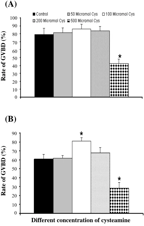
Fig. 1. (A) Rate of GVBD and (B) MII in different concentration of cysteamine, *P<0.05; cys, cysteamine.
width at poles. Measurements were carried out using a micrometer on the fluorescent microscope. Taking the middle of the imaginary line presented by the two polar plates as end points, the length of the spindle was determined. Spindle width was measured at the spindle equator. Spindle area was
measured
at the spindle equator. Spindle area wascalculated according to Sanfins et al. formula [20].
Statistical analysis. Statistical analyses of data
from five replicate trials were carried out with INSTAT view. Comparisons among groups of oocytes were performed using Chi-square and ANOVA with the Tukey-Kramer multiple comparison test as a post test. All values are expressed as the Mean ± SD. A probability of P<0.05 was considered statistically significant.
RESULTS
Effects of different doses of cysteamine on rate of
IVM. Figure 1A shows the rate of GVBD
development in the presence of different doses of cysteamine in TCM 199. In all cysteamine groups except with concentration of 500 µM, the rate of GVBD was improved. Rate of GVBD in 100 µM cysteamine was significantly higher than the control group (86% versus 72%, P<0.5). Rate of GVBD development in group with 500 µM cysteamine was significantly lower than control group (72% versus 45.2%, P<0.5). Figure 1B indicates the rate of MII development in the presence of cysteamine. Rate of MII was higher in the presence of cysteamine when compared to control group except with 500 µM cysteamine. Rate of MII oocyte in the presence of 100 µM cysteamine was higher than control significantly (P<0.05).
Variations in meiotic spindle shape and size. For
assessment of shape and size of spindle in MII oocytes cultured in different conditions, immuno-cytochemistry of microtubule was performed. Figure 2 indicates the morphology of MII spindle in different groups. Our analysis showed that spindle
in vivo control 50 µm
100 µm 200 µm 500 µm
Fig. 2. MII oocyte spindle in different concentration of cysteamine. Spindle size in 500 µM cysteamine is smaller than in vivo and its spindle in not normal. With 100 µM cysteamine spindle shape and size in MII oocyte was similar to in vivo.
100 90 80 70 60 50 40 30 20 10 0
R
a
te
o
f GV
BD
(%)
90 80
70 60 50
40
30 20
10 0
R
a
te
o
f GV
BD
(%)
Different concentration of cysteamine
76 Mohammadi Roushandeh & Habibi Roudkenar Iran. Biomed. J., April 2009
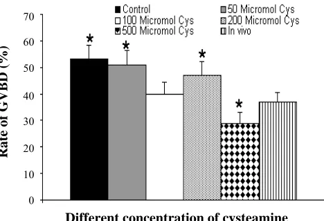
Fig 3. Mean of spindle area in different concentration of cysteamine. *P<0.05; cys , cysteamine.
area in all in vitro cultured oocytes except group treated with 500 µM cysteamine increased when compared to in vivo group. Shape and size of spindle in group with 100 µM cysteamine was close to in vivo group and difference between them was not significant (Fig. 3). Overall, the results indicate variations in meiotic spindle shape and size.
DISCUSSION
In this study, effect of different concentrations of cysteamine on IVM rate and cytoplasmic maturation of oocytes were investigated. GVBD and MII development increased in the presence of cysteamine in a dose dependant manner. In oocytes treated with 100 µM cysteamine, rate of in vitro maturation of mouse oocyte increased significantly. Several studies also reported similar results [25-27].
In 500 µM cysteamine, IVM rate decreased significantly when compared to control group. This means that the dose had toxic effects on oocytes development. Degeneration of oocytes in this group was higher than other groups (data not shown).
In mammals, oxidative stress interfere severely gamete viability and embryo development. A series of anti-oxidant enzymes and non-enzymatic processes protect gametes and embryos against ROS damage during oocyte maturation and early stage of development [28]. In ovine, GSH synthesis stimulated by cysteamine during IVM may improves embryo development by reducing intracellular peroxide levels [29]. In our previous study, in the presence of 100 µM cysteamine on IVM medium, GSH level in MII oocytes was increased. We also showed that highest level of GSH is produced in MII oocytes cultured with 100 µM cysteamine [30].
The GSH synthesis during oocyte maturation is reported to be a pre-requisite for sperm chromatin decondensation and hence for male pronucleus formation after sperm penetration of mouse and hamster oocytes [31].
Cytoplasmic GSH, defined as one of the indices of cytoplasmic maturation, plays an important role in protecting not only somatic cells but also mammalian gametes from oxidative stress [7]. The cumulus cells surrounding the oocytes are structurally and metabolically linked with the oocytes by gap junctional communications, and are reported to regulate GSH synthesis and accumulation in the ooplasm of pig and cow [32]. These reports suggested that cumulus cells are responsible for maintenance of cytoplasmic GSH concentrations through their gap junctional communications with the oocytes [32].
Immuno-cytochemistry of spindle in MII oocytes also showed that spindle area of MII oocytes in different culture condition was higher than in vivo group except group with 500 µM cysteamine. Sanfins et al. [20] showed that in vivo-derived oocytes have larger reductions in both spindle length and area than those of IVM oocytes. This finding is concurrent with the result obtained in our study. It has been proved that ROS play an important role on microtubule organization. The higher production of ROS by defective mitochondria may decrease the intracellular reduced GSH/oxidized GSH (GSSG, oxidative form of GSH) ratio. Each of these parameters acting separately or together may affect the dynamic equilibrium of microtubules (assembling/disassembling of tubulins) and/or microfilaments (assembling/disassembling of actin) [22, 23].
The in vitro culture environment differs from in vivo conditions in many aspects: the oxygen concentration is higher in vitro and in such conditions; there is higher ROS concentration in simple culture media. This could be attributed to the lack of suitable culture conditions that can dramatically impair oocyte quality. Tarian et al. [33] have demonstrated that a modification in standard culture medium composition significantly improves permission rate and the number of oocytes with normal spindle organization.
Therefore, cysteamine as an anti-oxidant acts as a scavenger of free radical in culture medium and improved microtubule organization in MII oocytes in a dose dependant manner and subsequently increase the rate of GVBD and MII during in vitro maturation of oocytes.
70
60
50
40
30
20
10
0
R
a
te
o
f GV
BD
(%)
Different concentration of cysteamine
Iran. Biomed. J., April 2009 Influence of Cysteamine on Meiotic Spindle Configuration 77
REFERENCES
1. Han, D., Zhao, B.T., Liu, Y., Li, J.J., Wu, Y.G., Lan,
G.C. and Tan, J.H. (2007) Interactive effects of low temperature and roscovitine (ROS) on meiotic resumption and developmental potential of goat
oocytes. Mol. Reprod. Dev. 75: 838-846.
2. Min, K.K., Mohammad, S.H., Hyun, J.O., Heru, Y.F.,
Goo, J., Hye, J.K., So, G.H., Jung Eun, E.P., Sung, K.K., and Byeong Chun, C.L. ( 2007) Glutathione
content of in vivo and in vitro matured canine oocytes
collected from different reproductive stages. J. Vet.
Med. Sci. 69: 627-632.
3. Brad, A.M., Bormann , C.L., Swain, J.E., Durkin,
R.E., Johnson , A.E. Clifford, A.L., and Krisher, R.L. (2003) Glutathione and adenosine triphosphate
content of in vivo and in vitro matured porcine
oocytes. Mol. Reprod. Dev. 64: 492-498.
4. Tatemoto, H., Ootaki, K., Shigeta, K. and Muto, N.
(2001) Enhancement of developmental competence
after in vitro fertilization of porcine oocytes by
treatment with ascorbic acid 2-O-alpha-glucoside
during in vitro maturation. Biol. Reprod.
65:1800-1806.
5. Agarwal A., Gupta, S., and Sharma, R.K. (2005)
Role of oxidative stress in female reproduction.
Reprod .Biol. Endocrinol. 3: 28 (Review).
6. Bedaiwy, M.A., Falcone, T., Mohamed, M.S., Aleem,
A.A., Sharma, R.K., Worley, S.E., Thornton, J., and Agarwal, A. (2004) Differential growth of human
embryos in vitro: role of reactive oxygen species.
Fertil. Steril. 82: 593-600.
7. Guérin, P., Mouatassim, E.S. and Ménézo, Y. (2001)
Oxidative stress and protection against reactive oxygen species in the pre-implantation embryo and
its surroundings. Hum. Reprod. Update 7: 175-189
(Review).
8. Kim, I.H., Van Langendonckt, A., Van Soom, A.,
Vanroose, M.G., Casi, A.L., Hendriksen, P.J. and Bevers, M.M. (1999) Effect of exogenous glutathione
on the in vitro fertilization of bovine oocytes.
Theriogenology 52: 537-547.
9. Kitagawa, Y., Suzuki, K., Yoneda, A., and Watanabe,
T. (2004) Effects of oxygen concentration and
antioxidants on the in vitro developmental ability,
production of reactive oxygen species (ROS), and DNA fragmentation in porcine embryos.
Theriogenology 62: 1186-1197.
10. Mohammad, S.H., Min, K., Goo, J., Hynt, J.O.,
Okjae, K., Jeong, J.K., Sung, K.K., Byeong, C.L. and
Woo, S.H. (2007) Effects of thiol compounds on in
vitro maturation of canine oocytes collected from
different reproductive stages. Mol. Reprod. Develop.
74: 1213-1220.
11. Ping, Z., Yan-Guang, W., Qing, L., Guo-Cheng, L.,
Gang, W., Da, G. and Jing-He, T. (2008) The interactions between cysteamine, cystine and cumulus cells increase the intracellular glutathione
level and developmental capacity of goat
cumulus-denuded oocytes. Reproduction 135: 605-611.
12. Gasparrini, B., Boccia, L., Marchandise, J., Palo,
R.D., Georgr, F., Donay, I., and Zicarelli, L. (2006)
Enrichment of in vitro maturation medium for buffalo
(Bubalus bubalis) oocytes with thiol compounds:
Effects of cystine on glutathione synthesis and
embryo development. Theriogenology 65: 275-287.
13. Furnus, C.C., de Matos, D.G. and Moses, D.F. (1998)
Cumulus expansions during in vitro maturation of
bovine oocytes: relationship level and its role on
subsequent embryo development. Mol. Reprod.
Develop. 51:76-83.
14. Gasparrini, B., Neglia, G., Palo, R.D., Campanile, G.
and Zicarelli, L. (2000) Effect of cysteamine during
in vitro maturation on buffalo embryo development. Theriogenology 54: 1537-1542.
15. Kobayashi, M., Lee, E.S., and Fukui, Y. (2006)
Cysteamine or beta-mercaptoethanol added to a defined maturation medium improves blastocyst formation of porcine oocytes after intracytoplasmic
sperm injection. Theriogenology 65: 1191-1199.
16. Balasubramanian, S. and Rho, G.J. (2007) Effect of
cysteamine supplementation of in vitro. Matured
bovine oocytes on chilling sensitivity and
development of embryos. Anim. Reprod. Sci. 98:
282-292.
17. Song. K. and Lee, E. (2007) Modification of
maturation condition improves oocyte maturation and
in vitro development of somatic cell nuclear transfer
pig embryos. J. Vet. Sci. 8: 81-87.
18. De Matos, D.G., Gasparrini, B., Pasqualini, S.R. and
Thompson, J.G. (2002) Effect of glutathione
synthesis stimulation during in vitro maturation of
ovine oocytes on embryo development and
intracellular peroxide content. Theriogenology 5:
1443-1451.
19. Rossi, G., Macchiarelli, G., Palmerini, M.G.,
Canipari, R. and Cecconi, S. (2006) Meiotic spindle configuration is differentially influenced by FSH and
epidermal growth factor during in vitro maturation of
mouse oocytes. Hum. Reprod. 21: 1765-1770.
20. Sanfins, A., Lee, G.Y., Plancha, C.E., Overstrom,
E.W. and Albertini, D.F. (2003) Distinctions in
meiotic spindle structure and assembly during in vitro
and in vivo maturation of mouse oocytes. Biol. Reprod. 69: 2059-2067.
21. Ueno, S., Kurome, M., Ueda, H., Tomii, R., Hiruma,
K. and Nagashima, H. (2005) Effects of maturation conditions on spindle morphology in porcine MII
oocytes. J. Reprod. Dev. 51: 405-410.
22. Won-Jun, C., M.D., Jashoman, B., Tommaso, F.,
James, B., Ashok, A. and Rakesh, K.S. (2007) Oxidative stress and tumor necrosis factor-a-induced alterations in metaphase II mouse oocyte spindle
structure. Fertil. Steril. 88: 1220-1231.
23. Juan, J., Tari, N.L., Francisco, J.V., Jorge, T. and
Antonio, C. (1998) Antioxidant therapy counteracts
78 Mohammadi Roushandeh & Habibi Roudkenar Iran. Biomed. J., April 2009
the disturbing effects of diamide and maternal ageing on meiotic division and chromosomal sergregation in
mouse oocytes. Mol. Hum. Reprod. 4: 281-288.
24. Hu, Y., Betzendahl, I., Cortvrindt, R., Smitz, J. and
Eichenlaub-Ritter, U. (2002) Effects of low O2 and
aging on spindles and chromosomes in mouse
oocytes from pre-antral follicle culture. Hum. Reprod.
16: 737-748.
25. Gasparrini, B., Boccia, L., Marchandise, J., Palo,
R.D., Georgeb, F., Donnay, I. and Zicarelli, L. (2006)
Enrichment of in vitro maturation medium for buffalo
(Bubalus bubalis) oocytes with thiol compounds: Effects of cystine on glutathione synthesis and
embryo development. Theriogenology. 65: 275-287.
26. Whitaker, B.D. and Knight, J.W. (2004) Exogenous
gamma-glutamyl cycle compounds supplemented to
in vitro maturation medium influence in vitro
fertilization, culture, and viability parameters of
porcine oocytes and embryos. Theriogenology 62:
311-322.
27. De matos, D.G., Nogueira, D.R, Cortvrinda, C.,
Herrera, T., Adriaenssens, R.S., Pasqualini, J., and Smitz, J. (2003) Capacity of Adult and Prepubertal Mouse Oocytes to Undergo Embryo Development in
the Presence of Cysteamine. Mol. Reprod. Dev. 64:
214-218.
28. Luberda, Z. (2005) The role of glutathione in
mammalian gametes. Reprod. Biol. 5: 5-17.
29. Mohammadi Roushandeh, A., Noori Mooghahi,.
M.H., Pasbakhsh,P., Abdolvahhab, M., Akbari, M.,
Shokrgozar, M.A., Sobhani, A., Amidi, F., Abbasi
Habibi, M. and Habibi Roudkenar, M. (2007)
Intracytoplasmic glutathione level in MII oocyte
during in vitro maturation of germinal vesicle: Effect
of cysteamine. DARU 15: 12-16.
30. Zuelke, K.A., Jones, D.P. and Perreault, S.D. (1997)
Glutathione oxidation is associated with altered microtubule function and disrupted fertilization in
mature hamster oocytes. Biol. Reprod. 57:
1413-1419.
31. Mohammadi Roushande, A., Pasbakhsh, P., Alizadeh,
Z. and Habibi Roudkenar, M. (2007) In vitro
maturation media, cysteamine concentration and gluthione level affect blstocysts development in
mouse. Iran. J. Reprod. Med. 5: 159-163.
32. Maedomari, N., Kikuchi, K., Ozawa, M., Noguchi, J.,
Kaneko, H., Ohnuma, K., Nakai, M., Shino, M., Nagai, T. and Kashiwazaki, N. (2007) Cytoplasmic glutathione regulated by cumulus cells during porcine oocyte maturation affects fertilization and embryonic
development in vitro. Theriogenology 67: 983-993.
33. Tarian, J.J., Vendrell, F.J. and Ten, J. (1996) The
oxidizing agent butyl hydroperoxide induces disturbances in spindle organization, c-meiosis, and
aneuploidy in mouse oocytes. Mol. Hum. Reprod. 2:
895-901.