Biological Properties of Blend Nanofibrous Scaffolds from
Poly(caprolactone)-Chitosan-Poly(vinyl alcohol)
Adeleh Gholipour-Kanani
*, Ali Samadikuchacksaraei, and Mohammadreza Fayyazi
Abstract- Natural-synthetic polyblend nanofibrous scaffolds have attracted numerous research interests in regenerative medicine due to their high cell compatibility and appropriate biological, physical and mechanical properties. In the current study, poly(caprolactone)-chitosan-poly(vinyl alcohol) (PCL: Cs: PVA) nanofibrous blend scaffolds were prepared and their biological properties were investigated. In our previous studies we found a high efficacy in healing on rat and dog models for PCL: Cs: PVA nanofibers. In this study, the cell compatibility of scaffold was examined using MTS method and its high biological properties in cell adhesion and proliferation were proved. The scaffold shelf life was also studied using FTIR and DSC methods as well as cell culture test. The results showed that after one-year post-fabrication, the chemical bonds, thermal behavior and cell viability of scaffolds did not change obviously, so the minimum expiration time of the scaffold was found to be about 1 year. The in vitro degradation was also investigated by tracking the rate of weight loss at different time points in PBS solution in a 37 ºC incubator under mild shaking. A 90% weight loss was observed for the scaffold after 6 weeks in PBS solution. As a result, the PCL: Cs: PVA nanofibrous web due to its high biological properties and appropriate shelf life and biodegradation time is a good candidate to be used in medical applications.
Keywords: nanofibrous scaffolds, chitosan, poly(caprolactone), scaffold degradation, shelf life
I. INTRODUCTION
I
n the recent decades, nanofibrous webs with various morphologies and also different orientations from completely random to completely parallel have been attracted many interests in different fields of study, especially in regenerative medicine. The main reason forA. Gholipour-Kanani
Department of Textile Engineering, Science and Research branch, Islamic Azad University, Tehran, Iran.
A. Samadikuchacksaraei and M. Fayyazi
Cellular and Molecular Research Center, Iran University of Medical Sciences, Tehran, Iran.
Correspondence should be addressed to A. Gholipour-Kanani e-mail: a.gholipour@srbiau.ac.ir
doing many of research works on nanofibrous webs in biomedical applications is because of the scaffold’s nano-scale, 3-D and porous network structures which can easily mimic the structure of native extra-cellular matrix (ECM) of different tissues [1].
Regardless of this versatile structure, the desired physical, mechanical and (bio)chemical properties of the webs can also be achieved by using a very wide range of polymers, biopolymers and their derivatives which have a large potential for converting to nanostructures using different techniques of nanotechnology.
High promising biomaterials potentially beneficial for using in different applications such as wound healing, drug delivery and tissue engineering can be obtained through combining high biological properties of nanofibrous structures with their unique features [1, 2]. A wide range of studies have reported on the fabrication of nanofibrous structures from blends of naturally synthesized polymers using electrospinning method. There are many literatures about the nanofibers electrospun from biopolymer-synthetic polymer blends such as collagen-PVA [3-5], chitosan-PVA [6-8], alginate-PEO [9, 10], chitosan-PCL [11-13], etc.
in mimicking the natural ECM and the other ones is the inherent biological properties and chemical structure of the precursors. So, the degradation rate of the scaffolds will affect these two parameters which have important roles in healing process using nanofibrous scaffolds.
II. MATERIALS AND METHODS
A. Materials and scaffold fabrication
Poly(caprolactone) (Mw 80 KDa) was purchased from Sigma-Aldrich. Chitosan was obtained from Chitotech Co., PVA, acetic acid and other chemical materials were purchased from Merck Company. PCL: Cs: PVA blend nanofibrous webs were prepared by electrospinning technique according to the authors’ previous studies and the results were reported elsewhere [14]. Briefly, the solutions of 5% chitosan in 80% acetic acid, 10% PVA in distilled water and 10% PCL in 90% acetic acid were prepared separately. The different solutions were blended slowly under stirrer at 40 ºC, in 2:1:1.33 mass ratio for PCL: Cs: PVA. The blend solution was electrospun with 15 kV applied voltage, 15 cm nozzle to collector distance, and 1 ml/hr extrusion rate. Morphological investigations were carried out using SEM micrographs taken by scanning electron microscopy (SEM, XL30-SFEG, FEI Philips).
B. Cell-compatibility of the scaffold
One of the most important criteria in selecting a material as biomaterial for biomedical applications is its high biocompatibility. In this way, MTS assay is a reliable method in investigating of cell-compatibility. Briefly, the designed protocol for PCL: Cs: PVA nanofibrous scaffold in MTS assay was as bellow:
Fibroblasts (9000 cells/well) were cultured in DMEM media culture on the scaffold for 3 days under CO2 incubator, 99% RH and 37 °C. The cells proliferation rate was compared with the fibroblasts cultured on standard plastic culture surfaces (TCP). In order validity of statistical studies, six samples were prepared. MTS test was performed according to the manufacturer’s (Sigma) instructions and the colorimetry was performed at 530 nm. To correct the possible absorption of MTS or formazan by the scaffold, the same weight of scaffold used for scaffolds plus fibroblasts samples were added to the fibroblasts samples at the time of adding MTS solution. A blank OD value was reduced from each sample’s reading.
C. Determination of the scaffold’s shelf life
The scaffolds were kept in a closed plastic bag under room temperature. Shelf life determination has been done using FTIR and DSC techniques as well as repeating the cell
culture test on the scaffold. The standard methods were carried out according to “Guidance for Industry: Stability Testing of New Drug Substance and Products: 2003” [17]. FTIR spectra of the web and DSC thermographs of the web in different time points were investigated for any changes in chemical bonds or thermal behavior. Fourier transforms infrared spectroscopy (FTIR) measurements were performed (Nicolet Magna-IR 560) using KBr method. Thermal behaviors were investigated using differential scanning calorimeter (Universal V3.8B TA Instruments). The samples were heated to 300 at a heating rate of 10 °C/min in air atmosphere. After one year preservation, cell viability was also investigated on PCL: Cs: PVA scaffold using fibroblast cell seeding (9000 cells/well) in DMEM as a culture medium for 3 days. The SEM image of the cells on the scaffold was captured to investigate the cell line on the scaffold.
D. In vitro degradation
The degradability tests were performed in phosphate buffered saline solution (PBS) adjusted to pH 7.4 at 37 °C. PCL: Cs: PVA nanofibrous webs were cut into 1 cm × 1 cm pieces, vacuum-dried at room temperature, and weighed accurately. They were then placed into 12-well culture plate in 2 mL of PBS at each well and were kept in a 37 ºC incubator under mild shaking [18, 19]. At each time point (1, 4, 6, 12, 14, 21, 28, 42, and 56 days), three samples were removed from PBS solution, rinsed with distilled water, and completely dried under vacuum at room temperature. The degradation rate was reported as the loss in the mass of electrospun mat using the formula “mass loss (%) = ((M0-Mt)/M0) × 100”, where M0 and Mt are the mass of electrospun mat before and after incubating in PBS solution, respectively.
III. RESULTS AND DISCUSSION
A. Cell-compatibility Results
MTS assay as a reliable method in cell-compatibility was carried out on the scaffold. The mechanism behind this assay is that metabolically active cells react with a tetrozolium salt in the MTS reagent to produce a soluble formazan dye. The cellular constructs were rinsed with PBS followed by incubation with 20% MTS reagent in serum free medium for 3 h. Thereafter, aliquots were pipetted in to 96 well plates and the samples were read in a spectrophotometric plate reader. The MTS assay result for PCL: Cs: PVA (2: 1: 1.5 mass ratio) is shown in Table I.
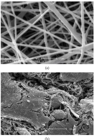
Cs: PVA nanofibrous scaffold shows high level of cell-compatibility. In order confirming the MTS result, cell morphology on the scaffold was examined under SEM. Fig. 1 shows the SEM images of the electrospun scaffold and the cell seeded scaffold 3 days after culturing. The resulted nanofibrous scaffold with bimodal morphology induces the native ECM structure of the skin. Furthermore, the chemical structure of the main biopolymer (chitosan) in the blend scaffold leads to inherent biological and antimicrobial properties attached to the unique nanofibrous structure. Regards, from Fig. 1b, the fibroblasts morphology on the PCL: Cs: PVA nanofibrous scaffold shows high cell-adhesion and also proliferation as it was expected.
B. Shelf life of the scaffold
Shelf life is the time range that a material could be used
without changing its chemical, physical or biological properties. One the other hand after this time the material would be expired and useless. Determination of a material shelf life would be done using different techniques. Investigation of any change in material chemical bonds using comparison of its FTIR spectra in different times is one of the techniques. The other one could be tracking the materials thermal behavior using DCS thermographs in different time points. After all, the cell viability of the scaffold was tested again after one year preservation to investigate the ability of maintenance of scaffold biological properties.
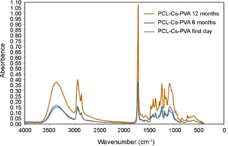
C. FTIR results
FTIR spectra of a material show different peaks in different wavenumbers due to its special chemical bonds. Any changing or shifting in peaks could be due to changing in bonds. When a material shows obvious changes or shifts, it means the nature of the material has been changed and the material is expired.
Fig. 2 exhibits the FTIR spectra of PCL: Cs: PVA nanofibrous scaffold in different time points post scaffold fabricating (first day, 6 months and 12 months). As it can be seen from figure one there is no difference in peaks of 6 and 12 months spectra with the first day one. From the result it could be concluded that the PCL: Cs: PVA nanofibrous scaffold has not expired after one year. So the shelf life of the scaffold would be at least one year.
Fibroblasts on ... Number of samples Colorimetric ReadingAverage OD (± SD) P-value*
PCL: Cs: PVA (2:1:1.5) 6 0.89917 (± 0.040519)
> 0.05
plastic surface 6 0.90211 (± 0.039028)
TABLE I
MTS RESULT FOR PCL: CS: PVA ELECTROSPUN NANOFIBROUS WEBS
(b)
Fig. 1. (a) The SEM images of electrospun PCL: Cs: PVA (2:1:1.5 mass ratio) nanofibers (magnification 5000x) under electrospinning conditions of 15KV applied high voltage, 15 cm distance of nozzle to collector and 1ml/h solution extrusion rate, and (b) The SEM images of fibroblasts morphology on PCL: Cs: PVA nanofibrous scaffold cultured in 72 h (magnification 1000x).
(a)
* Independent samples t test, SPSS 16.0 for Windows (Release 16.0.1)
D. DSC results
It is very difficult to determine experimentally the thermal stability of materials at ambient temperatures due to the fact that small changes of physicochemical properties occurring at the beginning of process proceed with very low rates. In addition, any attempt to determine the lifetime of a material is strongly dependent upon the ability to correctly identify the physical properties that are the test criteria. The process of the thermal stability investigation is seeking for an indication of material’s ability to retain a particular physical, chemical, mechanical or biological property above a certain level after exposure to elevated temperatures and/or extended periods of time. Fig. 3 shows the thermal curves of PCL: Cs: PVA nanofibrous scaffolds after first day, 6 months and one year post fabricating. Although there are some differences in first day curve compared with 6 and 12 months ones, but the whole thermal behaviors are in the same ways. The same thermal behavior after one year demonstrated that the scaffold has not expired yet and the shelf life of PCL: Cs: PVA scaffold is at least one year.
E. Cell-culture result after one year
The cell viability of the scaffold was tested after one year preservation. The test was carried out by seeding fibroblast cells (9×10⁴ cells/well) on 1-year aged PCL: Cs: PVA nanofibrous web as well as TCP (Tissue Culture Plate) as control. The cells were exposed the chitosan/PVA nanofibers for 3 days in CO2 incubator, 99% RH and 37 °C.
The SEM images of preserved scaffolds were shown before and after cell culture in Figs. 4a and 4b respectively. As it can be seen there is no significant morphological changes after one year preservation and most of the cells show high compatibility in exposure of the scaffold. Form the result, it can be concluded that the scaffold has maintained its biological properties during a year of aging.
F. Degradation of the scaffold
To simulate the degradation process, the hydrolysis in vitro under physiological conditions at pH 7.4 using PBS [18, 19] have been studied. The visual changes of the webs, after 6 days, two weeks and four weeks of incubation in PBS during in vitro degradation, are shown in Fig. 5. As it is clear, the physical integrity of the nanofibrous web was depressed after 2 weeks and the integrity was completely destroyed after 4 weeks post-examination. In this regard Ajalloueian et al. [18] investigated the in vitro degradation of nanofibrous mats (PLGA/chitosan) in comparison with PLGA nanofibers and they observed a higher mass loss for the polyblend nanofibrous mat (PLGA/chitosan) in all time points in comparison with PLGA. It was concluded that the result I due to hydrophilic properties of chitosan in the blend form.
Table II, shows the mass loss of the nanofibrous scaffolds in PBS under 37 ºC and shaking incubating at different times. A high mass loss for the nanofibrous web (PCL: Cs: PVA) in all time points was observed. According to Table II, the higher mass loss was occurred at the first time point. It means half of web weight was loss at the first day. It could be due to hydrophilic properties of two polymers in blend i.e. chitosan and PVA that leads to higher hydrolysis rate. After that the rate of mass loss was under a mild slope until 6 Fig. 3. DSC thermographs at different times: first day, 6 and 12 months
post-fabrication.
(b)
Fig. 4. PCL: Cs: PVA scaffolds in with PBS in a 37 ºC incubator under mild shaking after: (a) 6 days, (b) 2 weeks, and (c) 4 weeks.
weeks. The mass loss was constant till 8 weeks. It could be due to longer degradation time of PCL. These results suggest that the PCL: Cs: PVA nanofibrous web can be considered as potential scaffold for biomedical applications like skin tissue engineering where the rate of tissue formation can be around several weeks.
IV. CONCLUSION
The nanofibrous web from PCL: Cs: PVA (2:1:1.5) showed high level of cell-compatibility and cell-adhesion. This approach has some direct reasons. First one is, the engineered bimodal nanofibrous morphology of the web which mimics the native skin’s Extra-cellular matrix structure. The other ones is, presence of three biocompatible polymers in the blend form, in which chitosan as a well-known biopolymer with high biological properties, inherent antibacteriality, and promising effects on wound healing process, has an enormous role in bio-mimicking of collagen functions in connective tissue.
The shelf life of the scaffold was investigated using different techniques. The FTIR graphs with no significant shifting in important peaks, indicated that the materials nature has not been destroyed during one year testing. This result was confirmed by DSC technique, in which the thermal behavior of the scaffold was not changed after one year. So, the PCL: Cs: PVA nanofibrous scaffolds will not been expired for at least one year.
The degradability of the scaffold was also examined by immersing into PBS under 37 °C and shaking incubator. The results shows that the physical integrity of the scaffold was gone partially in two weeks and completely in 4 weeks. It was shown that almost 50% of the mass was loss in first day due to hydrophilic nature of chitosan and also PVA in the blend.
In conclusion, PCL: Cs: PVA nanofibrous web has high potential in biomedical applications specially in wound healing due to its high biological properties, good Sample no. Passed time First weight (g) Final weight (g) Mass loss (%)
1 1 day 0.0004 0.0002 50
2 4 days 0.0006 0.0002 66.67
3 6 days 0.0007 0.00022 68.57
4 12 days 0.0008 0.0002 75
5 2 weeks 0.0009 0.0002 77.77
6 3 weeks 0.0009 0.00015 83.3
7 4 weeks 0.001 0.00015 85
8 6 weeks 0.001 0.0001 90
9 8 weeks 0.0015 0.00015 90
TABLE II
MASS LOSS OF PCL: CS: PVA SCAFFOLDS IN PBS IN 37 ºC INCUBATOR UNDER SHAKING AT DIFFERENT TIMES
(c)
Fig. 5. PCL: Cs: PVA scaffolds in with PBS in a 37 ºC incubator under mild shaking after: (a) 6 days, (b) 2 weeks, and (c) 4 weeks.
(a)
cell-attachments and high level of cell-compatibility, its biodegradability, and also its promising shelf life.
V. ACKNOWLEDGMENT
The authors would appreciate Iran National Science Foundation (INSF).
REFERENCES
[1] J. Sharma, M. Lizu, M. Stewart, K. Zygula, Y. Lu, R. Chauhan, X. Yan, Z. Guo, E.K. Wujcik, and S. Wei, “Multifunctional nanofibers towards active biomedical therapeutics”, Polymers, vol. 7, pp. 186-219, 2015. [2] A.J. Hassiba, M.E. El Zowalaty, G. Nasrallah, T.J.
Webster, A.S. Luyt, A.M. Abdullah, and A.A. Elzatahry, “Review of recent research on biomedical applications of electrospun polymer nanofibers for improved wound healing”, Nanomedicine, vol. 11, no. 6, pp. 715-737, 2016.
[3] H.Y. Lin, W.C. Tsai, and S.H. Chang, “Collagen-PVA aligned nanofiber on collagen sponge as bi-layered scaffold for surface cartilage repair”, J. Biomater. Sci.
Polym. Ed., vol. 28, no.7, pp. 664-678, 2017.
[4] W. Song, D.C. Markel, S. Wang, T. Shi, G. Mao, and W. Ren, “Electrospun polyvinyl alcohol–collagen– hydroxyapatite nanofibers: a biomimetic extracellular matrix for osteoblastic cells”, Nanotechnology, vol. 23, no. 11, 2012.
[5] G. Abedi, A. Sotoudeh, M. Soleymani, A. Shafiee, P. Mortazavi, and M.R. Aflatoonian, “A collagen-poly(vinyl alcohol) nanofiber scaffold for cartilage repair”, J. Biomater. Sci. Polym. Ed., vol. 22, no. 18, pp. 2445-55, 2011.
[6] N. Charernsriwilaiwat and T. Rojanarata, T. Nqawhirunpat, and P. Opanasopit, “Electrospun chitosan/polyvinyl alcohol nanofiber mats for wound healing”, Int. Wound J., vol. 11, no. 2, 215-222, 2014. [7] R. Sharma, N. Singh, A. Gupta, S. Tiwari, S.K. Tiwari,
and S.R. Dgakate, “Electrospun hitosan-polyvinyl alcohol composite nanofibers loaded with cerium for efficient removal of arsenic from contaminated water”,
J. Mater. Chem. A, vol. 2, pp. 16669-16677, 2014.
[8] S. Fathollahipour, A.A. Mehrizi, A. Ghaee, and M. Koosha, “Electrospinning of PVA/chitosan nanocomposite nanofibers containing gelatin nanoparticles as a dual drug delivery system”, J. Biomed. Mater. Res. A, vol. 103, no.12, pp. 3852-3862, 2015.
[9] C.D. Saquing, C. Tang, B. Monian, C.A. Bonino, J.L. Manasco, E. Alsberg, and S.A. Khan, “Alginate-polyethylene oxide blend nanofibers and the role of the carrier polymer in electrospinning”, Ind. Eng. Chem.
Res., vol. 52, no. 26, pp. 8692–8704, 2013.
[10] C. Hu, R.H. Gong, and F.L. Zhou, “Electrospun sodium alginate/polyethylene oxide fibers and nanocoated yarns”, Int. J. Polym. Sci., vol. 2015, pp. 1-12, 2015. [11] K.T. Shalumon, K.H. Anulekha, C.M. Girish, R.
Prasanth, S.V. Nair, and R. Jayakumar, “Single step electrospinning of chitosan/poly(caprolactone) nanofibers using formic acid/acetone solvent mixture”,
Carbohyd. Polym., vol. 80, pp. 413–419, 2010.
[12] D. Semnani, E. Naghashzargar, M. Hadijanfar, F.D. Manshadi, S. Mohammadi, S. Karbasi, and F. Effaty, “Evaluation of PCL/chitosan electrospun nanofibers for liver tissue engineering”, Int. J. Polym. Mater.
Polym. Biomater., vol. 66, no. 3, pp. 149-157, 2017.
[13] K.T. Shalumon, K.H. Anulekha, K.P. Chennazhi, H. Tamura, S.V. Nair, and R. Jayakumar, “Fabrication of chitosan/poly(caprolactone) nanofibrous scaffold for bone and skin tissue engineering”, Int. J. Bio.
Macromol., vol. 48, no. 4, pp. 571-576, 2011.
[14] A. Gholipour-Kanani, S.H. Bahrami, and A. Samadikuchaksaraei, “Novel blend scaffolds from poly(caprolactone)-chitosan-poly(vinyl alcohol): physical, morphological and biological studies”, J.
Biomater. Tissue Eng., vol. 4, no. 2, pp. 245-252, 2014.
[15] A. Gholipour-Kanani, S.H. Bahrami, and M.T. Joghataei, “Tissue engineered poly(caprolactone)-chitosan-PVA nanofibrous scaffolds for burn and cutting wound healing”, IET- Nanobiotechnol., vol. 8, no. 2, pp. 123-131, 2014.
[16] A. Gholipour-Kanani, S.H. Bahrami, and S. Rabbani, “Effect of novel blend nanofibrous scaffolds on diabetic wound healing”, IET-Nanobiotechnol., vol. 10, no. 1, pp. 1-7, 2016.
[17] “Guidance for industry: stability testing of new drug substance and products” 2003, by U.S. Department of Health and Human Service and FDA.
[18] F. Ajalloueian, H. Tavanai, J. Hilborn, O. Donzel-Gargand, K. Leifer, A. Wickham, and A. Arpanaei, “Emulsion electrospinning as an approach to fabricate PLGA/chitosan nanofibers for biomedical applications”, BioMed Research International, Vol. 2014, pp. 1-13, 2014.
[19] M.P. Prabhakaran, E. Vatankhah, and S. Ramakrishna, “Electrospun aligned PHBV/collagen nanofibers as substrates for nerve tissue engineering” Biotechnol.