International Doctoral School in Biomolecular
Sciences
XXV Cycle
Genetically encoded division machinery
for cell free synthetic biology
Tutor
Sheref Samir Mansy
Armenise Harvard Laboratory of synthetic and reconstructive biology CIBIO (Centre for Integrative Biology)
Ph.D. Thesis of
Paola Torre
Armenise-‐ Harvard Laboratory of synthetic and reconstructive biology CIBIO (Centre for Integrative Biology)
ATPS aqueous two-‐phase systems
A3PS aqueous three phase systems
CFP cyan fluorescent protein
DOPG 1,2-‐dioleoyl-‐sn-‐glycero-‐3-‐[phospho-‐rac-‐(1-‐glycerol)]
FITC fluorescein isothiocyanate
GVs giant vesicles
His x histidine tag
kDa kilo Dalton
MW Molecular weight
MWCO Molecular weight cut off
PC phosphatidylcoline
PCR polymerase chain reaction
PEG polyethylene glycol
sfGFP super folder GFP
T/T transcription/translation
w/ o water in oil
YFP yellow fluorescent protein
The de novo construction of cellular life requires, in part, the assembly of components that confer the ability to replicate. Herein we describe efforts to reconstitute parts of the Escherichia coli cell division machinery inside of water-‐ in-‐oil emulsion compartments and synthetic phospholipid vesicles. The system was built with DNA and purified transcription and translation machinery housed in a compartment. A particular emphasis was placed on FtsZ, a protein that oligomerizes into a ring at the midcell and splits the cell into two. FtsZ does not contain a membrane interaction domain. In vivo, FtsZ interactions with the membrane are mediated by FtsA and ZipA. Therefore, the influence of FtsA on the behavior of FtsZ also was investigated. Fluorescently tagged constructs were used to facilitate evaluation by microscopy. The data showed that FtsZ readily assembles into rings in the presence of FtsA, thereby suggesting that the Fts system can be exploited for building a genetically encoded, self-‐replicating, cell-‐ like system. We also explored additional methods of dividing compartments, such as the use of aqueous two and three phase systems.
1.Introduction 1
1.1 E. coli cell division is an excellent model of cell replication. 4 1.2 The keystones of the divisome: FtsZ, ZipA and FtsA 7
1.3 Reconstitution of minimal cell division machinery in vitro. 8 1.4 Cell free-‐systems for protein synthesis. 9 1.5 Cell-‐like compartments: water-‐in-‐oil emulsion droplets 11 and phospholipid vesicles.
1.6 Water-‐ oil emulsion Aqueous Two Phase System (ATPS) 12
Chapter 2 Development of multi compartmentalized water-‐in-‐oil emulsion for the topography of genetically encoded system
2.1 Introduction 13
2.2 Materials and Methods 14 2.2.1 Chemical and Materials 14
2.2.2 ATPS and A3PS composition 14
2.2.3 Treatment of the w/o emulsion with SDS 15 2.2.4 Encapsulation of Aqueous Two phase System in
w/o emulsion 15 2.2.5 Encapsulation of in vitro transcription/translation
system in ATPS/emulsion and expression of m YPET 15 2.2.6 Preparation of A2PS and A3PS/emulsion samples
and PURE SYSTEM ATPS/Emulsion samples for confocal
microscopy 16 2.2.7 Instrumentation 17
2. 3 Results and Discussion 17 2.3.1 Encapsulation of ATPS in w/o emulsions 17
2.3.2 Treatment of w/o emulsions with SDS micelles 22
2.3.3 Expression of fluorescent proteins in w/o ATPS 25 2.3.4 Encapsulation of A3PS in w/o emulsions 28
Chapter 3 Reconstitution of a synthetic minimal divisome in w/o emulsions and in phospholipids vesicles
3.1 Introduction 30
3.2 Results and Discussion 31 3.2.1 Engineering of fluorescent genetic constructs for
the synthesis of a minimal divisome made of FtsZ
FtsA constructs 58 3.3.3 Transcription-‐translation reactions in vitro 58 3.3.4 Radioactive labeling of proteins expressed in vitro 61 3.3.5 W/o emulsions 62 3.3.6 Vesicles preparation 62 3.3.7 Instrumentation 63
Chapter 4 Reconstitution of a minimal divisome with purified proteins in compartments.
4.1 Introduction 64
4.2 Results and Discussion 65 4.2.1 In vivo expression and purification of FtsZ constructs 65 4.2.2 In vivo expression and purification of FtsA constructs 65 4.2.3 Encapsulation of FtsZ and FtsA constructs in w/o
emulsions 80 4.3 Materials and Methods 84
4.3.1 Bacterial strains and plasmids 84 4.3.2 Test of protein solubility 84 4.3.3 SDS-‐PAGE 85 4.3.4 Affinity chromatography protein purification 85 4.3.5 Dialysis 86 4.3.6 Protein concentration and storage 87 4.3.7 Instrumentation 87
Chapter 5 Conclusions and future directions
5.1 Conclusions 88
5.2 Future directions 90
Appendix 91
Chapter 1
1.Introduction
Projects that fall within the “Synthetic Biology Space” either construct in vivo or in vitro living or life-‐like systems.1,2,3. In other words, synthetic biology
deals with life or tries to mimic it. Those that are engaged with in vivo studies, engineer biomolecules and genomes and implement them in extant cells or organisms follow a “top down strategy”4. On the other hand, synthetic biologists
that decide to face in vitro studies, design biomolecules and genomes for the generation of synthetic cells from scratch embrace a “bottom up strategy”.5
Two frequent terms encountered in synthetic biology are “minimal” and “artificial”. A minimal cell is a cell that has the minimal requirements and parts to be considered alive. An artificial cell is made from artificial components6-‐8.
Whatever could be the design of the de novo cell, the synthetic biologist, as the word synthesis from the ancient Greek σύνθεσις (putting together) suggests, will put together well characterized biological parts and assemble them in different ways in order to obtain desired function. The biological parts are like a defined set of Lego bricks that could be assembled in different ways in order to get different functions (Fig 1).
cell replication, the genetically encoded system for the proteins FtsZ and FtsA (E. coli cell division proteins), the compartments used (phospholipid vesicles and water-‐oil emulsions), and finally a cytoplasm-‐like model (ATPS in water-‐oil emulsions).
Fig.1: The desire of the synthetic biologist. Defined parts assembled in different ways, for different desired functionality. Retrieved from Wikimedia.
Fig. 1.2 : An overview of the synthetic minimal cell assembled from the bottom-‐up. The aim is to reconstitute in cell-‐like compartments a transcription and translation system for the production of a minimal division machinery , made of FtsZ and FtsA. The system is assembled from the scratch, starting from simple chemical molecules such as
nucleotides, amino acids and simple biomolecules as ribosomes, tRNAs and enzymes. The surplus value is represented by the fact that has also an internal
compartmentalization, obtained by the addition of polymers.
1.1 E. coli cell division is an excellent model of cell replication.
There is no life without replication and without replication there is no evolution. Our aim is to mimic replication, both DNA replication and cell body division for a better comprehension of life and its diversity. We use E. coli as our cell model system.
Cell division in E. coli, as in other bacterial cells, is driven by the divisome8-‐9 a multi protein machinery system (Fig.1.3). The machinery is
composed of a dozen proteins and among these, a protein called FtsZ (filamentous thermo sensitive protein) plays a pivotal role. FtsZ is a structural homologue of the eukaryotic protein tubulin10,11 and undergoes a GTP-‐
dependent self-‐assembly into polymers which form the Z ring. The Z ring has a dynamic structure that generates a constriction force necessary for the cell to
divide and serves as a scaffold for the recruitment of the downstream components of the divisome.
Fig1.3: The Divisome of E. coli is a multi protein machinery system, composed by a dozen of proteins participating to the process of cell division. Between these proteins FtsZ and FtsA are playing a pivotal role. Vicente et al., EMBO reports 4, 655–60 (2003).
The process of cytokinesis in E. coli requires some proteins and some events to occur. Cytokinesis occurs after chromosome replication and
segregation. The Z ring is localized at the midpoint of the rod, perpendicular to the long axis of the cell. This specific positioning of the ring permits the division of the cell in two daughter cells (Fig 1.4). A precision so strict is guaranteed by the synergistic action of two protein systems able to spatially regulate the cell division: the Noc system and the Min system. The Noc system13, inhibits division
Fig. 1.4: E. coli cell division. After replication of the chromosome, the Z ring, formed by FtsZ polymerization, divides the cell in two exactly identical daughter cells. The strict positioning of the ring is guaranteed by the Noc system and the Min system. The Noc system avoids cell division in proximity of the chromosome, the Min system in the proximity of the cell poles.
The Min system is composed of MinC, MinD and MinE. MinC is the direct inhibitor of FtsZ polymerization and binds MinE that interacts with MinD, an ATPase membrane protein. MinE moves MinD from the cell poles to the middle of the rod, hydrolyzing the ATP bound to MinD. Min E is the protein responsible of the oscillation that regulates the positioning of the Z ring in the middle of the cell.
1.2 The keystones of the divisome: FtsZ, ZipA and FtsA
The spatial disposition of the Z ring is controlled by the Min and the Noc system, but the ring needs to be tethered to the membrane in order to generate the constriction force for cell division. FtsA and ZipA tether the Z ring to the membrane and in combination with FtsZ are considered the proto-‐ring elements.
ZipA is a trans-‐membrane protein, which tethers the Z ring to the membrane through interactions between its C terminus and the conserved C-‐terminal tail of FtsZ, made of 10 amino acids (DYLDIPAFLR) 15.. Given the role of ZipA in Z ring
tethering, it has been shown that a gain of function point mutation in FtsA (R286WFtsA) 16 can render E. coli independent of ZipA. Therefore, it appears
that FtsA is the primary tether of the Z ring to the membrane.
FtsA is a homologue of the eukaryotic actin17-‐ 18 and interacts with the
membrane through an amphipathic helix of 15 amino acids
(GSWIKRLVSWLRKEF) present at the C -‐terminus of the protein sequence. The ratio of FtsA versus FtsZ, plays a crucial role in cell division. This ratio is roughly 1:5, with approximately 700 molecules of FtsA and 3200 molecules of FtsZ per cell19.
1.3 Reconstitution of minimal cell division machinery in vitro.
To date it is possible to model simpler division systems in vitro despite the redundancy of the divisome and complexity of division in vivo. The imitation in vitro of cell replication focuses on two main aspects, the reconstitution of a minimal divisome and the development of cell-‐like compartments able to mimic the physicochemical but also mechanical process of division.
There are marvelous examples of bottom-‐up reconstitution of minimal cell division machinery in vitro. Erickson and colleagues engineered FtsZ-‐YFP-‐ MinD, a modified version of FtsZ , able to insert directly into the membrane of multilamellar liposomes20-‐21. This version of FtZ is able to polymerize and to
form Z-‐rings in tubular liposomes, causing indentations within the membranes. The Min protein system was reconstituted on supported lipid bilayers,
generating protein waves, correspondent to the oscillation produced in vivo22
and proto ring elements (FtsZ, FtsA and ZipA) of the Divisome were assembled in Giant Vesicles (GVs) 23(Fig 1.5).
Fig. 1-‐5: Structure of a Giant Vesicle having one lipid bilayer (unilamellar).
Reprinted with permission from Menger and Angelova, Acc. Chem. Res. 1998, 31, 789-797.
Copyright 1998American Chemical Society.
The design of minimal models does not take in account that replication is also a physicochemical process. There are several examples of vesicle division mechanisms that do not rely on protein activity and were built in vitro.
Membranes composed of three different lipids that phase separate into liquid ordered and disordered domains result in membrane curvature, budding and division by osmotic pressures24. While GVs with an encapsulated aqueous two
phase system have budding morphology that undergo division in hypertonic solution. In both cases there is a single cycle of division, because the
asymmetries are not retained in the daughter vesicles 25-‐26. The combination of
liquid ordered and disordered domains and the encapsulation of ATPS in GVs allow a second cycle27. In the Sugawara laboratory an alternative pathway was
developed that uses DNA replication to drive vesicle division28. A cationic
composition of the lipid membranes is combined with the replication of the DNA by PCR and then ionic interactions between the lipids and the DNA result in vesicle division. This system combines two processes crucial for the construction of cellular life, genomic replication and compartment division. In our system we privilege the feature of compartment division.
Our plan was to synthesize a genetically encoded minimal divisome with a cell-‐free system in multi-‐compartmentalized, cell-‐like compartments containing aqueous two-‐phase systems.
1.4 Cell free-‐systems for protein synthesis.
Cell-‐free systems are our toolbox for the synthesis of a minimal synthetic cell. We worked with transcription translation systems from cellular extracts and purified T/T as the PURESYSTEM.
We have availability of several sources for cell-‐free transcription-‐translation systems consisting of cell extracts from Escherichia coli (ECE), rabbit
biologically active protein that has to be produced. The factors to be considered are, of course, the yield of the protein needed, the origin and the complexity of the protein, the cost, and the downstream processing necessary30. Proteins for
our minimal cell division machinery system were produced by E. coli cell extract (Promega S30) supplied with a T7 RNA polymerase or by fully purified
transcription/translation (T/T) components from E. coli (PUREXPRESS® from
NEB).
The advantages of using a cellular lysate from E. coli is that it gives high yield of proteins, from hundreds of micrograms per milliliter to milligrams per milliliter in a batch reaction, depending on the protein of interest. Furthermore, in E. coli lysate are present factors that can help the folding of the proteins. The disadvantage is the presence of nucleases, meaning that higher amounts of DNA and circular DNA are needed. An example of E. coli cell lysate for the expression of a minimal protein system in vesicles is the synthesis of the bacterial actin-‐like proteins, MreB and MreC31. This example is significant because the synthesis of
MreB and MreC within vesicles shows the possibility to co-‐express different proteins at the same time in just one compartment using a cell-‐free system as source of transcription and translation machinery.
In contrast, using the PURESYSTEM (PURExpress® from NEB) we can count
upon a system made of fully defined, purified components, of which we know exactly the composition. The PURESYSTEM32 contains recombinant histidine-‐
tagged amino acyl tRNA synthetases, initiation factors, elongation factors, release factors, ribosome recycling factors, methionyl-‐tRNA transformylase, 70S
ribosomes, amino acids, rNTPs, and tRNAs. The mixture additionally includes T7 RNA polymerase, creatine kinase, myokinase, nucleoside-‐di-‐phosphate kinase, and pyrophosphatase. The PURESYSTEM is free of inhibitory substances such as nucleases, proteases, and enzymes that hydrolyze nucleotide triphosphate, so you can work with low quantity of DNA for a high level of expression. The PURESYSTEM has been successful for other groups, i.e. Yomo and colleagues used the PURESYSTEM for in vitro compartmentalization (IVC) in liposomes. 33,34.
1.5 Cell-‐like compartments: water-‐in-‐oil emulsion droplets and phospholipid vesicles.
The cell-‐like compartments used in the following experiments for the synthesis or reconstitution of a minimal divisome are water in oil emulsions (w/o emulsions) and phospholipid vesicles. W/o emulsions are heterogeneous systems, made of a disperdent phase (oil) and a dispersed phase (water). Water in oil emulsions have features of great stability, due to the presence of
surfactants in their composition, and great efficiency of encapsulation. Emulsions droplets can be as small as bacteria, with diameters of 1 µm and volumes of less
than a femtolitre, or can have droplets of diameters up to 100 µm and volumes of
nearly 1 nanolitre. Their high capacity (>1010 droplets in 1 mL of emulsion), the
ease of preparation, and their high stability over a broad range of temperatures, pH and salt concentrations render w/o emulsion ideal for compartmentalizing biochemical and genetic reactions35. W/o emulsions have been used for a long
time for direct in vitro evolution and recently as container for biochemical reactions. 34,35,36
Lipid vesicles are formed by self-‐assembly and are composed of bilayer membranes surrounding an aqueous core. The membrane is composed of double chain amphiphiles, most notably glycerophospholipids, but can also contain cholesterol, integral membrane proteins, and ion channels. Vesicles
spontaneously assembly in aqueous solution to form micron-‐scale
compartments. A myriad of molecules such as proteins, enzymes, and nucleic acids, as well as polymers, hydrogels, and small vesicles have been encapsulated during vesicle formation37-‐39. Additionally, several types of biochemical reactions
have been reconstituted inside of vesicles, including transcription and translation40.
1.6 Water-‐ oil emulsion Aqueous Two Phase System (ATPS).
ATPS is a good imitation of the cytoplasm. In living cells the presence of the cytoplasm provides compartmentalization, macromolecular crowding and small volume to the cell42,43 , features that play an important role, facilitating and
regulating intracellular reactions and moreover providing a heterogeneous distribution of molecules. This is the reason for which we think that just a boundary with an aqueous phase is not representative of a cell, and we considered logic the incorporation of a synthetic cytoplasm in cell-‐like compartment.
When two or more incompatible polymers are mixed at appropriate concentrations in aqueous solution, phase separation occurs resulting in two aqueous phases or microcompartments44-‐45. Immiscibility arises due to the high
molecular weight of the polymers combined with interactions (van der Waals, hydrogen bonding, and ionic forces) between the polymer segments and can be influenced by temperature, inorganic salts, and pH .
We encapsulated in emulsion aqueous two phase systems made of polymers such as dextran, polyethylen glycol (PEG) and aqueous three phase systems made of dextran, PEG and ficoll dispersed in water, developing multicompartment systems useful for protein localization.
Chapter 2
Development of multi compartmentalized water-‐in-‐oil emulsion for the topography of genetically encoded system.
2.1 Introduction
The synthesis of our minimal artificial cell requires a logical choice of proteins from the divisome and the development of compartments with physicochemical properties able to make division. Keating and colleagues, as mentioned before, developed GVs ATPS as models of non living cell-‐like compartments. GVs ATPS are models of cell apolarity, have a budding morphology after osmotic deflation, and are able to divide asymmetrically. Although the presence of the ATPS inside the GVs is able to mimic the cytoplasm46,47 , there are no biological features in these multi-‐
compartmentalized systems. The bottleneck is represented by the encapsulation of ATPS inside GVs. ATPS can be captured by GVs only through a phase
transition, a process not compatible with in vitro transcription/translation machinery and consequentially not compatible with building cellular mimics from the bottom-‐up. Phase diagrams help to determine the conditions necessary and the polymer concentrations needed to achieve immiscibility at different temperatures.
We demonstrated that w/o emulsion, due to their high stability and high efficiency of encapsulation of water phases, are good cell-‐like compartments for the development of genetically encoded systems. W/o emulsions are able not only to encapsulate directly ATPS in two phases, but also ATPS containing all the components necessary for in vitro transcription/translation reaction.
compartments and permit to have a more compartmentalized environment for the spatial localization of biological components.
2.2 Materials and Methods
2.2.1 Chemical and Materials
Polymers PEG 8,000 Da, PEG 5,000 Da, dextran 10,000 Da, ficoll 400,000 Da and PEG 5,000 Da (o-‐(2-‐aminoethyl)-‐o-‐methylpolyethylene) were purchased from Sigma-‐Aldrich. PEG 20,000 Da was purchased from Nektar. Mineral Oil (#M-‐5904) , Span 80 (Fluka) and Tween 80 (#P-‐8074) for the emulsion composition were purchased from Sigma-‐Aldrich. Sodium Dodecyl Sulphate (SDS) powder was purchased by Sigma-‐Aldrich. Alexa 647-‐conjugated dextran 10,000 Da and Alexa 488 for PEG labeling were purchased from Molecular Probes, Life Technologies. Tethramethylrhodamine isothiocyanate conjugated ficoll 40,000 Da was purchased from Sigma-‐Aldrich. Distilled water was purified to a resistivity of ≥ 18.2 MΩ with a Barnstead NANOPure Diamond system. Silicone spacers were from Molecular Probes (Life Technologies) and were used to enclose ATPS/emulsions on microscope slides for imaging.
2.2.2 ATPS and A3PS composition.
Three different ATPS compositions were encapsulated in mineral oil emulsions: 19.8% dextran 10 kDa and 2.8% PEG 8 kDa, 10 % dextran 10 kDa and 7% PEG 8kDa and18.5 % dextran 10 kDa and 5.5% PEG 8 kDa. The polymers were dissolved in distilled water purified to a resistivity of ≥ 18.2 MΩ and stirred on a magnetic stir plate until the solution became turbid.
solution into a bulk ATPS of 19.8% w/w dextran 10kDa, 2.8% w/w PEG 8 kDa . The final composition of A3PS is 19.8% w/w dextran 10kDa, 6% Ficoll 400 kDa and 2.8% PEG 8 kDa.
2.2.3 Treatment of the w/o emulsion with SDS.
A stock solution of 2 M SDS was prepared in 70% methanol. To 50 µL of
ATPS (19.8% dextran 10 kDa, 2.8% PEG 8 kDa) in an Eppendorf tube, the SDS was added to a final concentration of 0.1 µM, 1 µM, 10 µM, 25 mM, 100 mM.
2.2.4 Encapsulation of Aqueous Two phase System in w/o emulsion.
The oil phase was freshly prepared by mixing 4.5% (vol/vol) Span 80 and 0.5 % (vol/vol) Tween 80 in 0.95 mL of mineral Oil. This mixture was vortex until the Span 80 was completely dissolved in the mineral oil. 0.05 mL of bulk ATPS or bulk A3PS were added to the oil phase and vigorously vortex until the dispersion became cloudy.
2.2.5 Encapsulation of in vitro transcription/translation system in ATPS/emulsion and expression of m YPET
The mixture for protein production was prepared in a test tube by adding 250 ng of plasmid DNA encoding mYPet to 20 μL of the in vitro
transcription/translation system. For the in vitro transcription/translation system we used the PURESYSTEM (NEB, New England Biolabs). The reaction was performed on ice in order to prevent unnecessary reaction and then the reaction was encapsulated in ATPS, gently mixing in an Eppendorf tube 25 µL of ATPS
(19.8 % dextran 10 kDa, 2.8 % PEG 8 kDa) and 25 µL of reaction. The reaction
2.2.6 Preparation of A2PS and A3PS/emulsion samples and PURE SYSTEM ATPS/Emulsion samples for confocal microscopy.
The dextran phase in A2PS/emulsion samples was labeled with Alexa 647 conjugated dextran 10 kDa or with Alexa 647 conjugated dextran 40kDa with a final concentration of 4 mg/mL. The PEG phase in A2PS/emulsion samples was labeled with Alexa 488 conjugated PEG 20 kDa with a final concentration of 7.2 mg/mL. The dextran phase in A3PS/emulsion samples was labeled with Alexa 647 conjugates dextran 10 kDa or with Alexa 647 conjugated dextran 40 kDa with a final concentration of 0.8 mg/mL. The PEG phase was labeled with Alexa 488 conjugated PEG 20 kDa or PEG 5 KDa with a final concentration of 3.6 mg/mL. Finally the Ficoll phase, in A3PS emulsion, was labeled with
tethramethylrhodamine isothiocyanate at a final concentration of 12 mg/mL. The expression of mYPet, that has an excitation wavelength at 517nm and an emission at 530 nm, inside ATPS/emulsion was evaluated by confocal
microscopy exploiting the fluorescent properties of the protein.
A2PS, A3PS and PURE SYSTEM /ATPS emulsion samples were observed in sample chambers, constructed by placing a 20 x 5-‐mm silicon spacer
(Molecular Probes) onto a microscope slide.
2.2.7 Instrumentation.
Confocal images were obtained with a Leica TCS SP5 laser scanning confocal inverted microscope using a 63x oil objective equipped with
galvanometric stage and a 63.3x /1.4 NA HCX PL APO oil -‐ immersion objective. Z step size was 0.3 µm or where mentioned 0.1 µm. Ar, He and Ne laser lines were
reduce crosstalk between different signals below 5%. Multichannel images were analyzed in ImageJ (NIH.gov).
2.3 Results and Discussion
2.3.1 Encapsulation of ATPS in w/o emulsions.
In general the construction of a phase diagram is of crucial importance for the encapsulation of ATPS in GVs and for the understanding of polymer
compositions to use. In order to define the conditions for the encapsulation of ATPS inside of water-‐in-‐oil emulsions we tried to build phase diagrams at
different temperatures using the method of cloud point titration. In this method, one aqueous polymer solution (PEG 8 kDa) is added drop wise to the other aqueous polymer solution (dextran 10 kDa) until the solution turns opaque, an indication that the polymer solution is entering in the two-‐phase region. Water is then added to the point at which the solution turns clear. The weights of the polymer solutions are recorded at each step, and the procedure is repeated. A graph of the polymer weight percent compositions is constructed, and the compositions at which the solution is one or two-‐phases is determined by a binodial curve. At polymer weight percentage below the binodial, the solution exists as a single phase while above the line, phase separation will occur. In Fig. 2.1 is reported a phase diagram at 4 °C for our system made of PEG 8 kDa and dextran 10 kDa. The phase diagram reported does not show the typical profile of a binodial curve, but was useful to verify some points above the curve, if arise in two phase, and a couple below the curve, if arise in one phase.
Fig 2.1: Phase diagram at 4°C for dextran 10 kDa and PEG 8 kDa. The profile of the curve does not correspond to the profile of a binodial curve. In general, a solution of 20% w/vol dextran 10 kDa was titrated with a solution 30% dextran w/vol and added drop wise to the other aqueous polymer solution (dextran 10 kDa) until the solution turns opaque, an indication that the polymer solution is entering in the two-‐phase region. Water is then added to the point at which the solution turns clear. The weights of the polymer solutions are recorded at each step, and the procedure is repeated, until to obtain 19 points. The phase of some points above the curve (red triangles) and the phase of some points below the curve (green triangles) was verified.
We encapsulated in w/o emulsion by vigorous vortexing an ATPS composition of 19.8% dextran 10 kDa and 2.8% w/w PEG 8 kDa, which in bulk solution appeared in two phase. The emulsion was able to encapsulate the ATPS directly in two phase and the droplets appear surrounded by mineral oil
(Fig.2.2). In addition the droplets show a budding morphology.
1
2
3
4
5
6
7
8
9
10
10
12
14
16
18
20
22
24
w/w P
EG 8 k
D
a
w/w Dextran 10 kDa
4°C
` 2.2: A composition 19.8% dextran 10 kDa and 2.8 % PEG 8 kDa is encapsulated in w/ o emulsion. The dextran occupy the major part of the volume of the droplets .The droplets show a budding morphology. The rhodamine is spread in the oil. W/o emulsions ATPS are false colored, in green for PEG 8 kDa labeled with PEG 20kDa-‐Alexa 488(A), in blu for Dextran 10 kDa labeled with Dextran 20kDa-‐Alexa 647(B), in red for the mineral oil labeled with Rhodamine-‐HCl(D). Panel C is a merge of all the three channels.
We observed that the composition of the emulsion used, 4.5% (vol/vol) Span 80 and 0.5 % (vol/vol) Tween 80 in 0.95 mL of mineral oil provided a good efficiency of encapsulation. In fact, with a modification of the percentage in volume of the surfactants to 3% (vol/vol) Tween 80 and 2% (vol/vol) Span 80, the emulsion lost the ability of retaining the two phases (Fig 2.3). We observed many droplets containing just the dextran phase or the PEG phase
Fig 2.3: w/o emulsion ATPS with a modified surfactant composition. The efficiency of encapsulation of the emulsion of the two phases is reduced The droplets are not
compartmentalized anymore, but retain in their inner volume, the PEG or the dextran. False color in red for Dextran labeled with Dextran 20kDa-‐Alexa 647, and green for PEG labeled with PEG 20kDa-‐Alexa 488.
The droplets had an interesting budding morphology (Fig.2.4) without the presence of any osmotic deflation and we observed that the polymers composition influenced the morphology of the droplets.
In contrast, another composition encapsulated (5.5% w/w PEG 8kDa and 18.5% w/w Dextran 10 kDa), existing in two phases according to the phase diagram, showed a non budding morphology (Fig. 2-‐4, B), but clearly the droplets had two compartments. In both the cases the PEG occupy less volume compared to the dextran.
Fig 2.4: ATPS encapsulated in water-‐oil emulsions. Not all the composition give rise to the same morphology. Two different ATPS composition were realized in bulk solution: 19.8% dextran 10 kDa and 2.8% PEG 8 kDa, 5.5% w/w PEG 8kDa, 18.5% w/w Dextran 10 kDa. In panel A,
correspondent to a composition 19.8% w/w dextran 10 kDa and 2.8%w/w PEG 8 kDa water-‐oil emulsion show a budding morphology. Scale bar is 10 µm. In panel B water-‐oil emulsion show a non budding morphology. Scale bar is 10 µm. On the left DIC images. Fluorescence images have been false colored: PEG is labeled with PEG 20kDa-‐Alexa 488 and is green. Dextran is labeled with dextran 20kDa-‐Alexa 647 and is red.
A
B
The same polymer composition, dextran 10 kDa, 10% w/w and PEG 8 KDa 7% w/w, previously encapsulated in GVs is easily encapsulated in w/o emulsion directly in two phase without any phase transition and similarly assumed a budding morphology (Fig 2-‐5). The advantage of our system is that we were able to encapsulate the polymers directly in two phase without undergo to phase transition, that in the case of the ATPS composition in GVs, was
occurring at 43°C.
Fig 2.5: ATPS encapsulated in water-‐oil emulsions. The ATPS composition encapsulated is dextran 10 kDa, 10% w/w and PEG 8 KDa 7% w/w. This composition was previously
encapsulated in giant vesicles. W/o/ATPS show a budding morphology. Scale bar is 25 µm. On the left DIC images are shown. Fluorescence images have been false colored: PEG is labeled with PEG 20kDa-‐Alexa 488 and is green. Dextran is labeled with Dextran 20kDa-‐Alexa 647 and is red.
2. 3.2 Treatment of w/o emulsions with SDS micelles.
The budding morphology showed by certain ATPS composition lead us to test the efficiency of the droplets to retain two phases. We treated a w/o ATPS (19.8% dextran 10 kDa, 2.8% PEG 8 kDa) with SDS micelles in order to see changing of morphology. We noticed that in absence of SDS or in the presence of a low amount of SDS, such as 0.1 mM and 1 mM, the w/o emulsions keep the compartmentalization in two phase and the budding morphology (Fig.2.6-‐Fig 2.7). Increasing the concentration of the SDS in the sample, we observed droplets containing just one of the two phases in their inner volume, suggesting that the droplets were dividing. In some samples treated with 25 mM SDS, we noticed droplets directed to a process of fission (Fig.2.8)
Fig. 2.6: SDS causes changing in the morphology of the w/o ATPS droplets. In absence of the SDS (panel A) the droplets have the ability of retain two phases in their inner volume. The major amount of droplets of dextran, false colored in red and labeled with dextran 20 kDa-‐Alexa 647, is due to the fact that the ATPS composition used forecasts an higher amount of dextran versus PEG ( 19.8 % dextran 10 kDa and 2.8 % PEG 8 kDa). In presence of 0.1 µM (panel B) and 1 µM (panel C) the droplets are still retaining their internal compartmentalization. The images on the top are acquired in bright-‐field, the images on the bottom are merge of red (dextran) and green (PEG), labeled as mentioned other times.
Fig.2.7: SDS causes changing in the morphology of the w/o ATPS droplets. The behavior observed suggests the possibility that the droplets, in presence of micelles of SDS can divide. Increasing concentration of SDS progressively, it is possible to see that the droplets loose their ability to retain the two phases. Bright field and merged images (dextran false colored in red, PEG false colored in green).
Fig 2.8: Fission of w/o emulsion ATPS in presence of 25 mM of SDS. Together with the loosing of ability of retaining two phases, we noticed that some droplets were engaged in a process of fission. This fact strongly motivated us to consider that division was occurring.
2.3.3 Expression of fluorescent proteins in w/o ATPS.
Multi compartmentalized water-‐in-‐oil emulsion with their features of easy preparation, have the big advantage of direct encapsulation of ATPS, showing ideal characteristics to guest in vitro T/T. Ideally an in vitro T/T, housed in compartments like these, should be able to produce proteins and the proteins once produced could distribute in one of two compartments. We successfully expressed mYPET in w/o emulsions, using the PURESYSTEM. The protein once produced preferred mostly the dextran phase (Fig 2.9). mYPet has an excitation wavelength at 517 nm and an emission at 530 nm, and its
expression was evaluated by confocal microscopy.
Fig 2.9: Expression by PURE SYSTEM of A206KYPet in w/o/ATPS. A PURE SYSTEM reaction expressing mYPet, was mixed with 25 µL of ATPS, then encapsulated in w/o emulsion and incubated for 2 h at 37°C.The protein prefers the Dextran phase. On the left BF image on the right fluorescence image false colored in gold. Scale bar is 10 µm. The expression of mYPet (Ex=517nm, Em=530nm) inside ATPS/emulsion was evaluated by confocal microscopy exploiting the fluorescent properties of the protein.
We aim to build genetically encoded systems in multi compartmentalized w/o emulsions for spatially localized biological components. In particular we want to build a genetically encoded system expressing FtsZ and FtsA and localize them in these compartments. In Fig.2.10 we show expression of YFP-‐ FtsZ-‐FtsA inside w/o ATPS emulsions. We can notice expression of YFP-‐FtsZ-‐ FtsA with a distribution in the dextran phase with some polymers distributing at the interface.
Fig 2.10:YFP-‐FtsZ-‐FtsA is expressed by PURESYSTEM in w/o emulsions ATPS after 2 h. Is possible to notice some aggregation of FtsZ proto filaments in the dextran phase.
Top, from the right, DIC image of a w/o emulsion ATPS expressing a YFP-‐FtsZ-‐FtsA by
PURESYSTEM. False coloration green is for the expression of YFP-‐FtsZ-‐FtsA (YFP=YPet, Ex=517 nm, Em=530 nm), false coloration is blu for Dextran 10 kDa labeled with Dextran 20 kDa -‐Alexa 647.
2.3.4 Encapsulation of A3PS in w/o emulsions.
The exciting possibility of spatially localizing proteins once produced, lead us to develop water-‐in-‐oil emulsions containing Aqueous Three Phase Systems (A3PS). Aqueous Three Phase Systems are made of three polymers:
dextran 10 kDa, PEG 8 kDa and Ficoll 400 kDa. The third phase is generated simply by adding a solution of 30% w/w of Ficoll 400 kDa to an ATPS with a composition of 19.8% w/w Dextran and 2.8% w/w PEG 8 kDa. Water-‐in-‐oil emulsions, also in this case, showed to be ideal for the direct encapsulation of A3PS in three phases (Figure 2.11). The third phase is represented by the Ficoll 400 and is the dark phase between the dextran phase (Dextran 40 kDa-‐
Alexa647) and the PEG phase (PEG 20 kDa-‐ Alexa 488), (Fig 2.11).
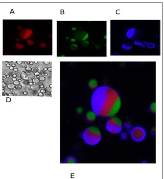
Fig 2.11: A3PS are directly encapsulated inside water-‐oil emulsion. A3PS are produced by adding ficoll 400 kDa, to a bulk ATPS made of 19.8% dextran 10 kDa and 2.8% PEG 8 kDa. On the left DIC image. Fluorescent images are false colored in red for Dextran 40kDa-‐Alexa 647, in green PEG 20kDa-‐Alexa 488 . In the middle of this two phase there is the ficoll 400 kDa, labeled with ficoll 40 kDa tethramethylrhodamine isothiocyanate . Scale bar is10 µm.
In order to confirm the presence of the third phase between the Dextran and the PEG, we labeled the Ficoll 400 kDa using ficoll 40 kDa
tethramethylrhodamine isothiocyanate. The emulsion is able to retain effectively in the aqueous compartment three polymers that separate according to their physicochemical properties and their molecular weight (Fig.2.10).
Fig 2.10: Is possible to label the third phase constituted by the ficoll 400 kDa in w/o A3PS. The A3PS composition is of 19.8% dextran 10 kDa, 2.8% PEG 8 kDa, 6 % ficoll 400 kDa.
DIC images(panel D). Fluorescent images are false colored, in red for the Ficoll 400 kDa labeled with Ficoll 40 kDa tethramethylrhodamine isothiocyanate (panel A), in green for PEG labeled with PEG 20 kDa-‐ Alexa 488 (panel B), in blu for Dextran labeled with Dextran 40kDa-‐Alexa 647. Scale bar is 25 µm.
Chapter 3
Reconstitution of a synthetic minimal divisome in w/o emulsions and in phospholipids vesicles
3.1 Introduction
Cell body division is a feature of life still difficult to be reproduced in a laboratory. There are advancements, as discussed before, that demonstrate the possibility to reconstitute the main features of cell body division in cell-‐like compartments and the difficulty in mediating compartments division. Herein we describe our efforts to reconstitute a genetically encoded system of minimal division machinery both in phospholipids vesicles and in w/o emulsions. We developed a genetically encoded system made of FtsZ and FtsA. FtsZ and FtsA , in w/o emulsions and in phospholipids vesicles, assemble together and form ring-‐ like structures.
The assembly of our genetically encoded system required the engineering of some fluorescent constructs to monitor the expression of FtsZ and FtsA in the compartments and the choice of a T/T system for their optimal expression. Ericksson and colleagues, as discussed in the first chapter, demonstrated that a FtsZ-‐YFP-‐MinD, purified and encapsulated in multilamellar tubular liposomes21 ,
is able to reproduce the typical phenomena of cell division. FtsZ, in this construct, comprise amino acids from 1-‐366 of E. coli sequence and at the C-‐ terminus has a yellow fluorescent protein (YFP-‐Venus) followed by an
amphiphatic helix made of 15 aminoacids (FIEEEKKGFLKRLFGG). It is important to notice that the amphiphatic helix is the membrane targeting sequence (MTS) of MinD and is similar to the amphiphatic helix of FtsA sequence.49 Although
FtsZ-‐YFP-‐MinD forms polymers in liposomes that generate membrane indentations, the Z-‐ring does not divide the liposome into daughter compartments.