Spring 2019
Can key protein interactions be disrupted in PTEX to inhibit its
Can key protein interactions be disrupted in PTEX to inhibit its
function?
function?
(Maria) Elena Y. Bartemes
Iowa State University, [email protected]
Follow this and additional works at: https://lib.dr.iastate.edu/creativecomponents Part of the Parasitology Commons
Recommended Citation Recommended Citation
Bartemes, (Maria) Elena Y., "Can key protein interactions be disrupted in PTEX to inhibit its function?" (2019). Creative Components. 134.
https://lib.dr.iastate.edu/creativecomponents/134
Can key protein interactions be disrupted in PTEX to inhibit its
function?
Elena Bartemes
Iowa State University - BMS
Creative Component Spring 2019
Abstract: PTEX is the Plasmodium translocon of exported proteins of the malaria parasite
(Plasmodium sp.) and functions to export proteins out of the parasite vacuolar membrane, remodeling the host erythrocyte. Remodeling enables the parasite to access vital nutrients and
avoid immune defense of the host, enabling parasite survival and leading to malaria disease.
A key protein interaction in PTEX involves 12 residues of component EXP2, which form an
augmented β-sheet with component HSP101. Theoretically, interrupting this essential
interaction by introducing a peptide corresponding to these 12 residues of EXP2 to compete
with endogenous EXP2 would fatally damage the protein transport mechanism. If utilized
therapeutically, this peptide could inhibit function of PTEX, thereby protecting host
Plasmodium parasites are responsible for malaria, the infectious disease that claimed over 435,000 lives in 219 million cases worldwide in 20176. Plasmodium is transmitted between
human and mosquito hosts/vectors with asexual human liver and blood stages and sexual
mosquito states. Malaria disease occurs in the human host during the blood-stage of the infection when P. falciparum survives by invading and reproducing within human erythrocytes, then
exporting hundreds of proteins to remodel the cell1. Plasmodium sporozoites reside in mosquito salivary glands and are introduced to host via mosquito during saliva deposit during bite. Sporozoites travel through bloodstream to the liver, infecting hepatocytes where they can replicate asexually. Asexually replicated sporozoites are termed merozoites. Merozoites are released into the bloodstream and infect host erythrocytes. During erythrocytic cycle,
Plasmodium is encased within a vacuole, namely the parasitophorous vacuole. Parasite
[image:3.612.98.509.187.518.2]progresses through developmental stages within the parasitophorous vacuole, eventually forming a schizont to rupture and return merozoites to bloodstream. Gametocytes develop from
merozoites following each cycle of erythrocyte infection. Gametocytes are the sexual form of parasite and undergo five morphological stages prior to be taken as blood meal. Stage five gametocytes are taken by female Anopheles mosquito during blood meal and enter midgut of new host for sexual reproduction. Gametocytes differentiate into male and female microgametes and macrogametes, respectively. Fertilization forms zygote, maturing into ookinete that
penetrates gut wall and develops to oocyst. Maturation of oocyst into sporozoites leads to release and migration to salivary gland. Life cycle repeats as mosquito takes another blood meal and deposits sporozoites1 (Figure 1).
To survive within the erythrocyte, a parasite needs access to an external
environment from which to hijack nutrients. Through a complex and active mechanism, parasites enter erythrocytes and create a parasitophorous vacuole where they are protected from host immune system. Proteins
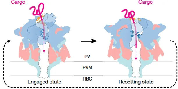
[image:4.612.258.564.239.392.2]necessary for parasite survival are unable to diffuse across the parasitophorous vacuolar membrane (PVM), and require the Plasmodium translocon of exported proteins (PTEX) for transport into the host cytosol1,4. The parasite exports proteins that remodel the red blood cell, enabling nutrient use and waste disposal. Erythrocyte remodeling as well as parasite metabolism require this export of protein with other small molecules across PVM3. Proteins traffic through the parasite secretory pathway to the vacuole and are recognized and transported across the PVM by PTEX1,4. PTEX is a recently discovered protein translocon located at the luminal face of the parasite vacuole membrane. PTEX requires ATP and unfolding of protein to cross PVM via PTEX (Figure 2). It is unknown if proteins are unfolded prior to entering PTEX or if PTEX facilitates unfolding within its components. PTEX operates in “hand-over-hand” mechanism, changing conformation to “pull” unfolded protein through pore and out to erythrocyte cytosol. Engaged state of PTEX is actively pulling unfolded protein through pore and then condenses into a slightly smaller resetting state, releasing to allow for protein to move through pore, then
condenses again to continue along protein and ultimately thread protein through pore (Figure 3).
Most proteins are then free to refold with exit into erythrocyte cytosol.
PTEX is a triad of core components – AAA+ ATPase heat shock protein 101 (HSP101), exported protein 2 (EXP2), and PTEX1504.
[image:5.612.66.547.114.261.2]These three proteins are essential for the survival of the parasite, as their function allows for parasite protein and nutrient availability. The three core components of PTEX form a stable complex within both parasitophorous vacuole membrane and parasite plasma membrane. HSP101 is made up of six individual pseudo-symmetrical subunits, while EXP2 and PTEX150 are made up of seven individual pseudo-symmetrical subunits (Figure 4).
[image:5.612.310.559.350.677.2]Figure 4: PTEX is a triad of three components each with their own subunits creating a pseudo-symmetrical interlocking conformation. Nature 2018
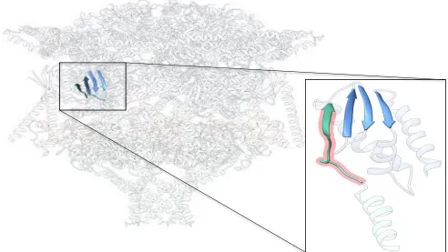
Interactions essential for PTEX function include a C-terminal domain of EXP2 and β-sheet of HSP101 with EXP2 promoter. This interaction forms an augmented β-β-sheet within each component subunit, for six total interactions within PTEX. These interactions are key for the stable assembly of the PTEX core complex. Total knockdown of EXP2 produced lethal defect in
P. falciparum growth and export1, but an EXP2 knockdown is rescued by the introduction of mutant EXP2 complete with 54 residues. However, a mutant lacking final 12 residues does not rescue parasite with EXP2 knockdown4. This shows the final 12 residues of C-terminus EXP2 within augmented β-sheet are the essential feature of interaction. Prior to, and following 54 residues, amino acids are not directly responsible for PTEX function4 (Figure 5). Because there are six subunits of HSP101 and seven subunits of EXP2 within each PTEX, six interactions between HSP101 and EXP2 are taking place among the translocon in psudo-symmetrical increments.
[image:6.612.69.561.397.674.2]Disruption of the 12 crucial residues of EXP2 with HSP101 augmented β-sheet would inhibit function of PTEX. In order to create disruption, an exogenous EXP2 peptide will be introduced to compete with endogenous EXP2. This peptide would operate as a block,
preventing formation of augmented β-sheet. Without augmented β-sheet, EXP2 and HSP101 would have no interaction among any PTEX component subunits, inhibiting function of translocon, and thereby preventing any protein export. Eventually, this competitive inhibition would theoretically result in parasite death following lack of resources. Within a conditional system, parasite death can be linked to conditional expression of exogenous EXP2 peptide.
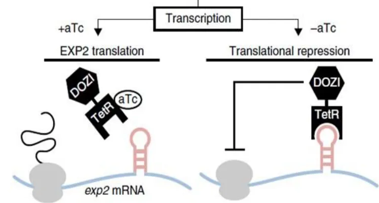
To transfect exogenous EXP2 peptide into parasitic line, sequence must be introduced into plasmid and transformed into DNA. One plasmid received two different inserts in two separate experiments to test hypothesis of disruption of endogenous EXP2 to competitively inhibit PTEX. Plasmid pLN-HSP101-SP-mRuby3 utilized for PCR mutagenesis to insert EXP2_222-233 and EXP2_222-233 scrambled. This plasmid was chosen for presence of mRuby3 fluorescence protein and signaling peptide. The signaling peptide has been previously shown to appropriately flag proteins for export by PTEX, guaranteeing interaction between exogenous insert and endogenous PTEX machinery. Fluorescent tag mRuby3 is useful as partner with EXP2 peptides as the EXP2 peptide is only approximately 50 residues and small peptides have risk of becoming lost within parasitic cytosol. mRuby3 tag will operate both as a well-folded chaperone protein for potential export through PTEX and as florescent tag following transfection into parasite line. Scrambled version of EXP2 is proof of concept of specific 12 amino acid sequence required for interaction. The scrambled EXP2 peptide would be theoretically unable to compete with endogenous EXP2 because it could not form required augmented β-sheet, rescuing parasite. Placement into plasmid pLN-HSP101-SP-mRuby3 is deemed “The Easy Way” due to commonly used plasmid and comparative “The Hard Way”. The Hard Way involves a long, linear plasmid not often used but contains a conditional repression system. This plasmid, psn154_pfcrt_HA-FKBP-dCas9-FLUC+hyp12 flanks is 30kb and contains the TetR-DOZI-aptamer system as the conditional knockout system (Figure 6). With this
approach, parasite survival and death can be observed as a result of the introduced exogenous
peptide expression, “turning off and on”, respectively. Turning the conditional system “on” will
lead to the expression of competitive EXP2, effectively killing the parasite due to PTEX
dysfunction.
The TetR (Tet-repressor protein) DOZI (development of zygote inhibited)5 aptamer
conditional knockout system uses the addition of anhydrotetracycline (aTc)2 as conditional
TetR-DOZI, ceasing repression of RNA polymerase. Without the addition of aTc, EXP2 will not
be translated due to RNA polymerase inhibition and the exogenous EXP2 peptide will not be
available to compete with endogenous EXP2. Lack of competition allows parasite within
erythrocyte to normally function via PTEX and continue to proliferate. The presence of this
system will allow direct observations of the successful competitive interaction of exogenous
EXP2 over endogenous EXP2.
T
This is an ongoing experiment in the Beck Lab and will hopefully produce results within
the coming weeks. The expected result following transfection is no proliferation of EB1
(non-scrambled) and viable EB2 (non-(non-scrambled) within pLN-HSP101-SP-mRuby3 plasmid. The EB1
strain is not expected to grow because it will be unable to remodel the erythrocyte to hijack vital
nutrients and proteins. If the observed result is as expected, research will move forward with
“hard way” plasmid containing conditional knockout system.
Quick Change Lightning Multi Site Mutagenesis PCR with primers EB1 (non-scrambled) and EB2 (scrambled) were performed to introduce EXP2_222-233 non-scrambled insert and EXP2_222-233 scrambled insert to pLN-HSP101-SP-mRuby3. PrimeStar GXLDNA polymerase PCR reaction protocol was used to produce plasmid for E. coli mutagenesis and transformation.
Primers used were 100 base pairs long and approximately 25 µg per OD. Primer EB1 was used
[image:8.612.111.485.200.400.2]for non-scrambled insert and primer EB2 used for scrambled insert. PCR mutagenesis used quick
Figure 6: Schematic of TetR-DOZI-aptamer repression system to be used in the future of the study. TetR-DOZI-aptamer will allow the “on/off switch” with application of aTc to media. Nature
change lightning buffer, water, quick solution, primers, and quick change lightning enzyme
blend in addition to parent plasmid DNA, according to protocol. DpnI digest was applied to PCR
mix following PCR cycle. The two new plasmids, non-scrambled and scrambled (EB1 and EB2, respectively), undergo transformation into XL 10Gold ultra-competent Escherichia coli with
β-MercaptoEtOH (β-ME). β-ME reduces disulfide bonds to deactivate nucleases so DNA is
conserved. 100μL of plasmid DNA with inserts are grown on Ampicillin-resistant agar plates for 24 hours. Following 24-hour incubation and successful growth, colonies were picked and mini-preps were performed to isolate DNA and prove presence of insert within plasmid via Sanger
sequencing. Mini-prep procedure was utilized multiple times because electrophoresis gel often
did not show significant evidence of presence of insert within plasmid. Gel electrophoresis
process digested plasmids cut by specific enzymes to locate newly introduced insert. EB1 digest
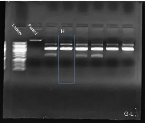
utilized NheI and AflII enzymes, and EB2 utilized PacI enzyme. Colonies A-F of EB1 and EB2
were not successfully transformed. Colonies G-L of EB1 was not successfully transformed,
however, EB2-H showed evidence, upon gel electrophoresis, of successful transformation. DNA
for sequencing was submitted to Iowa State DNA Facility and sequencing matched, proving
presence of insert within plasmid (successful transformation – Figure 7). EB2-G, I-L colonies
were all unsuccessful. Colonies M-Q, 1-10, and AA-AN of EB1 were also unsuccessful.
Fortunately, colony AO was clearly positive for successful transformation on gel electrophoresis,
and Sanger sequencing via Iowa State DNA Facility confirmed correct sequence (Figure 8).
These two colonies were midi-prepped to increase concentration of DNA to prepare for parasite
[image:9.612.184.430.506.713.2]transfection.
During the course of isolating colonies and digesting to find confirmed transformed plasmid,
several adjustments and corrections were made. The first four digests (EB1, EB2 E-F and EB1,
EB2 G-L) used the incorrect parent. The parent used was the “Hard Way” though was thought to
be the “Easy Way”. Additionally, EB1 A-F were originally digested with BseGI and AflII but
did not show positive result so chose to use NheI in place of BseGI for future digests.
Newly synthesized DNA containing desired insert (EB1-AO and EB2-H) transfected into
known plasmodium falciparum strain with blasticidin (BSD) to be observed. If EB1 parasites are
not successful and do not proliferate, “Hard Way” will be carried out to prove the presence of
exogenous EXP2 peptide is the reason for result. Transfection carried out via electroporation
with BioRad Gene Pulser II. Electroporation is the technique in which an electric current is
applied to cells to briefly open cell membranes via pores to introduce new DNA or chromosomes
into bacteria. Plasmid DNA prepared with 1X and 2X cytomix to replicate erythrocyte cytosol
condition and increase transfection chance of success. Parasites were transfected at equivalent of
~5% parasitemia of 2% hematocrit culture in ring stage. Cells suspended in media and treated
with BSD selection after 24 hours for better conditions.
This research and further research in similar capacity could be utilized therapeutically to
[image:10.612.212.402.70.305.2]produce a drug that introduces exogenous peptide to interrupt function of PTEX. Unfortunately,
this drug would only treat patients already infected with malaria. Additionally, the drug would
only be targeting Plasmodium while in erythrocyte stages, and would have no effect on any
parasite within hepatocytes, presenting a narrow and hard to track therapeutic window. Malaria
is a devastating disease and recently progress in the form of decreasing cases and deaths per year
has stagnated. Any promising research is worth studying to help rid the world of this destruction
References
1. de Koning-Ward, Tania F, Dixon, Matthew W.A., Tilley, Leann, and Gilson, Paul R. (2016). Plasmodium species: master renovators of their host cells. Nature Reviews Microbiology. (8):494-507.
2. Ganesan, S. M., Falla, A., Goldfless, S. J., Nasamu, A. S., & Niles, J. C. (2016).
Synthetic RNA-protein modules integrated with native translation mechanisms to control
gene expression in malaria parasites. Nature communications, 7:10727.
3. Garten, M, Nasamu AS, Niles JC, Zimmerberg J, Goldberg DE, Beck JR. (2016). EXP2 is a nutrient-permeable channel in the vacuolar membrane of Plasmodium and is essential for protein export via PTEX. Natural Microbiology. (10):1090-1098
4. Ho, C. M., Beck JR, Lai M, Cui Y, Goldberg DE, Egea PF, Zhou ZH. (2018). Malaria parasite translocon structure and mechanism of effector export. Nature 561(7721):70-75.
5. Mair, G. R., Braks, J. A., Garver, L. S., Wiegant, J. C., Hall, N., Dirks, R. W., … Waters,
A. P. (2006). Regulation of sexual development of Plasmodium by translational
repression. Science (New York, N.Y.), 313(5787), 667–669.
6. World Health Organization. (2018). World Malaria Report 2018. World Health