*Corresponding author:Santosh Kumar Goje ISSN: 0976-3031
Research Article
COMPARATIVE EVALUATION OF SALIVARY PARAMETERS BEFORE AND
DURING ORTHODONTIC TREATMENT
Archie Bhavsar., Santosh Kumar Goje* and Jay Patel
Department of Orthodontics, K.M. Shah Dental College and Hospital Sumandeep Vidyapeeth,
Piparia, Vadodara
DOI: http://dx.doi.org/10.24327/ijrsr.2017.0807.0541
ARTICLE INFO ABSTRACT
Background and Objectives: To evaluate and compare the salivary calcium, phosphate, amylase, total protein concentrations, flow rate, pH and buffering capacity before and during orthodontic treatment.
Methods: Salivary samples were collected from 40 individuals in the age group of 15-25 years having malocclusion and comparison of salivary calcium, phosphate, amylase, total protein concentrations, flow rate, pH and buffering capacity was done 45 days after beginning the orthodontic treatment.
Results: Salivary alpha-amylase, calcium, and phosphate concentrations were higher in patients undergoing orthodontic treatment as compared to those patients before starting treatment.
Interpretation and Conclusion: The salivary parameters such as pH, buffering capacity, flow rate were significantly reduced in patients undergoing orthodontic treatment whereas parameters such as total protein concentration, salivary calcium, phosphate and amylase were increased.
INTRODUCTION
Patients generally seek orthodontic treatment for aesthetics, health of periodontal and dental tissues, and occlusion, as well as for reasons of self esteem. In order to treat malocclusion it is of utmost importance to ascertain the changes in the oral environment during therapy1. Fixed orthodontic intervention therapy poses challenge for the patients in terms of oral hygiene maintenance leading to plaque accumulation, causing inflammation in the gingival tissues as well as demineralisation of teeth and white spot formation. Saliva is an oral fluid that can be collected by non invasive means and it has beneficial effects to the oral health by virtue of its flow, buffering capacity, pH and other various factors. 2 & 3 Individuals seeking orthodontic treatment may have altered salivary pH, buffering capacity 1 & 4 ultimately leading to variation in amylase, calcium and phosphate contents.1 Alpha- amylase of saliva cleaves alpha (1-4) glycoside linkage in starch and glycogen which helps in dissolution as well as removal of starch containing food debris that gets accumulated surrounding teeth as well as oral mucosa.5 It can further interact with several types of streptococci enhancing bacterial adhesion. Pellicle formulated on the orthodontic modules is a harbour of salivary proteins as well.6 Salivary fluid is a good source of calcium and phosphate ions which induces dissolution of
calcium hydroxyappatite which is the key inorganic component of teeth.7, 8 & 9 Such ions have a vital role for post eruptive maturation of enamel, thus allowing remineralisation of incipient carious lesion.10,11&12 As scarcity of studies relating changes in salivary parameters and demineralisation of enamel during orthodontic treatment was observed this study was planned with the null hypothesis that there is no difference between salivary parameters at baseline and 45 days after orthodontic treatment.
MATERIALS AND METHODS
This prospective observational study was planned in the Department of Orthodontics and Biochemistry. Study was started only after receiving Ethics committee approval. A total of 40 participants having an age range of 15-25 years, & willing to undergo orthodontic treatment were selected, their follow up were conducted till their next appointment which had a minimum time interval of 45 days after start of orthodontic treatment. All the participants were given participants information sheet regarding study.
Participants having any systemic or some local disease that affects secretions of saliva as well as participant who require special health care needs such as the ones who were medically indigent, participants with smoking habit, poor oral hygiene,
International Journal of
Recent Scientific
Research
International Journal of Recent Scientific Research
Vol. 8, Issue, 7, pp. 18630-18634, July, 2017
Copyright © Archie Bhavsar et al, 2017, this is an open-access article distributed under the terms of the Creative Commons Attribution License, which permits unrestricted use, distribution and reproduction in any medium, provided the original work is properly cited.
DOI: 10.24327/IJRSR CODEN: IJRSFP (USA)
Article History: Received 15th April, 2017 Received in revised form 25th May, 2017
Accepted 23rd June, 2017 Published online 28th July, 2017
Key Words:
active caries or periodontal disease, previous orthodontic treatment and those who did not wish to provide informed consent were not included in the study. All subjects were treated with a straight wire technique using MBT, 0.022-inch-slot brackets (3 M Unitek, Monrovia, California, USA) on both the maxillary and the mandibular arches. Before the beginning of treatment, all patients underwent meticulous professional oral hygiene at the Department of Periodontology. Oral hygiene instructions were given after fixed appliance placement.
Salivary Analysis
Salivary sample collection was carried out by using sterile test tubes twice, before and 45 days after starting the orthodontic treatment. The participating individuals were instructed to avoid eating/drinking anything for at least one hour before collection of saliva and were asked to rinse the mouth thoroughly 10 min before collection of saliva. All the salivary samples were collected in the morning session between 10 am to 11-30 am. Total of 4-5ml of saliva was collected and stored in the eppendorf tubes.
The pH was measured using pH meter after which the salivary sample was titrated with 0.1N HCL which determined the buffering capacity.13& 14 Also the flow rate was calculated by measuring the amount of saliva (ml) collected per minute. The lower lip was dried, and the time(s) taken for a salivary droplet to form was recorded.
The collected salivary samples were stored at 4 ºC till they were subjected to analysis. Care was taken that the analysis of the saliva sample was carried out on the same day. The entire saliva sample collected was subjected to centrifugation at 5000 rpm for 5 min. The Estimation of the parameters such as total protein, calcium, phosphates and amylase was done using auto- analyser and observations were recorded in Microsoft excel sheet which was subjected for statistical analysis.
Descriptive statistics included mean, standard deviation and Standard error mean. Values were determined for each of the test groups. T-Test for pair wise comparisons for both groups was performed.
Significance for all the statistical tests was predetermined at a p-value of 0.05 or less.
RESULTS
In the present study, equal number of male and female participants were involved, 20 each, with mean age of 17 years. Since no statistically significant differences were found between males and females, data were combined for sex.
All the salivary parameters such as pH, buffering capacity, flow rate showed statistically significant differences before and 45 days after starting fixed orthodontic treatment. Similarly statistically significant differences were found for salivary calcium, phosphate and amylase levels in both control group and study group.
DISCUSSION
Saliva performs a multiplicity of roles within the oral cavity, and like many things in life, its importance is usually not appreciated until it is absent. Saliva has an important role to protect the oral health. 10 and 15
Flushing (clearance and exposure) of the soft and hard tissues, as well as salivary constituents, such as pH-regulatory components, inorganic acids and carbohydrates, affecting microbial growth and colonization are important functions carried out by saliva. 10, 15&16 Resting/ unstimulated, stimulated whole saliva secretion and glandular salivary secretion are routinely employed methods for the collection and analysis of saliva.17 Unstimulated whole saliva mimics basal salivary flow rate and is the secretion that provides protection to oral tissues and is present for about 14 hours a day.1 Factors such as diet, gender, age as well as stress related conditions can also alter flow rate of saliva. So the method of saliva collection was chosen to be unstimulated in participants having similar dietary habits being permanent residents.
Dawes et al 18 reported wide range of flow rate for unstimulated salivary samples but it averages 0.3-0.4 ml per min. Also the evidence of hypo-salivation was considered if it is less than 0.1 ml per min.
Table 1 The mean scores, standard deviations and standard error for salivary parameters before orthodontic treatment (control group) and 45 days after orthodontic treatment (study group).
Salivary Parameters
Mean Std.
Deviation Std. Error Mean
Control Study Group Control Study Group Control Study Group
pH 7.0180 6.4261 0.23758 0.27064 0.03710 0.04227
Buffering Capacity 5.7023 5.1861 0.21202 0.16515 0.03311 0.02579
Flow rate 0.4754 0.3968 0.06201 0.06784 0.00968 0.01059
Total Protein 0.2417 0.6624 0.07553 0.10156 0.01180 0.01586
Calcium 3.6088 7.4244 0.93053 1.25756 0.14532 0.19640
Phosphate 4.2927 18.2439 2.44176 5.26679 0.38134 0.82253
Amylase 50731.70 67932.43 29426.08 32370.69 4595.58 5055.45
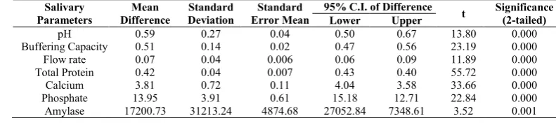
Table 2 Paired t-test for salivary parameters for study and control group.
Salivary Parameters
Mean Difference
Standard Deviation
Standard Error Mean
95% C.I. of Difference
t Significance
(2-tailed)
Lower Upper
pH 0.59 0.27 0.04 0.50 0.67 13.80 0.000
Buffering Capacity 0.51 0.14 0.02 0.47 0.56 23.19 0.000
Flow rate 0.07 0.04 0.006 0.06 0.09 11.89 0.000
Total Protein 0.42 0.04 0.007 0.43 0.40 55.72 0.000
Calcium 3.81 0.72 0.11 4.04 3.58 33.66 0.000
Phosphate 13.95 3.91 0.61 15.18 12.71 22.84 0.000
The bicarbonate system is responsible for about 85% of the buffering capacity in the pH range of 7.2 to 6.8. When the salivary flow rate increases, the concentration of bicarbonate ions also increases.18- 21
Significant decrease in salivary pH and buffering capacity was reported by Teixira et al1. And Kanaya et al 22 which was in accordance to our present study whereas Chang et al 23 reported increased buffering capacity in study group. In 1978 Neil 24 reported optimum buffering capacity range which is 3-30 mg per 100 ml. The buffering capacity is crucial for dental erosion, caries and micro biota ecology as a pH below 5.5 creates an environment of demineralisation and supports bacteria dysbiosis characterised by acidophilic and aciduric bacteria.25 and 26
This decreased pH and buffering capacity makes the tooth more prone to demineralise and caries hence preventive measures should be taken.
One important aspect of saliva is its contribution to remineralisation of the teeth as well as preventing demineralisation by buffering. 27 After each intake of food, the pH in dental plaque drops and remains at that state until soluble carbohydrates are cleared from the oral cavity, and the acid
produced from bacteria are neutralized (buffered). The magnitude and the time period below the tooth tissue,
critical pH are determined by the amount of acid produced by bacteria, by the buffer capacity of the saliva, and saliva flushing.
Helmerhost and Oppenheim28 reported 309 proteins which comprised major salivary proteins amounting to 95% acidic and basic proline rich protein along with amylase and other minor amounts of glycol protein, agglutinin, cystatine, hystatins, and statherins were also observed.
In our study mean salivary total protein was 0.247 before starting fixed orthodontic treatment while it was increased to 0.66 during orthodontic treatment. This finding was coinciding with the study of Teixira et al1 who reported increase in total protein level 0.189 in experimental group of orthodontic patients as compared to control group. Similar increase in total salivary protein level with active caries than caries free
individuals was reported by Tulunoglu et al 29 and Preethi et al.30 Salivary calcium and phosphate helps in post eruptive
maturation of enamel and aids in precipitation/ dissolution of hydroxyappatite of enamel.
In our study, there was increase in salivary calcium upto7.42 in study group who were the participants undergoing orthodontic treatment as compared to control group wherein salivary calcium was 3.60 for the participants before orthodontic treatment. This was in contradiction to the finding of Teixira et al1 who reported lower concentration of calcium ions in saliva in experimental group as compared to control group.
Increased Salivary calcium concentration may be due to demineralisation of teeth and fixed orthodontic treatment. Salivary calcium influences the precipitation or dissolution of hydroxyapatite of enamel. 31and 32
Calcium ion concentration is highly dependent on pH and salivary flow rate. It was reported that individuals who have low calcium concentration in saliva have reduced pH and hence
they are more prone to demineralisation than those having higher concentration of calcium. As reported by Jenkins, 8 as the flow rate increases the calcium ion concentration of saliva also increases. Saliva containing calcium and phosphate acts as a source of essential ion31, 33 & 34. Under such conditions, process of remineralisation predominates than demineralisation. Plaque receives calcium and phosphorous from the residual saliva, thereby initiating the cycle of caries. Phosphate ions serve to maintain salivary pH and these ions are present in the form of PO43- ions. So when the pH becomes highly acidic this ion gets converted into HPO42- and then H2PO4- thus it acts as one of many salivary buffers. So saliva is not saturated with phosphate, chances for enamel dissolution and release of phosphate ion increases.35
In our study, mean salivary phosphate was 4.29 before orthodontic treatment while it was increased to 18.24 during the orthodontic treatment. This high salivary phosphate concentration can lead to dissolution of hard tissue in the oral cavity in the presence of saliva. Thus the balance between remineralisation and demineralisation depends on salivary calcium and phosphate concentration so it is important that calcium and phosphate be saturated in saliva so as to have its effect on demineralisation and remineralisation. So when the teeth are constantly bathed in saliva, it acts as a lubricant, buffer and ion source of calcium and phosphate which are important for remineralizing initial carious lesion.
Saliva assists in digestion through amylase and also serves as a reservoir for ions (calcium, phosphorous) for remineralisation. And further through balance between pH and buffering factors influence the redistribution of ions between enamel demineralisation and remineralisation.
Salivary amylase levels in our study were raised significantly in study group 67932.43 whereas it was 50731.70 in control group. This was congruent with study of teixeria et al1 who also reported increase in salivary amylase in experimental group as compared to control group.
Amylase is the enzyme which is of salivary origin which clears carbohydrate debris. It plays a leading role in colonisation as well as metabolism of streptococci, enhancing formation of dental plaque and caries.36
Acquired pellicle contains amylase and hence is available to act as receptor for adhesion of micro-organisms to tooth surface. Patients seeking orthodontic treatment may be more prone to demineralization as the mean salivary and buffering capacity was diminished in experimental group.
Hence preventive measures such as hygiene, diet advising, topical fluoride application and prophylaxsis should be accounted.
CONCLUSION
Salivary alpha-amylase, calcium, and phosphate concentrations were higher in patients undergoing orthodontic treatment as compared to those patients before starting treatment.
However, clinical interpretation of the results obtained in the present study should be made carefully as it involved only few of the host factor components of the multifactorial process due to wearing of fixed orthodontic appliances.
This study is of importance to orthodontist as fixed orthodontic appliances are a part of routine orthodontic treatment for correction of malocclusion but at the same time, it may be associated with difficulty in maintaining good oral hygiene, and hence further may lead to tooth demineralisation. This study also evaluates the role played by a host protective factor, ‘saliva’, in the protection of teeth by maintaining balance between demineralisation and remineralisation through various ions such as calcium, phosphorous, amylase and basic salivary parameters. The salivary factors evaluated here may prove to be useful measures of dental health experience in orthodontic patients and lead to target preventive measures appropriately.
Since the proportion of the salivary contents is more important in susceptibility or resistance to tooth demineralisation, further evaluation of factors related to immunity of host will be of at most importance for salivary parameters evaluations in participants undergoing orthodontic treatment.
Summary
The present study was conducted on 80 salivary samples of 40 participants, with 40 saliva samples before starting orthodontic-treatment and 40 saliva samples, 45 days after starting fixed orthodontic appliance treatment. The participants were aged between 20-30 years. The main objective of this study was to evaluate and compare the salivary calcium, phosphate, amylase, total protein concentrations, flow rate, pH and buffering capacity before and during Orthodontic treatment with fixed appliances.
Salivary parameters
1. Mean salivary pH, buffering capacity and salivary flow rate were reduced in participants who were undergoing fixed orthodontic appliance treatment.
2. Salivary total proteins, calcium, phosphates and amylase levels were raised in the participants who were undergoing fixed orthodontic appliance treatment as compared to the same participants before starting orthodontic-treatment. 3. Paired t-test for all the salivary parameters was found to be
statistically highly significant when compared before and during orthodontic treatment. (p<0.001)
References
1. Teixeira HS, Kaulfuss SMO, Ribeiro JS, Pereira BR, Brancher JA, Camargo ES. Calcium, amylase, glucose, total protein concentrations, flow rate, ph and buffering capacity of saliva in patients undergoing orthodontic treatment with fixed appliances. Dental Press J Orthod. 2012; 17(2):157-61.
2. Jingyi Liu, Yixiang Duan. Saliva: A potential media for disease diagnostics and monitoring. Oral oncol 2012, 48: 569-577
3. 3. Sausan Al Kawas, Zubaidah H.A. Rahim, David B. Ferguson. Review: Potential uses of human salivary protein and peptide analysis in the diagnosis of disease. Archives of oral biology 2012 (57); 1-9
4. Steinberg D and Eyal S. Initial bioilm formation of streptococcus sobrinus on various orthodontic appliances. Journal of Oral Rehabilitation 2004(31): 1041-45.
5. Jacobsen K, Lyche Melvaer K, Hensten-Pettersen A. Some properties of salivary amylase: A survey of the literature and some observations. J Dent Res 1972, 51 (2): 381-88.
6. Scannapieco FA, Torres GI, Levine MJ. Salivary amylase promotes adhesion of oral streptococci to hydroxyapatite. J Dent Res.1995; 74: 1360-66.
7. Shafer WG, Hine MK, Levy BM. A text book of Oral Pathology. 6th Ed. Rajendran and Sivapathasundharam, Elsevier 2009, 409-473.
8. Jenkins GN. Physiology and Biochemistry of the Mouth, 4th ed. Oxford: Blackwell Scientific Pub. 1978, 284-359. 9. Nikiforuk G. Understanding dental caries - Etiology and mechanisms, Basic and Clinical Aspects. Vol. 1: 1st Ed. Newyork: Karger 1985, 236-260.
10. Anderson P, Hector MP, Rampersad MA. Critical pH in resting and stimulated whole saliva in groups of children and adults. Int J Pediatr Dent 200, 11: 266-273
11. Larsen M.J., Pearce E.I.F. Saturation of human saliva with respect to calcium salts. Archives of Oral Biology 2003; 48: 317-22
12. Prabhakar A.R., Shubha A.B, Mahantesh T. Estimation of calcium, phosphate and alpha-amylase concentrations in stimulated whole saliva of children with caries status: A Comparative study. Malaysian Dent J 2008; 29(1):6- 13. Birkhed D, Heintze U. Salivary secretion rate, buffer capacity and pH. In: Tenovuo JO. Human saliva: clinical chemistry and microbiology. Boca Raton: CRC Press; 1989. v. 1, p. 25-74.
14. Newbrun E. Preventing dental caries: current and prospective strategies. J Am Dent Assoc. 1992; 123(5):68-73.
15. 15. Silvia Chiappin, Giorgia Antonellia, Rosalba Gatti, Elio. De Palo. Invited critical review: Saliva specimen: A new laboratory tool for diagnostic and basic investigation. Clinica Chimica Acta 2007; (383): 30-40. 16. Edgar M, Dawes C, O’Mullane D (eds.). Saliva and Oral
Health. 3rd ed. London: British Dental Association. 2004.
17. Fenoll-Palomares C, Muñoz-Montagud JV, Sanchiz V, Herreros B, Hernández V, Mínguez M, Benages A. Unstimulated Salivary flow rate, pH, and buffer capacity of saliva in healthy Volunteers. Rev Esp Enferm Dig 2004; 96: 773-783.
18. Dawes C. Salivary flow patterns and the health of hard and soft oral tissues. J Am Dent Assoc.2008; 139:172-90.
19. Dowd FJ. Saliva and dental caries. Dent Clin North Am. 1999; 43(4):579-97.
20. Edgar WM. Saliva: its secretion, composition and functions. Br Dent J.1992; 172(8):305-12.
21. Ericson T, Mäkinen KK. Saliva-formation, composition and possible role. In: Thylstrup A, Fejerskov O. Textbook of cariology. Copenhagen: Munksgaard; 1986. p. 28-45
and microbial flora with the placement of Edgewise appliance. Orthod Waves; 2007; 66(2):27-32.
23. Chang HG, Walsh LJ, Freer TJ. The effect of orthodontic treatment on salivary flow, pH, buffer capacity, and levels of mutans streptococci and lactobacilli. Aust Orthod J. 1999; 15(4):229-63. 24. Neil JG. J Oral Pathol Med. 4th ed. 1978. The
Physiology and Biochemistry of the Mouth; pp. 284-359.
25. Schuurs A. Pathology of the hard dental tissues. West Sussex: Wiley-Blackwell: 2013
26. Marsh PD. Microbiology of dental plaque biofilms and their role in oral health and Caries. Dent Clin North Am 2010:54:441-454
27. Selwitz RH, Ismail AI, Pitts NB. Dental caries. Lancet 2007:369:51-59
28. Helmerhorst EJ, Oppenheim FG. Saliva: A dynamic Proteome. J Dent Res. 25 2007; 86:680-693
29. Tulunoglu O, Demirtas S, Tulunoglu I. Total antioxidant Levels of saliva in children related to caries, age, and Gender. Int J Paediatr Dent. 2006;16:186
30. Preethi BP, Reshma D, Anand P (2010). Evaluation of flow rate, pH, buffering capacity, calcium, total proteins and total antioxidant capacity levels of saliva in caries free and caries active children: An in vivo study. Indian J. Clin. Biochem. 25(4):425-428.
31. Shahrabi M.A, Nikfarjam J.A, Alikhani A.A, Akhoundi N.B, Ashtiani M., Seraj B. A comparison of salivary calcium, phosphate, and alkaline phosphatise in children with severe, moderate caries, and caries free in Tehran’s kindergartens. Indian Soc Pedod Prevent Dent 2008; 74-77
32. Karshan M. Factors in saliva correlated with dental caries. J Dent Res 1939; 18:395-407.
33. Ashley FP, Wilson RF. The relationship between calcium and human saliva and dental plaque. Archs Oral Boil 1978; 23:69-73.
34. Shaw L, Murray JJ, Burchell CK, Best JS. Calcium and phosphorus content of plaque and saliva in relation to dental caries. Caries Res 1983, 17: 543-548.
35. Gandhy M, Damle SG. Relation of salivary inorganic phosphorus and alkaline phosphatase to the dental caries status in children. J Indian Soc Pedod Prev Dent 2003, 21: 135-138.
36. Jacobsen K, Lyche Melvaer K, Hensten-Pettersen A. Some properties of salivary amylase: A survey of the literature and some observations. J Dent Res 1972, 51(2): 381-388